Hair-bearing thin skin
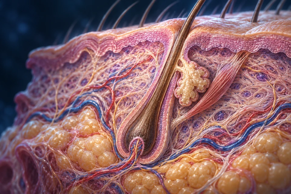
Most body regions have a thinner epidermis and may carry hair follicles and associated sebaceous glands. The main block above represents this broad organisation.
Basal → spinous → granular → cornified regions
CHAPTER / A LIVING INTERFACE
A protective surface. A living organ.
Renewing cells, connective support, vessels, nerves and appendages link protection with sensation and heat exchange. Follow the surface into its living structures.
01 / SURFACE → SUPPORT → UNDERLYING TISSUE
Skin proper includes epidermis and dermis. The underlying hypodermis connects it to deeper tissues. A hair canal, sweat duct and blood vessel are separate routes, not interchangeable openings.

Surface → renewing epithelium · The avascular epidermis limits water loss and unwanted entry. Living cells below continually supply the surface barrier.
Gold outlines locate a teaching context; they do not measure receptor activity, blood flow or sweat rate. Short names remain visible before selection.
The epidermal barrier is avascular. Dermal circulation supports its nutrition across tissue spaces while also serving the follicle, glands and connective tissue. The lower fat-rich region provides context rather than a third layer of skin proper.
Teaching references: NCBI · Normal integument anatomy · NCBI · Dermal connective tissue · OpenStax5.1 · Skin layers and selected cell roles
02 / ONE ORGAN, REGIONAL VARIATION
The same general compartments vary by body site. A fingertip should not be illustrated by adding a dense crop of follicles to thick skin.
Most body regions have a thinner epidermis and may carry hair follicles and associated sebaceous glands. The main block above represents this broad organisation.
Basal → spinous → granular → cornified regions
Thick skin has a much more substantial cornified layer, an additional clear stratum lucidum and no hair follicles or sebaceous glands. Eccrine glands remain important.
A different surface architecture, not just enlarged hair-bearing skin
“Thick” does not mean every deeper tissue is thicker. Ridges and papillae also vary; the illustrations are not thickness measurements or an exact map of a particular person.
Teaching references: NCBI · Epidermal organisation · OpenStax5.2 · Hair, nails and glands · Duke · Identified human thin-skin and fingertip H&E records
03 / TISSUE → LAYER → CELL
The basal region supports renewal. Above it, viable keratinocytes mature before becoming flattened cornified cells. A capillary below the boundary does not make the epidermis vascular.
Flattened cornified cells and surrounding lipids form the outer barrier.
Corneocytes are terminally differentiated rather than living nucleated surface cells. The artwork does not independently resolve real lipid lamellae.
Renewal begins near the epithelial base.
Basal keratinocytes contribute new cells. Other selected cell roles are explained separately; pigment does not identify every cell subtype.
Blood remains in the dermal compartment.
The epithelial cells above are nourished across tissue spaces. This tiny channel is not a vessel running within the stratum basale.
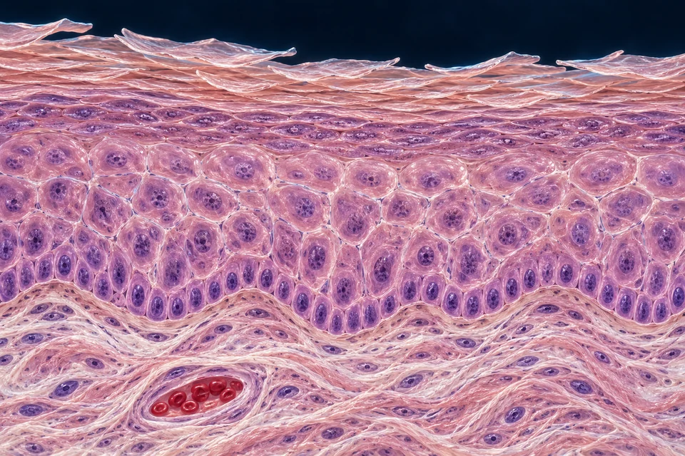
Upper viable cells change as they approach cornification.
Granular cells participate in barrier formation. Coloured granules are illustrative, not a molecular identification or measured layer thickness.
Keratinocytes remain connected as they mature above the base.
The spinous region lies between basal and granular layers. Cell junctions help tissue cohesion; no individual generated junction is validated as a real desmosome.
A supporting boundary separates epithelium from connective tissue.
The pale rim locates the basement-membrane region at teaching scale; its exact layers and dimensions are not independently resolved.
The expanded plate shows broad thin-skin layer order. Its granular transition and basal boundary are locators, not a promise that each generated granule, junction or cell is a faithfully measured histological feature.
Teaching references: NCBI · Epidermal organisation · NCBI · Cornified barrier structure · Duke · Identified human thin-skin and fingertip H&E records
04 / CONTINUOUS RENEWAL
New cell production, differentiation and shedding overlap across the tissue. Highlight a step to follow the process; the complete explanation remains visible.
Basal keratinocytes → new epithelial cells
New cells contribute to the viable epithelium. Not every basal cell divides at the same moment.
Upward progression → changing proteins and barrier materials
Keratinocytes differentiate through viable layers. This is not passive stacking of identical spheres.
Terminal differentiation → flattened corneocytes + extracellular lipids
A resilient surface emerges through cellular transformation, not a layer of living red-cell-fed plates.
Surface cohesion changes → release of superficial corneocytes
Renewal continues beneath ongoing shedding. Rates differ by site and context; no universal cycle length is assigned.
Supporting surface-renewal sequence, not exact cell lineage tracking or a measured time course. All steps are visible before highlighting.
Teaching references: NCBI · Epidermal organisation · NCBI · Cornified barrier structure
05 / SELECTED CELL ROLES
Location and function help distinguish selected populations. Molecular identity cannot be assigned from the colour of an AI-generated cell.
Across the epidermis
Build epithelial structure and change as they move toward a cornified barrier.
A surface corneocyte is not the same living state as a basal keratinocyte.
Basal epidermal neighbourhood
Make melanin and transfer pigment-containing material to neighbouring keratinocytes.
Pigment in a keratinocyte does not make it a melanocyte.
Selected viable epidermal regions
Contribute to immune surveillance and antigen handling.
No generated pale cell is assigned this identity by colour alone.
Selected basal epidermal sites
Participate in sustained fine-touch information.
A Merkel complex is not an encapsulated dermal corpuscle.
Connective tissue below the epidermis
Produce and maintain extracellular matrix.
They are not the epithelial cells that form the nail or hair shaft.
Teaching references: OpenStax5.1 · Skin layers and selected cell roles · NCBI · Dermal connective tissue · University of Leeds · Melanocytes and pigment transfer
06 / A SELECTIVE INTERFACE
Flattened corneocytes, extracellular lipids and deeper viable epithelial junctions contribute different kinds of protection. Surface organisms and local immune activity add further context.
Keratin-rich surface cells provide resilient material. Their compact arrangement is more than a decorative stack of tissue-coloured tiles.
Extracellular lipids help limit water escape and unwanted penetration. Fine lipid sheets are not independently resolved in the generated plate.
Junctions in viable layers and local immune mechanisms complement the cornified barrier. Normal skin still permits water loss and selective exchange.
Sweat ducts and follicles are specialised routes through the surface; they do not mean that all external substances freely enter the circulation. This is normal physiology, not a skin-treatment or product claim.
Teaching references: NCBI · Cornified barrier structure · NLM · Epidermal barrier overview · OpenStax5.3 · Normal skin functions
Each explanation remains available below. Opening a question selects its anatomical context above.
07 / PIGMENT PRODUCTION AND TRANSFER
Melanocytes produce melanin-containing material that reaches neighbouring keratinocytes. Its amount, type, distribution and handling contribute to visible skin colour.
A melanocyte produces melanin within specialised intracellular organelles.
Pigment-containing material passes to nearby keratinocytes; a pigment-bearing cell need not be the pigment-producing cell.
Intracellular placement can help shield nuclear material. The illustration does not verify each tiny brown packet.
Turnover and pigment handling affect appearance over time. Skin colour cannot be reduced to rigid racial categories or one universal cell-count difference.
Supporting transfer sequence, not a molecular animation. No promise of complete UV protection or advice to increase sun exposure.
Teaching references: University of Leeds · Melanocytes and pigment transfer · OpenStax5.1 · Skin layers and selected cell roles
08 / CONNECTIVE SUPPORT AND CIRCULATION
Superficial papillary tissue supports the epithelial boundary; deeper reticular tissue contains substantial collagenous and elastic support. Vessels, nerves and appendages share this environment without becoming one common channel.
Fibroblasts maintain connective material. Collagen resists tension while elastic components help recoil; real fibre orientation is more complex than the fine generated texture.
Blood supplies tissue and exchanges heat. Lymphatic pathways return interstitial fluid and participate in immune transport; they are distinct from sweat ducts and hair canals.
Connect with circulation →Teaching references: NCBI · Dermal connective tissue · NCBI · Normal integument anatomy · NCBI · Sweat gland organisation
09 / AN APPENDAGE WITH A LIVING ROOT
An active matrix surrounds the connective papilla at the bulb. Epithelial sheaths, a sebaceous connection and an oblique smooth muscle belong to the wider follicular unit.
Nested epithelial tissue surrounds the developing hair.
Inner and outer sheaths have distinct roles. This broad locator does not label each microscopic sheath layer or claim that the sheath alone makes the hair shaft.
Living matrix cells around the papilla produce the developing hair.
The enlarged lower end is an active growth compartment. The artwork locates the region, not exact proliferative or melanocyte boundaries.
Connective tissue enters the bulb from below.
This is a follicular papilla, distinct from the dermal papillae along the epidermal boundary. Local support and signalling influence the surrounding matrix.
Vessels support living tissue outside the keratinized shaft.
Blood can supply the follicular surroundings and papilla. Individual generated branches and loops are not a verified vascular map.

Compact keratinized material extends toward and beyond the surface.
Its exposed length consists of non-living cells; growth is supplied by the follicle below. The shaft does not contain a blood supply.
Lobulated cells release sebum into the upper follicular canal.
Sebaceous secretion differs from the separate eccrine sweat route. Fine ducts are illustrative; the main visible joining relationship is the teaching scope.
A small smooth-muscle band links follicle and superficial dermis.
Contraction changes hair position. In humans, raised hairs offer limited insulation compared with a dense fur coat; this is not the shivering muscle system.
Hair colour involves pigment incorporated during production. Cutting an exposed shaft does not cut a nerve inside it. Follicles are living structures even when shaft production changes during the cycle.
Teaching references: NCBI · Normal hair organisation and cycling · OpenStax5.2 · Hair, nails and glands
10 / GROWTH, REGRESSION AND RELEASE
The realistic plate shows one active-growth context. These supporting comparisons explain changing activity without presenting a single frozen picture as every phase.
Active matrix production → shaft elongation
The lower follicle supports ongoing hair formation. A long shaft alone does not reveal its current microscopic state.
Production reduces → lower follicle remodels
A brief transition changes the growing lower compartment. The whole skin does not disappear or become an empty hole.
Reduced shaft production → retained club hair
The follicle remains a living unit. Rest does not mean every surrounding cell is dead or inactive.
Old hair can be shed as another cycle develops
Exogen describes release; it can overlap other cycle events. This is not a rigid fourth clock stage that always waits for all other events.
Supporting cycle comparison beside the realistic follicle plate. Different follicles are not synchronized; scalp and body sites differ. No universal phase duration or daily shedding quota.
LIVING FOLLICLE / CHANGING PRODUCTION / DURABLE FIBRE
A strand of hair is the product, not the whole living structure. Explore the tissues that remain, the lower production region that changes, and the cells that form fibre and its surrounding sheath.
The upper epithelial framework and renewal context persist while the lower production compartment changes. Matrix is epithelial; the dermal papilla is separate connective tissue.

The full comparison’s bracket locates approximate upper renewal context, not a sharp stem-cell boundary. The three preparations compare tissue organization, not an exact time sequence or identical individual cells. Cropped views enlarge unchanged windows of the same original plates. Use the detail control to inspect the tissue boundaries.
Look closely: follow the outer epithelium, then locate the lower matrix and the papilla. Which is epithelial, which is mesenchymal, and which production compartment is absent in the resting comparison?
Evidence: Ohyama M et al. Characterization and isolation of stem cell-enriched human hair follicle bulge cells. Journal of Clinical Investigation. 2006;116:249–260. · Hawkshaw NJ, Hardman JA, Alam M, Jimenez F, Paus R. Deciphering the molecular morphology of the human hair cycle: Wnt signalling during the telogen–anagen transformation. British Journal of Dermatology. 2020;182:1184–1193. · Wu S et al. Single-cell transcriptomics reveals lineage trajectory of human scalp hair follicle and informs mechanisms of hair graying. Cell Discovery. 2022;8:49. · Zorn-Kruppa M et al. Tight junction barriers in human hair follicles: role of claudin-1. Scientific Reports. 2018;8:12800. · Hsu YC, Pasolli HA, Fuchs E. Dynamics between stem cells niche and progeny in the hair follicle. Cell. 2011;144:92–105. · Panteleyev AA. Functional anatomy of the hair follicle: the secondary hair germ. Experimental Dermatology. 2018;27:701–720. · University of Leeds Histology Guide. Skin: Hair.
Read this explanation and its limits ↓The upper epithelial framework and renewal context persist while the lower production compartment changes. Matrix is epithelial; the dermal papilla is separate connective tissue.
A hair follicle is living tissue that produces a largely non-living fibre. The visible shaft is therefore not the whole follicle. During growth, dividing epithelial cells in the lower bulb form a matrix around a separate connective-tissue dermal papilla. The papilla is mesenchymal tissue, not a cluster of hair-making epithelial cells. These tissues interact across a boundary; papilla cells do not turn into the shaft.
The upper follicle provides a more persistent epithelial framework. A renewal-cell-rich region is associated with the bulge, near the attachment of the arrector pili muscle below the sebaceous gland. In human scalp it is not necessarily a conspicuous bulging lump. Its cell markers and extent should not be drawn as one universal, sharply bounded patch.
In a resting follicle, the epithelial region between the retained club and papilla is often called the secondary hair germ. It is not a mature anagen matrix. Human tissue studies identify this region and changing activity within it, but do not establish every lineage relationship demonstrated in mice. “Persistent” describes continuity of the renewal system, not a promise that every individual cell remains unchanged forever.
Evidence: Ohyama M et al. Characterization and isolation of stem cell-enriched human hair follicle bulge cells. Journal of Clinical Investigation. 2006;116:249–260. · Hawkshaw NJ, Hardman JA, Alam M, Jimenez F, Paus R. Deciphering the molecular morphology of the human hair cycle: Wnt signalling during the telogen–anagen transformation. British Journal of Dermatology. 2020;182:1184–1193. · Wu S et al. Single-cell transcriptomics reveals lineage trajectory of human scalp hair follicle and informs mechanisms of hair graying. Cell Discovery. 2022;8:49. · Zorn-Kruppa M et al. Tight junction barriers in human hair follicles: role of claudin-1. Scientific Reports. 2018;8:12800. · Hsu YC, Pasolli HA, Fuchs E. Dynamics between stem cells niche and progeny in the hair follicle. Cell. 2011;144:92–105. · Panteleyev AA. Functional anatomy of the hair follicle: the secondary hair germ. Experimental Dermatology. 2018;27:701–720. · University of Leeds Histology Guide. Skin: Hair.
Renewed growth depends on communication between epithelial cells and their surroundings. Human observations and mouse experiments provide different kinds of evidence, not one universal switch.
Renewal depends on communication between epithelial cells and their surroundings, including the papilla. A resting structure becomes an actively growing lower follicle; it is not simply an old shaft switched back on.
The evidence comes in different forms. In human scalp sections, researchers can compare where signalling-related genes are expressed in resting and early-growing follicles. These observations implicate changes in WNT-related communication, but an expression pattern alone cannot prove the first cause of restart.
Mouse experiments go further: researchers have tracked activation in the hair germ and bulge, and tested papilla involvement by targeted interventions. These experiments support coordinated epithelial–mesenchymal control. They do not establish one universal molecular switch, an identical sequence in every human follicle, or a treatment recipe.
Evidence: Hawkshaw NJ, Hardman JA, Alam M, Jimenez F, Paus R. Deciphering the molecular morphology of the human hair cycle: Wnt signalling during the telogen–anagen transformation. British Journal of Dermatology. 2020;182:1184–1193. · Greco V et al. A two-step mechanism for stem cell activation during hair regeneration. Cell Stem Cell. 2009;4:155–169. · Rompolas P et al. Live imaging of stem cell and progeny behaviour in physiological hair-follicle regeneration. Nature. 2012;487:496–499.
Matrix descendants specialize into the shaft and its inner sheath. The fibre becomes keratin-rich while the sheath remains a distinct structure around it.
The matrix amplifies the supply of epithelial cells during growth. As descendants leave the proliferative region, they specialize into different structures. The shaft and its surrounding inner root sheath are related outputs, not the same tube under two names. The outer root sheath is the more external epithelial compartment.
Most of the shaft is cortex, covered by a thin cuticle. A central medulla may be present, discontinuous or absent; a hollow or cellular centre is not compulsory. The inner root sheath surrounds the developing shaft within the follicle and does not accompany it as a full-length external coat. Its organization is distinct from the shaft cuticle.
Maturing shaft cells accumulate structural proteins, including hair keratins and keratin-associated proteins. Their internal organization changes as a resistant fibre forms. Nuclei and organelles are dismantled during this maturation, but they do not all disappear at one instantaneous line. The exposed hair is not living tissue with a blood supply. This specialized differentiation is not merely identical cells being pushed upward, and it is not interchangeable with making interfollicular epidermal corneocytes.
Pigment can be incorporated while the shaft is being formed; pigment-cell biology is not the subject of this course.
Evidence: Wu S et al. Single-cell transcriptomics reveals lineage trajectory of human scalp hair follicle and informs mechanisms of hair graying. Cell Discovery. 2022;8:49. · Langbein L et al. The catalog of human hair keratins II: expression of the six type II members in the hair follicle and the combined catalog of human type I and II keratins. Journal of Biological Chemistry. 2001;276:35123–35132. · Langbein L et al. K6irs1 K6irs2 K6irs3 and K6irs4 represent the inner-root-sheath-specific type II epithelial keratins of the human hair follicle. Journal of Investigative Dermatology. 2003;120:512–522. · Jones LA, Harland DP, Jarrold BB, Connolly JE, Davis MG. The walking dead: sequential nuclear and organelle destruction during hair development. British Journal of Dermatology. 2018;178:1341–1352. · Alibardi L. Ultrastructural localization of hair keratins, high sulfur keratin-associated proteins and sulfhydryl oxidase in the human hair. Anatomical Science International. 2017;92:248–261. · van den Berg C, Khumalo NP, Ngoepe MN. Quantifying whole human hair scalp fibres of varying curl: A micro-computed tomographic study. Journal of Microscopy. 2025;297:227–251; online 2024. · University of Leeds Histology Guide. Skin: Hair.
No. Much of the temporary lower epithelial production structure regresses, while living renewal structures and the papilla remain.
During catagen, productive activity in the lower follicle ends and much of its transient epithelial structure is removed or remodelled. Human studies detect programmed cell death in lower matrix and outer-sheath regions. That does not mean every outer-sheath cell dies, or that the entire follicle disappears.
The lower follicle shortens while the upper epithelial framework, renewal capacity and dermal papilla remain available for another cycle. The papilla also changes shape and position relative to the shortening epithelium. The old shaft becomes a retained club rather than remaining attached to a functioning, full-sized production bulb.
Mouse lineage experiments show that some epithelial descendants survive regression and contribute to later renewal compartments. They help explain how a system can rebuild without preserving every cell, but the exact mouse cell history must not be presented as a directly observed human lineage map.
Catagen regression is different from shaft keratinization: one remodels a temporary living production structure; the other makes the durable fibre. Neither should be depicted as an injury or explained simply as blood supply being switched off.
Evidence: Jones LA, Harland DP, Jarrold BB, Connolly JE, Davis MG. The walking dead: sequential nuclear and organelle destruction during hair development. British Journal of Dermatology. 2018;178:1341–1352. · Soma T et al. Analysis of apoptotic cell death in human hair follicles in vivo and in vitro. Journal of Investigative Dermatology. 1998;111:948–954. · Van Neste D, Leroy T, Conil S. Exogen hair characterization in human scalp. Skin Research and Technology. 2007;13:436–443. · Zorn-Kruppa M et al. Tight junction barriers in human hair follicles: role of claudin-1. Scientific Reports. 2018;8:12800. · Hsu YC, Pasolli HA, Fuchs E. Dynamics between stem cells niche and progeny in the hair follicle. Cell. 2011;144:92–105.
Yes. A retained club is old fibre held by living epithelium. Resting, renewed production and release are related but different events.
A club hair is an old keratinized fibre with a club-shaped root end. It is not a living matrix, a dermal papilla, or a whole follicle pulled out of the skin. Living epithelium can surround and retain it after its production has ended.
Telogen describes a resting state of the follicle; exogen describes release of the old fibre. Those are related but different events. Retention can overlap with renewed production, as the existing P0041 comparison shows. Do not impose a compulsory “shed first, then restart” sequence or claim that the new hair always mechanically pushes the old one out.
Human studies distinguish firmly retained clubs from loosely retained or shed fibres. Protease activity around club roots is one investigated part of release, not a complete, established timer for every human follicle. This evidence does not justify advice about products or how to extract hairs.
Evidence: Van Neste D, Leroy T, Conil S. Exogen hair characterization in human scalp. Skin Research and Technology. 2007;13:436–443. · Bhogal RK, Mouser PE, Higgins CA, Turner GA. Protease activity localization and inhibition in the human hair follicle. International Journal of Cosmetic Science. 2014;36:46–53. · Zorn-Kruppa M et al. Tight junction barriers in human hair follicles: role of claudin-1. Scientific Reports. 2018;8:12800.
Living renewal structures support a new lower production compartment. Its epithelial output forms fibre, and selective regression later reduces that compartment without erasing the whole system.
The upper follicle preserves a renewal context. Epithelial–mesenchymal communication helps establish renewed lower growth. The matrix generates differentiating epithelial outputs: the shaft and its inner sheath. Shaft cells mature into resistant fibre. Later, the transient lower production compartment regresses while living renewal structures and papilla remain. An old club can be retained, a new production cycle can begin, and release occurs with variable timing.
This is a connected mechanism, not a fixed clock. Use two questions throughout: Which compartment is changing? What does the arrow represent? A tissue boundary, a cell-descendant relationship, a signalling interaction and an old fibre leaving the canal are four different things. The existing cycle summaries orient the learner; the two new detailed views should make those distinctions visible.
Evidence: Ohyama M et al. Characterization and isolation of stem cell-enriched human hair follicle bulge cells. Journal of Clinical Investigation. 2006;116:249–260. · Hawkshaw NJ, Hardman JA, Alam M, Jimenez F, Paus R. Deciphering the molecular morphology of the human hair cycle: Wnt signalling during the telogen–anagen transformation. British Journal of Dermatology. 2020;182:1184–1193. · Wu S et al. Single-cell transcriptomics reveals lineage trajectory of human scalp hair follicle and informs mechanisms of hair graying. Cell Discovery. 2022;8:49. · Langbein L et al. The catalog of human hair keratins II: expression of the six type II members in the hair follicle and the combined catalog of human type I and II keratins. Journal of Biological Chemistry. 2001;276:35123–35132. · Langbein L et al. K6irs1 K6irs2 K6irs3 and K6irs4 represent the inner-root-sheath-specific type II epithelial keratins of the human hair follicle. Journal of Investigative Dermatology. 2003;120:512–522. · Jones LA, Harland DP, Jarrold BB, Connolly JE, Davis MG. The walking dead: sequential nuclear and organelle destruction during hair development. British Journal of Dermatology. 2018;178:1341–1352. · Alibardi L. Ultrastructural localization of hair keratins, high sulfur keratin-associated proteins and sulfhydryl oxidase in the human hair. Anatomical Science International. 2017;92:248–261. · Soma T et al. Analysis of apoptotic cell death in human hair follicles in vivo and in vitro. Journal of Investigative Dermatology. 1998;111:948–954. · Van Neste D, Leroy T, Conil S. Exogen hair characterization in human scalp. Skin Research and Technology. 2007;13:436–443. · Zorn-Kruppa M et al. Tight junction barriers in human hair follicles: role of claudin-1. Scientific Reports. 2018;8:12800. · Greco V et al. A two-step mechanism for stem cell activation during hair regeneration. Cell Stem Cell. 2009;4:155–169. · Rompolas P et al. Live imaging of stem cell and progeny behaviour in physiological hair-follicle regeneration. Nature. 2012;487:496–499. · Hsu YC, Pasolli HA, Fuchs E. Dynamics between stem cells niche and progeny in the hair follicle. Cell. 2011;144:92–105.
None. This removes fibre, not the matrix or papilla. The visible shaft is not the whole follicle.
No. Papilla and matrix belong to different tissue compartments. Communication is not lineage conversion.
Human niche morphology and marker distributions are not one universal polygon; marker enrichment alone is not direct proof of each cell's in-vivo fate.
No. It is human observational molecular evidence. Mouse interventions provide a different, species-bounded kind of evidence.
No. They are distinguishable differentiated outputs. In the new production view, follow each independently.
No. Absence or discontinuity is compatible with normal shaft structure.
No. It selectively regresses a transient lower structure while a renewal system remains.
No. They occur in different contexts and have different tissue outcomes.
No. Retention and renewed production can overlap. P0041 already illustrates this distinction.
No. Label the model and the inference; do not convert it into a human observation.
Source checks: 23 October 2026. Human observations, cultured human tissue, human scalp xenografts and mouse experiments are distinguished. An unread correction to the 2016 cycling guide prevents using that paper alone for new fine substage claims; independent sources support this bounded course. No concrete error in the retained poster was established. No physician review or exhaustive publication-integrity certification is claimed.
Evidence: Primary human tissue characterization with xenograft and culture components. Human anagen scalp; some label-retention localization used human scalp xenografts on nude mice.
Actual access: Publisher full text read.
Permitted use: Human renewal-cell-enriched upper outer-sheath region; human morphology differs from a conspicuous mouse bulge.
Limits: Study-specific regional localization is not a universal human boundary. Xenograft label retention is not untouched in-situ scalp observation. Enrichment or colony formation is not proof of every cell’s in-vivo stemness. Do not import mouse CD34 localization.
Integrity: No correction or retraction was identified in the inspected material and limited targeted searches. This is not an exhaustive integrity clearance.
Evidence: Primary morphology guide with human in-situ and xenograft evidence. Human scalp biopsies and human scalp xenografts on mice, explicitly distinguished.
Actual access: Full manuscript PDF text read. Direct PMC returned a browser check; some alternative repository links were inaccessible. Source-figure pixel inspection is not claimed.
Permitted use: Context for existing cycle anatomy only when corroborated independently.
Limits: Do not uniquely qualify new fine substages, precise boundaries, proportions or timing from this source while the correction is unread. Early-anagen xenograft morphology is not automatically unperturbed human scalp morphology.
Integrity: KNOWN ERRATUM H02E, PMID 27140062 / DOI 10.1016/j.jid.2016.02.023. Existence verified; correction content unread. Not presumed cosmetic. No concrete retained-poster error established.
Evidence: Published correction notice. Not applicable.
Actual access: Existence and bibliographic identity verified through indexed PubMed record; correction text not accessed.
Permitted use: Integrity flag only.
Limits: The correction text could not be accessed; its substance remains unknown. The original guide is not used alone to qualify new fine substage anatomy.
Integrity: UNRESOLVED CONTENT. Do not infer what was corrected or treat the original as fully cleared.
Evidence: Primary human molecular morphology. Human telogen and early-anagen scalp follicles; in-situ expression localization.
Actual access: Publisher and institutional abstracts read. Author-affiliated manuscript indexed excerpts supported details; full manuscript access did not succeed.
Permitted use: Human stage-associated expression evidence in epithelial and mesenchymal regions, including the secondary-germ context.
Limits: Association is not intervention or proof of the first trigger. No universal switch, exact cell lineage, fixed timing or therapeutic efficacy conclusion. R01 predates this human work.
Integrity: No correction or retraction was identified in the inspected material and limited targeted searches. This is not an exhaustive integrity clearance.
Evidence: Primary human single-cell and immunofluorescence study. Human discarded scalp tissue and isolated predominantly anagen follicles.
Actual access: Publisher full text read; direct PMC access was unavailable.
Permitted use: Distinct matrix, shaft and inner/outer-sheath identities; human structural differentiation context.
Limits: Pseudotime is inferred, not live lineage tracing. Does not resolve all human phases or prove a universal seven-lineage map. Companion-layer interpretation must not be made categorical from one dataset. Graying and interventions excluded.
Integrity: No correction or retraction was identified in the inspected material and limited targeted searches. This is not an exhaustive integrity clearance.
Evidence: Primary human spatial protein/gene localization. Human hair follicles studied with in-situ hybridization and immunohistochemistry.
Actual access: PubMed abstract read; full article not accessed.
Permitted use: Distinct early matrix/cuticle and later cortex differentiation patterns.
Limits: Do not recreate exact molecular maps or translate legacy nomenclature into unverified marker labels. Abstract supports broad spatial differentiation, not every microscopic cell identity.
Integrity: No correction or retraction was identified in the inspected material and limited targeted searches. This is not an exhaustive integrity clearance.
Evidence: Primary human localization study. Human follicular inner root sheath; in-situ hybridization and immunofluorescence.
Actual access: Indexed primary PubMed abstract inspected; direct page yielded only footer content. Full paper not accessed.
Permitted use: IRS is differentiated epithelium distinct from shaft; supports distinct sheath structure.
Limits: Do not derive exact cell counts, precise full-layer boundaries or present legacy keratin names as a required learner taxonomy. Sub-layer anchors require actual visual resolution and further qualification.
Integrity: No correction or retraction was identified in the inspected material and limited targeted searches. This is not an exhaustive integrity clearance.
Evidence: Primary human microscopy and maturation study. Healthy human scalp biopsy, follicular-unit extraction and plucked-follicle samples, including ultrastructure.
Actual access: Publisher full Methods, Results and Discussion read.
Permitted use: Maturation entails structural protein accumulation and sequential loss of living machinery, not one simultaneous organelle-loss line.
Limits: Small specimen set; no universal distance/time scale. Proposed ROS and BNIP3L mechanisms are not all established causal events. Do not repeat sensational title metaphors or equate shaft maturation with catagen apoptosis.
Integrity: No correction or retraction was identified in the inspected material and limited targeted searches. This is not an exhaustive integrity clearance.
Evidence: Primary human ultrastructural localization. Human developing hair shaft.
Actual access: Publisher and PubMed abstracts read; subscription full text not accessed.
Permitted use: Keratin-associated material contributes to compact shaft structure during differentiation.
Limits: Not authority for exact molecular-network geometry or a complete causally proven enzyme sequence. No quantitative bond diagram is required.
Integrity: No correction or retraction notice was seen in the inspected primary material. No exhaustive or separate database integrity certification is claimed.
Evidence: Primary human tissue and organ-culture study. Human follicles in vivo and in vitro, explicitly separate.
Actual access: PubMed abstract read; full paper not accessed.
Permitted use: Catagen involves apoptosis in lower epithelial compartments rather than whole-follicle death.
Limits: TUNEL/DNA changes require context. Culture TGF-beta effects do not establish a universal normal-human trigger. No every-ORS-cell-dies inference.
Integrity: No correction or retraction notice was seen in the inspected primary material. No exhaustive or separate database integrity certification is claimed.
Evidence: Primary human scalp fibre study. Human retained telogen and loose/exogen hairs.
Actual access: Publisher abstract read; full article not accessed.
Permitted use: Retained clubs differ from released/loosely retained fibres; exogen is not simply another name for telogen.
Limits: Not a universal timing experiment. No extraction instructions, force thresholds or shedding quotas. Does not prove that a new shaft must push out the old.
Integrity: No correction or retraction notice was seen in the inspected primary material. No exhaustive or separate database integrity certification is claimed.
Evidence: Primary mixed human observation and nonhuman experimental model. Naturally shed human hairs/protease assays; pig-skin extraction model for part of the intervention work.
Actual access: PubMed abstract read; indexed PMC methods excerpts inspected; direct PMC retrieval blocked.
Permitted use: Proteases around human club roots are one investigated part of release.
Limits: Do not call pig extraction results human causal proof. Does not identify a complete exogen timer. No inhibitor efficacy, product guidance or hair-retention advice.
Integrity: No correction or retraction notice was seen in the inspected primary material. No exhaustive or separate database integrity certification is claimed.
Evidence: Primary human fibre microscopy study. Human scalp fibres in different curl groups; optical and microCT observations.
Actual access: Publisher full text read.
Permitted use: Human medulla can be absent, fragmented or continuous.
Limits: MicroCT failure to resolve a cavity alone is not proof of no medulla. No ethnicity taxonomy, universal dimensions or new 3D asset derived.
Integrity: No correction or retraction notice was seen in the inspected primary material. No exhaustive or separate database integrity certification is claimed.
Evidence: Primary human tissue study with contextual cycle synthesis. Human follicles across stages; original epithelial barrier/localization assays.
Actual access: Publisher full text read.
Permitted use: Independent human epithelial topology supports living tissue around clubs and compartment distinctions.
Limits: Papilla persistence is contextual synthesis, not a new longitudinal lineage experiment in this paper. Do not import a rigid early-anagen shedding sequence. Barrier interventions and disease comparisons are out of scope.
Integrity: No correction or retraction notice was seen in the inspected primary material. No exhaustive or separate database integrity certification is claimed.
Evidence: Primary mouse activation experiments. Mouse follicles and experimental manipulation.
Actual access: Indexed primary manuscript Methods/Results text inspected. Direct PMC browser check and author-hosted PDF retrieval did not provide a full directly opened text.
Permitted use: Functional mouse evidence for coordinated activation and different epithelial responses.
Limits: Do not assert exact human order, universal WNT/FGF/BMP switch or treatment relevance. No timing extrapolation.
Integrity: No correction or retraction was identified in the inspected material and limited targeted searches. This is not an exhaustive integrity clearance.
Evidence: Primary mouse live imaging and ablation experiments. Mouse hair follicles in vivo.
Actual access: Indexed PubMed abstract and primary manuscript Results excerpts inspected. Publisher direct content inaccessible.
Permitted use: Mouse experimental support for mesenchymal involvement in growth initiation.
Limits: This is not a human intervention study, not proof of one chemical trigger, and not a claim that any papilla change makes hair permanently impossible.
Integrity: No correction or retraction was identified in the inspected material and limited targeted searches. This is not an exhaustive integrity clearance.
Evidence: Primary mouse cell-history and niche experiments. Mouse pulse-chase and lineage-related experiments.
Actual access: Author-hosted full manuscript text read; indexed primary sections also inspected.
Permitted use: Mouse evidence that persistent renewal architecture can include surviving/reorganized descendants rather than unchanged individual cells.
Limits: Do not turn a mouse fate map into directly observed human cell history. Marker positivity and proliferation history do not by themselves imply identical stem potential.
Integrity: No correction or retraction was identified in the inspected material and limited targeted searches. This is not an exhaustive integrity clearance.
Evidence: Invited scholarly review, not primary experiment. Primarily mouse model literature with explicit human comparisons.
Actual access: Publisher full relevant sections read.
Permitted use: Caution against equating mouse experimental lineage compartments with human morphology.
Limits: Predates H03 human molecular work. Do not claim there is no human secondary-germ evidence, or use review language to negate later primary observations.
Integrity: No correction or retraction notice was seen in the inspected primary material. No exhaustive or separate database integrity certification is claimed.
Evidence: Academic histology teaching reference, not primary research. Representative human histology teaching context.
Actual access: Full teaching page text read.
Permitted use: Limited spatial orientation alongside primary studies.
Limits: Do not import adjacent oversimplified physiological statements, rigid shed-then-grow ordering or pigment discussion. No images copied or reproduction rights acquired.
Integrity: Teaching reference; no formal research integrity claim applicable. Anatomical claims are independently corroborated where used.
Two original AI-assisted detailed teaching illustrations, editable labels and a supporting tissue-level comparison. Not microscopy, a measured time series, exact human lineage tracing or clinical assessment. No alopecia diagnosis, product recommendation, drug advice or regrowth promise. Existing anatomy, cycle teaching and P0041 remain unchanged.
Teaching references: NCBI · Hair growth physiology · NCBI · Normal hair organisation and cycling
11 / THREE OUTPUT CONTEXTS
The whole-skin and follicular plates show separate anatomical destinations. Gland names do not prove that all secretions are made or released in the same way.
Secretory coil → duct → independent surface pore
The duct modifies the primary fluid, including salt recovery. Follow its anatomical route in the enlarged gland below. Final composition varies with flow and context; no universal ion percentage is assigned.
Lobulated gland → upper hair canal → surface
Most join follicles. Sebum release involves loss of mature secretory cells, unlike simply transporting a watery sweat output.
Selected regional glands → associated follicular route
Distribution is restricted rather than body-wide. Human apocrine glands are not the main whole-body cooling mechanism; no unverified pheromone claim.
Teaching references: NCBI · Sweat gland organisation · OpenStax5.2 · Hair, nails and glands · NLM · Human sweating and evaporation
12 / NERVOUS, VASCULAR AND GLANDULAR COORDINATION
Skin blood flow and sweat are coordinated responses, not guarantees that more fluid or redder skin always means effective cooling. Local conditions and central regulation both matter.
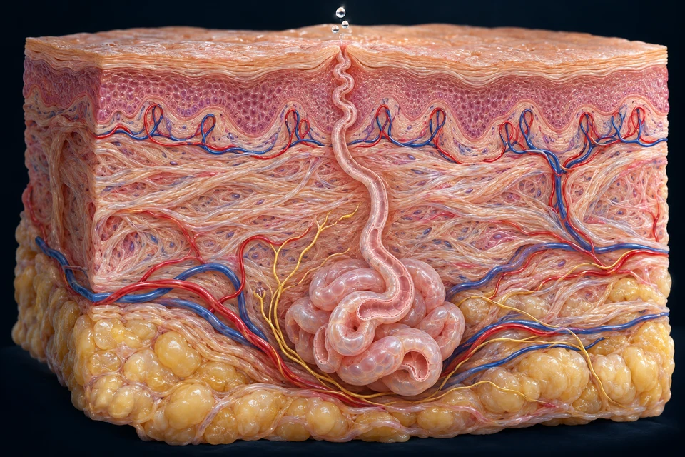
The coil forms primary sweat. Its surrounding blood supply and tissue provide the environment for secretion, without a direct blood-to-duct opening.
The coil forms primary sweat. Its surrounding blood supply and tissue provide the environment for secretion, without a direct blood-to-duct opening.
Sodium and chloride are reabsorbed as fluid travels toward the pore. Salt recovery and water movement are not interchangeable; final composition varies with flow.
A surface droplet is not yet evaporative heat loss. Cooling depends on water becoming vapour, with humidity, airflow and clothing changing how readily that occurs.
Gold arrows show outward travel toward the pore; outlines locate the selected job. These are different jobs along one route, not three on/off gland types. Broad anatomical labels are editable; fine generated tissue is not a cell-by-cell observation.
Sweat and blood take separate paths. The coil secretes fluid; the duct recovers salt; surface water removes heat when it evaporates. Vascular changes alter heat delivery to the skin rather than pumping blood into a gland lumen.
Mechanism references: Human sweat-gland physiology · Sweat composition and ductal transport · Facts checked 18 October 2026; AI-assisted, not physician review.
Warmer internal blood → skin circulation → exchange with surroundings
Cutaneous vasodilation can increase heat transfer when the environment permits it. Both withdrawal of constrictor drive and active neural vasodilation matter; one copied on/off nerve line is incomplete.
Secretory coil → duct → surface sweat → evaporation
Evaporation removes heat. Sweat that stays liquid or drips away does not provide the same cooling; humidity and airflow change how effectively evaporation occurs.
Cutaneous vasoconstriction → reduced surface blood flow
Reducing heat exchange helps conserve heat. Skeletal-muscle shivering is a different mechanism; raised human hairs provide limited insulation.
Source-supported supporting response map, not measured flow, sweat rate or temperature data. Human thermoregulatory eccrine control is predominantly sympathetic cholinergic; acetylcholine here does not imply parasympathetic control.
Teaching references: NLM · Human sweating and evaporation · NLM · Adult human cutaneous heat exchange · NLM · Neural control of skin blood flow and sweating
13 / TOUCH IS MORE THAN ONE RECEPTOR
Receptors transform mechanical or other input into neural information. Location, receptive fields and adaptation help distinguish selected responses; a decorative yellow shape cannot verify subtype.
The hair-free epithelial surface lies above the dermal sensory contexts.
No hair follicle or sebaceous gland is added here. This enlarged view is not a measured thick-skin layer census.
A Meissner-type corpuscle occupies a superficial dermal papilla.
The broad location is source-supported; exact lamellae and nerve branches are illustrative. Changing light contact is explained separately.
Connective support projects toward the epithelial boundary.
Dermal papillae differ from the connective papilla inside a hair bulb. Fine cell and fibre identity is illustrative.

A Pacinian-type corpuscle lies in deeper dermal/subcutaneous support.
Concentric lamellae provide an enlarged teaching cue, not an exact count or measured microscopic profile. Vibration-related responses are discussed below.
Sensory endings connect to neural routes below the surface.
These selected fibre paths are illustrative. They do not establish every human receptor subtype or represent one universal wire for touch, pain and temperature.
| Context | Typical location | Useful input | Limit |
|---|---|---|---|
| Merkel-related complexes | Selected epidermal base | Sustained local pressure, edges and shape information | Often taught with slowly adapting type-I touch responses. |
| Meissner corpuscles | Superficial papillae of glabrous skin | Changing light contact and low-frequency movement | Not inserted as the universal receptor of every hairy-skin region. |
| Pacinian corpuscles | Deeper dermal/subcutaneous context | Vibration and rapid changes | The capsule filters mechanical input; a static dot is not a measured receptive field. |
| Hair-follicle afferents | Around selected follicular regions | Hair movement and changing contact | An exposed shaft carries the mechanical movement, not nerves inside the hair. |
| Free nerve endings | Epithelial and connective tissue contexts | Selected temperature, pain and other cutaneous information | One unencapsulated shape does not identify every sensory modality. |
Slowly and rapidly adapting responses describe how activity changes during sustained stimulation. Ruffini-related stretch accounts and precise human subtype assignments need care; this page does not pretend that every drawn ending establishes an exact human receptor map.
Follow neural processing context →Teaching references: NCBI · Selected cutaneous mechanoreceptors · OpenStax5.1 · Skin layers and selected cell roles · Duke · Identified human thin-skin and fingertip H&E records
SENSORY DEPTH / LOCATION → POPULATION → PERCEPTION
A receptive field is a region where stimulation changes a sensory neuron's activity. Multiple responding afferents help describe an edge, a moving contact or the shape of a held object.
Supporting functional model of responding regions. Circles are receptive fields, not anatomical corpuscles, skin layers or measured human territories.
Smaller fields and closely spaced inputs can support distinguishing nearby features. Overlapping activity is interpreted across a population, not by one isolated receptor.
A larger field covers more skin. Nearby contacts can affect the same afferent; that does not by itself determine what a person will report.
Field size, density, skin mechanics, central processing, attention and the task all matter. This is not a two-point test, threshold calculator or clinical sensory assessment. A field need not be circular or have a uniform response.
Human fingertip glabrous skin supports fine spatial discrimination. Hair-bearing forearm fields have a different organisation; the same receptor cartoon cannot be copied everywhere.
The realistic skin plates above provide tissue context. This functional comparison deliberately adds no invented subtype to their generated endings.
Cortical sensory representation relates to the information being processed; it is not a body drawn to physical scale. Learning, task and central networks also shape interpretation.
Explore cortical anatomy and sensory integration →Teaching references: NLM · Human glabrous-skin receptive-field study · NLM · Human hand touch-afferent properties · NLM · Sensory neurons of touch, human/animal qualifications
SENSORY DEPTH / CONTACT → CHANGE → ACTION
Selected afferents emphasise maintained indentation; others strongly signal changing contact. Their combined activity helps guide handling without turning each receptor into one exclusive percept.
Supporting qualitative response comparison. Both rows use the same maintained model contact; activity marks are invented teaching symbols, not recordings, impulse counts or firing rates.
Selected slowly adapting afferents keep signalling during a maintained contact, although their activity can change with time.
Selected rapidly adapting afferents emphasise changes at contact onset and release. Vibration can repeatedly drive activity even while contact continues.
Adaptation describes a response over time, not conscious attention alone. A sustained stimulus does not imply that every afferent stops signalling. The model does not assign exact human corpuscle identity, latency or frequency.
Skin stretch and contact complement muscle-spindle, tendon and joint information during movement. Touch is not identical to proprioception, and no single receptor measures the whole limb.
Muscle feedback and coordinated action →The dorsal-column pathway is important for discriminative touch and position information from the body. Other ascending routes also carry somatic information; this is not one universal wire for all touch, pain and temperature.
Follow peripheral nerves and central relationships →Slowly adapting type-II responses are often paired with Ruffini endings in diagrams, but exact correspondence in human glabrous skin remains uncertain. Do not label every spindle-shaped generated ending as proven human Ruffini anatomy.
Selected normal mechanisms, not diagnostic testing, a measured neural trace or a fixed human receptor census.
Teaching references: NLM · Sensory neurons of touch, human/animal qualifications · NLM · Human hand touch-afferent properties · NCBI · Dorsal-column mechanosensory relationships · NLM · Human fingertip Ruffini identification uncertainty
14 / PRODUCTION → ATTACHMENT → FREE EDGE
The matrix beneath the proximal fold is the principal nail-production zone. The attached plate lies above the bed and reaches the distal free edge; it does not grow from the fingertip inward.
A skin fold shelters the plate root and matrix.
The underside contributes the cuticle that adheres to the dorsal plate. Its small generated edge is not treated as a measured or surgically verified seal.
The protected proximal region produces the hard nail plate.
It lies deep to the proximal fold and root. Growth is not generated at the free edge; the full microscopic matrix boundary is not resolved by the image.
The fingertip bone provides a deeper anatomical landmark.
The nail unit lies above the bone, not within it. Trabecular texture and the precise relationship to every surrounding vessel or tendon are not verified.

A compact keratinized plate extends toward the fingertip.
Its shape and translucency vary. Nail cells advance distally over an attached bed; exact growth rates are not assigned here.
Underlying tissue supports and attaches the advancing plate.
The bed is distinct from the principal matrix production zone. It is not a large open cavity or a second nail plate.
The hyponychium lies beneath the plate near its free edge.
It connects to fingertip epithelium and helps form a barrier where plate separates from bed. Fine boundaries are illustrative.
The outlined region is enlarged from the same artwork. Matching keys keep close teaching locations separate; no new anatomical detail is invented.
New plate material forms proximally beneath the fold. The underside of the proximal fold contributes the cuticle.
The hard keratinized plate moves distally over attached supporting tissue. The bed is not the entire production matrix.
The hyponychium meets the plate underneath near the point of separation. Nails protect fingertips and support manipulation.
Supporting growth relationships. Fine matrix extent, seal morphology and growth rates are not independently measured; no biopsy, manicure or treatment instructions.
Teaching references: NLM · Normal nail unit relationships · NLM · Nail matrix, bed and proximal-fold cuticle · OpenStax5.2 · Hair, nails and glands
15 / NORMAL REPAIR COORDINATION
When tissue is disrupted, cells and extracellular material coordinate protection and rebuilding. A normal mechanism overview belongs here; clinical wound assessment and treatment remain later-phase work.
Locate epithelial continuity first, then compare the living tissue underneath. The repair preparations and retained native reference are different selected fields, not three timed frames or a personal healing forecast.
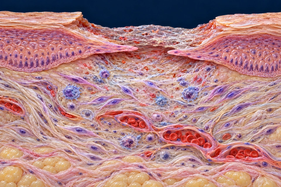
CoverKeratinocytes restore an epithelial surface.
RebuildCells, vessels and matrix form supporting tissue.
RemodelStructure continues changing after coverage.
Gold selects a tissue context. When present, cyan points toward the selected epithelial fronts’ movement direction; it is not a measured speed, blood stream or universal symmetric closing pattern. Cell identities and molecular activity are not established by image colour.
Thin epithelial fronts extend beneath the limited provisional cover. They have not yet made the same continuous, differentiated barrier as the intact edges. The two selected fronts explain a relationship, not universal symmetry or a timed patient sequence.
Normal tissue mechanisms: NLM · Epithelialization in wound healing · NLM · Skin acute wound-healing processes · NLM · Dynamics of the skin immune system
Haemostasis, inflammatory coordination, tissue formation and remodelling are useful organising terms. They are not four sealed rooms or switches that every cell changes on the same day.
Different locations within one repair field can be doing different work. The existing four-process summary remains below; use the preparations to locate the tissue that those words describe.
Normal tissue mechanisms: NLM · Physiology, wound healing · NLM · Principles of tissue repair
Early fibrin-rich material helps stabilise a disrupted region and provides a temporary setting for cellular activity. It differs from the collagen-rich matrix that accumulates and reorganises later.
The thin cover in the selected view is not labelled as a mature epidermis. Its displayed strands are not an assay of fibrin composition, and no instruction to remove or manipulate a wound surface is implied.
Normal tissue mechanisms: NLM · Principles of tissue repair · NLM · Skin acute wound-healing processes
Recruited and resident immune cells participate in signalling and material handling. Macrophages can clear debris and interact with epithelial, stromal and endothelial cells; their activity is more varied than one fixed inflammatory-to-repair switch.
The rounded profiles supply tissue context. Specific cell identity and functional states require evidence beyond a generated shape or colour. The separate Blood and Lymph lessons explain cell families without turning this scene into an immunostained specimen.
Normal tissue mechanisms: NLM · Dynamics of the skin immune system
Keratinocytes migrate and proliferate to restore epithelial coverage. Surviving epithelial sources depend on injury depth and location; margins and retained appendage-associated populations can contribute in appropriate settings.
Coverage is followed by continuing stratification, differentiation and restoration of barrier relationships. A thin cell tongue is not the same as fully mature cornification or an unchanged native basement-membrane architecture.
Normal tissue mechanisms: NLM · Epithelialization in wound healing · NLM · Cellular repair in humans and other mammals
Granulation tissue brings a provisional extracellular environment together with stromal activity and developing microvascular support. It is not a collection of keratinocytes underneath the epidermis, and its name does not mean that the tissue is infected.
Epithelial fronts advance over an appropriate tissue bed while deeper repair develops. The examples separate those compartments without imposing one universal order or a complete map of every cell.
Normal tissue mechanisms: NLM · Principles of tissue repair · NLM · Skin acute wound-healing processes
Fibroblasts synthesise and organise extracellular material, including collagen. Their interactions with matrix and other cells contribute to the mechanical environment beneath the epithelial cover.
Some repair-associated cells acquire contractile features. The spindle profiles are not all certified myofibroblasts: molecular identity cannot be established by a purple oval nucleus, and the matrix is not produced by a tiny mechanical rope machine.
Normal tissue mechanisms: NLM · Skin acute wound-healing processes · NLM · Scar formation: cellular mechanisms
Endothelial cells participate in new vessel formation during repair. Oxygen and nutrient supply help sustain the metabolically active tissue; changing vascular organisation is part of the coordinated response.
The selected lumens contain red cells and remain in connective tissue. The illustration does not show every sprout, prove perfusion rates or imply that the avascular epidermis develops blood vessels within its viable layers.
Normal tissue mechanisms: NLM · Skin acute wound-healing processes · NLM · Cellular repair in humans and other mammals
Repair-associated contractile cell–matrix interactions can reduce the extent of a tissue gap. Keratinocyte migration supplies an epithelial covering. These processes can cooperate without being the same mechanism.
Their relative contributions vary with tissue and experimental setting. Rodent skin contraction must not be presented as measured human closure; no broad subcutaneous muscle sheet is inserted into these general human preparations.
Normal tissue mechanisms: NLM · Cellular repair in humans and other mammals · NLM · Scar formation: cellular mechanisms
Matrix production and turnover change the organisation of repaired connective tissue. Cellular activity and vascular features also change as a highly cellular repair bed matures.
The collagen-rich preparation makes support visible but does not identify collagen types by colour, measure fibre strength or provide a rigid replacement calendar. Continuing reorganisation need not restore the original fibre architecture or every specialised structure.
Normal tissue mechanisms: NLM · Scar formation: cellular mechanisms · NLM · Skin acute wound-healing processes
Repair restores continuity and useful function, but scar-like replacement can differ from native skin. In human histological comparisons, normal scars can have a flatter epithelial–dermal interface and fewer native appendage features than unscarred skin.
The selected later example teaches that distinction. It does not diagnose a scar, guarantee that every appendage is lost after any injury, or claim complete regeneration from a continuous epidermal cover. Injury depth and context matter.
Normal tissue mechanisms: NLM · Human scar and unscarred-skin histology · NLM · Cellular repair in humans and other mammals
No. The advancing epithelial fronts and the temporary cover are distinct. Continued epithelial organisation and barrier maturation have their own roles.
No. It describes living repair tissue involving cells, extracellular material and vascular support. Infection assessment is outside this lesson.
No. Cell–matrix contraction and epithelial resurfacing can cooperate, but they involve different structural relationships.
No. The underlying matrix, appendages and interface can differ from native tissue; these images do not measure a patient’s function or recovery.
Sources checked 21 October 2026. Original generated preparations are separate from acquired tissue observations. Human, animal and experimental findings are not interchangeable. AI-assisted checks are not physician review; fine generated ultrastructure and patient-specific outcomes are not certified. This normal-mechanism lesson gives no wound-care instructions.
Platelet and coagulation activity can help limit blood loss where vessels are disrupted.
Local signals recruit and guide immune activity while debris is managed.
Epithelial coverage, vascular growth and fibroblast matrix production can overlap.
Matrix organisation and tissue properties continue changing; repaired tissue need not regain every original appendage.
Supporting overlapping-process comparison, not a rigid day-by-day schedule or diagnostic classification. No graphic wound photography or clinical advice.
Teaching references: NCBI · Overlapping tissue repair phases · NLM · Local cells and normal repair coordination
16 / OBSERVATION AND RELATED SYSTEMS
Different cutting angles can show a coiled tube as several profiles or omit part of a follicle. Tissue-scale observation complements a realistic teaching model without turning the model into microscopy.
Duke describes human thin skin in 0058_L and human fingertip thick skin in UCSF180. Their layer, gland and receptor contexts can be compared; the adjacent monkey lip record is not substituted for human skin.
Read the external specimen descriptions →Record descriptions checked; slide pixels not acquired or presented here. No microscopy diagnosis claimed.
Skin helps initiate vitamin-D production; later liver and kidney steps connect it with mineral regulation. Digestive and urinary surfaces have different epithelial structures and purposes.
This chapter supplies normal cutaneous foundations. A complete cross-organ epithelial atlas, precise human receptor mapping, specimen micrographs and clinical imaging/disease comparisons remain separately staged.
Teaching references: Duke · Identified human thin-skin and fingertip H&E records · OpenStax5.3 · Normal skin functions
EDITORIAL RECORD
Broad skin compartments, avascular epidermis, layer order and renewal, selected cell roles, barrier/pigment relationships, follicular/nail organisation, gland destinations, thermal/sensory principles and overlapping normal repair.
AI-assisted visual/content production. Fine generated cellular, vascular, neural, glandular and surface details remain illustrative / not independently verified. Human medical review: not performed. Educational only; not diagnostic, clinical or surgical material.
Revision: skin-normal-2026-10-23-hair-renewal-v1. Stable local key: ahb_ch_skin_hair_nails_english_v1. No invented canonical CMS/atlas entity or 3D dataset. Six retained text-free Golden-family anatomical masters and their thirty original labels are preserved. One new eccrine-gland preparation adds six independently editable anatomical labels and outward direction arrows. Superficial and deep sensory contexts source-supported; individual generated receptor lamellae, fibre paths and relative scale remain illustrative, not microscopy. Migration physical-phone acceptance: Pending, non-blocking.