Not a reservoir
The center is living tissue. It does not collect urine or drain cortical secretion through a gland duct.
CHAPTER / LOCATION TO RESPONSE
Two regions. Several control routes.
A steroid-producing cortex surrounds a neural–endocrine center. Different inputs regulate distinct products, while blood connects these small glands with tissue-specific responses.
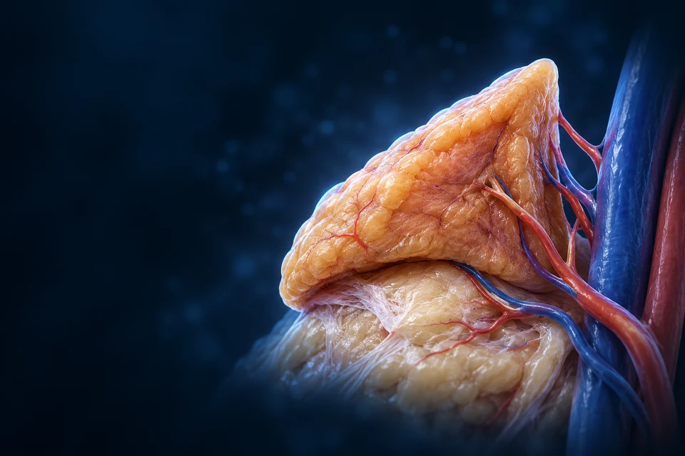
01 / DISTINCT ORGANS, CLOSE NEIGHBORS
These paired retroperitoneal glands lie near the upper medial kidneys. Right and left shape and venous drainage differ. Location links the organs visually; hormone signalling links their functions.

The paired glands sit beside the kidneys. Their hormones enter blood and act at responsive tissues; no adrenal duct drains into a kidney.
Teaching references: NCBI Bookshelf · Suprarenal anatomy · OpenStax17.6 · Adrenal overview
Explore renal hormonal regulation →02 / OUTSIDE TO INSIDE
A capsule encloses steroid-producing cortex and a neuroendocrine medulla. Different developmental origins help explain the contrast: cortical tissue is mesoderm-derived; medullary chromaffin cells arise from neural crest.

The center is living tissue. It does not collect urine or drain cortical secretion through a gland duct.
Cortical blood can reach medulla; direct medullary arterial supply also exists. A simple outside-to-inside arrow would not show every supply route.
Teaching references: NCBI Bookshelf · Suprarenal anatomy · Endotext · Adult adrenal cortex
03 / A TISSUE GRADIENT
Outer compact clusters give way to broad radial cords and a denser inner network. The pattern is useful for orientation, but actual sections vary and fine boundaries are not perfect colored rings.
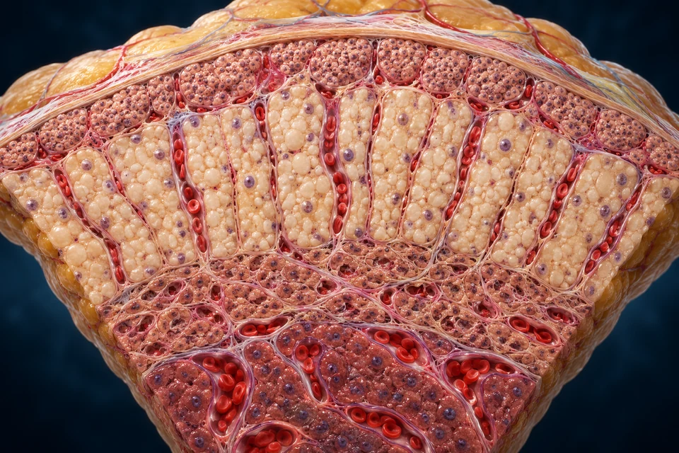
The outer cortical clusters principally produce aldosterone. Angiotensin II and extracellular potassium are major regulators. These cells do not filter blood like a kidney glomerulus.
Gold emphasis selects a tissue region. It is not a measured zone thickness, a hormone concentration or proof of individual cell identity.
Teaching references: University of Leeds · Adrenal tissue organization · Duke University · Human versus monkey adrenal specimens
04 / FOUR SELECTED REGION–OUTPUT RELATIONSHIPS
These are principal adult human teaching associations, not a claim that each zone has one exclusive molecule or that every cell can be identified from an illustration.
Aldosterone
Outer clusters; principal control is angiotensin II and potassium.
Cortisol
Pale radial cords; ACTH supports glucocorticoid output.
DHEA / DHEAS and related precursors
Denser inner cords; this is not a second gonad or a universal puberty clock.
Epinephrine / norepinephrine
Chromaffin cells respond to preganglionic sympathetic input.
Teaching references: Endotext · Adult adrenal cortex · NCBI Bookshelf · Adrenal physiology
05 / SUBSTRATE VERSUS STORED OUTPUT
Cortical steroid synthesis uses cholesterol and enzyme steps distributed between mitochondria and smooth endoplasmic reticulum. Chromaffin catecholamines are tyrosine-derived amines stored in granules before release.
The cortical preparation locates the cells that make steroids. Cholesterol substrate and mitochondrial/smooth-ER enzyme steps differ from a granule store of finished catecholamines. Molecular reactions are explained below, rather than assigned to arbitrary dots in the artwork.
Enzyme patterns help determine which products a zone can make. Storage of cholesterol substrate is different from storing a large ready-made steroid supply.
Stored products allow a different release mechanism. A nerve impulse does not create a new steroid pathway or open a gland duct.
These production steps explain the different native tissue preparations. They are not a complete enzyme network, an anatomical duct or a measured secretion trace.
Cholesterol supply provides substrate for steroid formation. Synthesis involves controlled movement into mitochondria and enzyme reactions in mitochondrial and smooth-ER compartments. In contrast, chromaffin granules hold catecholamines that can be released by regulated exocytosis. The illustrations locate the two tissue contexts; they do not identify molecular pathways simply from cell color.
Teaching references: Endotext · Adult adrenal cortex · NCBI Bookshelf · Steroid and catecholamine production · Human steroidogenesis review
Cholesterol delivery and enzyme expression determine access to steroid pathways. A lipid droplet is substrate context, not a bag of ready-made cortisol.
A rendered nucleus or granule cannot establish enzyme identity, hormone content or secretion rate. The tissue image supports the broad relationship; the mechanism explains the distinction.
Teaching references: NCBI Bookshelf · Steroid and catecholamine production · Human steroidogenesis review
06 / A NEURAL–ENDOCRINE INTERFACE
Sympathetic preganglionic fibers release acetylcholine onto chromaffin cells. Cellular activation can trigger calcium-dependent exocytosis. The secretory cell and endothelial boundary remain separate from the blood lumen.

Preganglionic sympathetic fibres release acetylcholine at chromaffin cells. The nerve ends at a cellular interface; it is not a tube that opens into blood.
Teaching references: NCBI Bookshelf · Steroid and catecholamine production · NCBI Bookshelf · Adrenal physiology
Return to neural output and spinal connections →07 / NOT ONE MASTER SWITCH
The hypothalamic–pituitary axis is important, but it does not explain all adrenal output. Angiotensin and potassium, pituitary ACTH and preganglionic input must remain distinct.
The outer clusters locate the main aldosterone-producing context. The renin–angiotensin system and potassium input are distinct from a pituitary ACTH chain.
Sodium handling and potassium excretion affect the signals that drove release.
ACTH can influence output, but it is not the principal everyday controller of aldosterone. Aldosterone is not the same as ADH.
Cortisol feeds back on hypothalamic and pituitary activity.
Sleep/wake timing, pulses and context influence output. This is not a constant stress meter or an exact daily chart.
Neural activity, secretion and clearance affect the response.
This is not an ACTH chain. Tissue receptor type and context determine the downstream response.
The three causal routes supplement the native tissue illustrations. All remain readable before highlighting; they are not measured hormone values, a complete vascular map or clinical advice.
Outer cortical mineral output responds principally to angiotensin II and potassium. ACTH supports the cortisol-producing cortex, whereas a preganglionic nerve can activate the medulla. A hormone can reach many tissues through blood without every tissue having the same receptor, enzyme capacity or response.
Teaching references: NCBI Bookshelf · Adrenal physiology · Endotext · Adult adrenal cortex · NCBI Bookshelf · Aldosterone physiology
Teaching references: NCBI Bookshelf · Adrenal physiology · NCBI Bookshelf · Aldosterone physiology · NCBI Bookshelf · Cortisol physiology
Compare pituitary target axes →08 / Gland → responsive nephron
Responsive distal-nephron cells increase sodium reabsorption and support potassium secretion. Channel/pump activity, tubular delivery and physiological context matter; this is not an unlimited salt-retention switch.
The original gland and control lessons stay intact. Connect the outer cortical source to the detailed epithelial mechanism, then separate the sodium, potassium and water consequences.
Cholesterol is a starting substrate; aldosterone is an output made through enzyme steps.
Zona-glomerulosa tissue supplies the principal aldosterone-producing context. The final synthetic capacity depends on aldosterone synthase, CYP11B2. A lipid-rich cell is not a bag of pre-made aldosterone, and a rendered cell cluster is not an enzyme assay.
Read the image: Locate the compact outer clusters below the capsule, then distinguish the broad cortisol-producing region underneath. The illustration preserves gross zones, not a uniform CYP11B2-positive band in every adult.
Mechanism evidence: Endotext · Aldosterone biosynthesis and normal regulation · Endotext · Normal aldosterone production and control
Substrate access and enzyme capacity differ from stored catecholamine release. A tissue zone is not an enzyme assay.
The selected short arrow is one boundary transfer. It does not draw a single Na+/K+ exchanger joining urine to blood.
Aldosterone concentration alone cannot predict a fixed sodium, potassium or water output.
Mechanism evidence: Endotext · Aldosterone biosynthesis and normal regulation · Collecting-duct principal-cell transport and regulation · Palmer · Regulation of potassium homeostasis
The adrenal cortex has regional biochemical specialisation. Aldosterone production depends on cholesterol delivery, mitochondrial and endoplasmic-reticulum reactions, and terminal aldosterone-synthase capacity. Steroid production differs from releasing pre-packed catecholamine granules.
CYP11B2 helps distinguish this synthetic route from cortisol production. Adult human expression can be patchy within the subcapsular region. Gross cell arrangement teaches location; it does not establish which individual illustrated cells express an enzyme.
Mechanism evidence: Endotext · Aldosterone biosynthesis and normal regulation · Endotext · Normal aldosterone production and control
Renin is released by specialised renal vascular cells in response to integrated pressure, macula-densa and sympathetic signals. Renin acts on angiotensinogen; subsequent processing forms angiotensin II. ACE activity is not confined to a single universal lung-only checkpoint.
Angiotensin II acts through AT1-receptor signalling, while extracellular potassium changes glomerulosa-cell electrical conditions and calcium entry. Both influence steroid production. A direct potassium input does not first require a pituitary command or a change in renin.
Mechanism evidence: Endotext · Aldosterone biosynthesis and normal regulation · Endotext · Normal aldosterone production and control
The mineralocorticoid receptor is not intrinsically blind to cortisol. In selected epithelial tissues, 11β-HSD2 reduces local cortisol access by converting it to cortisone. This is different from making aldosterone or increasing its concentration in blood.
MR-dependent regulation changes a cell’s capacity to transport ions. Tissue-specific metabolism, receptor abundance and the availability of downstream machinery all matter. The normal renal mechanism should not be extended to every MR-expressing organ as an identical response.
Mechanism evidence: Endotext · Aldosterone biosynthesis and normal regulation · Aldosterone, SGK1 and renal ion channels
MR-dependent gene regulation includes SGK1. One well-studied route involves SGK1 modifying the ENaC-inhibitory ubiquitin ligase Nedd4-2, reducing channel removal and supporting apical sodium-channel availability. Other regulatory proteins and processes also participate.
This is a regulation-of-machinery story, not direct aldosterone passage through ENaC. Channel abundance, probability of opening and the electrochemical driving force are distinct variables. The tissue image provides cellular context rather than a reconstruction of the molecular complex.
Mechanism evidence: Aldosterone, SGK1 and renal ion channels · Aldosterone regulation of ENaC surface availability
The apical membrane faces tubular fluid; the basolateral membrane faces tissue. Sodium entering through apical channels can be exported through the basolateral sodium–potassium pump. Potassium supplied to the cell can leave through separate apical pathways.
Aldosterone-sensitive transport is distributed across late distal and connecting/collecting segments; it is not the same in every nephron cell. The retained principal-cell preparation illustrates useful polarity. It does not make acid-secretory intercalated cells identical to principal cells.
Mechanism evidence: Collecting-duct principal-cell transport and regulation · Palmer · Regulation of potassium homeostasis
Electrogenic sodium absorption helps create a lumen-negative voltage that favours potassium secretion. Distal sodium delivery and tubular flow therefore influence what an available transport system can accomplish. A hormone signal does not guarantee a fixed potassium output.
ROMK and BK are different potassium pathways. Flow-dependent and aldosterone-independent regulation also contribute. Whole-body potassium balance additionally involves shifts between cells and extracellular fluid; a secretion cartoon cannot substitute for that broader physiology.
Mechanism evidence: Palmer · Regulation of potassium homeostasis · Collecting-duct principal-cell transport and regulation
Vasopressin regulates water-channel availability in responsive collecting cells. Aldosterone regulates an overlapping but different transport system for sodium, potassium and related epithelial functions. Neither hormone alone explains the full medullary gradient.
The amount of water recovered depends on permeability, driving gradients and delivery. Sodium reabsorption should not be drawn as an obligatory equal-volume water arrow under every condition. The existing kidney water course explains the separate wall-to-tissue-to-blood route.
Mechanism evidence: Collecting-duct principal-cell transport and regulation · Endotext · Aldosterone biosynthesis and normal regulation
A volume-related renin–angiotensin response and a direct potassium-driven adrenal response can engage different upstream and distal-nephron conditions. Changes in upstream sodium handling help explain why aldosterone does not always impose one fixed ratio of sodium conservation to potassium excretion.
Feedback can reduce the signals that stimulated secretion, but regulation remains distributed across renal delivery, circulation and cellular transport. This normal-function course supplies no blood-pressure targets, hormone-test interpretation, treatment threshold or salt/potassium intake advice.
Mechanism evidence: Palmer · Regulation of potassium homeostasis · Endotext · Normal aldosterone production and control
No. These similar names refer to different tissues: adrenal steroid-producing clusters versus a renal capillary filter.
No. ENaC is an apical channel; the ATP-driven pump is a different basolateral transport protein.
No. Extracellular potassium can directly stimulate responsive adrenal cells.
No. Water permeability, osmotic conditions and delivery remain separate determinants.
Normal physiology · source checks 21 October 2026. Qualified wordless gland, cortical and epithelial illustrations with original structural coordinates and separate editable terminology. Not acquired microscopy, a hormone assay, fine molecular certification, physician review or personal laboratory/dietary/treatment advice.
ADH regulates water permeability in responsive collecting-duct cells. Aldosterone influences ion handling. Water consequences also depend on permeability and gradients; the two hormones are not interchangeable.
Follow water concentration mechanisms →In relevant renal tissues, 11β-HSD2 converts cortisol to cortisone, helping protect mineralocorticoid receptors from cortisol action. A receptor’s name alone does not establish all tissue selectivity.
Teaching references: NCBI Bookshelf · Aldosterone physiology · Endotext · Normal renal mineralocorticoid selectivity
09 / NORMAL METABOLIC COORDINATION
It contributes to metabolic availability, immune regulation and cardiovascular responsiveness. Its normal output is pulsatile and related to sleep/wake timing, with feedback to the hypothalamus and pituitary.
Follow a peptide message to a steroid-producing cortex, then distinguish synthesis, blood delivery and local action. Keep these scales connected without turning them into one anatomical duct.
Three different messages: hypothalamic CRH/AVP → anterior-pituitary ACTH → cortical cortisol. Feedback: cortisol restrains upstream drive. Aldosterone and medullary catecholamines keep their distinct principal controls.
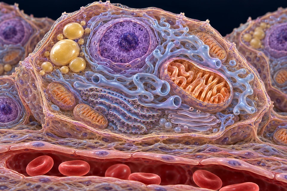
Gold locates the selected broad compartment. It does not show hormone flow rate, receptor abundance or a change in organelle size. Regional brain, cortical tissue and cell cutaway are different observation scales.
StAR helps make cholesterol available to the inner mitochondrial membrane. CYP11A1 begins steroid synthesis there by producing pregnenolone. Further steps use smooth-ER and mitochondrial enzymes; a lipid droplet is not a granule of completed cortisol.
Mechanism evidence: Early steroidogenesis · intracellular cholesterol trafficking · Human steroidogenesis · molecular and physiological review · Plasma steroid-binding proteins · review · Glucocorticoid receptor and tissue sensitivity · review
Hypothalamic neurons supply CRH and vasopressin-related control of pituitary ACTH. Hypophysial portal blood connects the hypothalamic release region with anterior-pituitary cells. It is a local vascular route, not an axon carrying ACTH into the adrenal.
Corticotroph ACTH reaches the cortex through systemic blood. Inhibitory glucocorticoid feedback acts on more than one level of the system. Neural and immune inputs can also influence this network; the three named hormones do not describe every regulator.
Mechanism evidence: Endotext · Stress endocrine physiology · Endotext · ACTH action on the adrenals
MC2R is a G-protein-coupled receptor through which ACTH stimulates adrenal steroid production. MRAP contributes to receptor trafficking and function. cAMP and protein kinase A connect reception with downstream steroidogenic processes.
MRAP’s importance was established through human genetic observations and cell experiments. The coloured cell surface in this illustration is not a measurement of receptor abundance, and no inherited disorder is diagnosed from its appearance.
Mechanism evidence: Endotext · ACTH action on the adrenals · Metherell et al. · MRAP and the ACTH receptor
Cholesterol can be obtained from circulating lipoproteins and cellular synthesis, with cholesteryl esters providing a mobilisable intracellular store. Lipid-droplet context therefore represents a source of substrate, not a packet of cortisol waiting to fuse with the membrane.
Delivery to mitochondrial steroidogenic machinery requires regulated intracellular handling. StAR promotes cholesterol access to the inner mitochondrial membrane. This is not a bulk stream of lipid pouring through an open tube, and the drawing does not assign a unique transporter to a visible membrane strand.
Mechanism evidence: Early steroidogenesis · intracellular cholesterol trafficking · Human steroidogenesis · molecular and physiological review
Mitochondrial CYP11A1 converts cholesterol into pregnenolone. In human cortisol synthesis, subsequent reactions involve smooth-ER enzymes, including CYP17A1 and CYP21A2, before mitochondrial CYP11B1 carries out the final 11β-hydroxylation of 11-deoxycortisol.
Nearby ER and mitochondrial membranes remain separate organelles even when they cooperate closely. The route is a set of biochemical transformations, not a hormone duct. Smooth tubular ER and opened mitochondrial cristae provide structural context; no enzyme can be identified merely from the generated texture.
Mechanism evidence: Human steroidogenesis · molecular and physiological review · Endotext · Adult adrenal cortex · Early steroidogenesis · intracellular cholesterol trafficking
Human cortisol secretion is coordinated with daily timing and sleep/wake organisation. Superimposed ultradian pulses add shorter fluctuations. Circadian and ultradian describe different timescales, not two different cortisol molecules.
A single level cannot reconstruct the preceding secretion pattern or every tissue exposure. No universal clock time, pulse interval or personal “stress score” is supplied here. The normal system is dynamic rather than a constant output tap.
Mechanism evidence: Circadian secretion and tissue glucocorticoid actions · review · Glucocorticoid receptor and tissue sensitivity · review
Much circulating cortisol is reversibly bound to corticosteroid-binding globulin, CBG, or albumin. An unbound fraction exchanges with tissues. Binding influences transport, clearance and availability; a total amount includes more than that immediately unbound pool.
Binding proteins are not red blood cells. The blood channel in the picture locates a transport compartment without showing molecular complexes or assigning a universal bound percentage. This lesson does not interpret a blood test or recommend a clinical assay.
Mechanism evidence: Plasma steroid-binding proteins · review · Glucocorticoid receptor and tissue sensitivity · review
Glucocorticoid receptor signalling influences gene regulation through interactions with DNA, transcriptional machinery and other regulatory proteins. Receptor variants, cofactors, chromatin context and exposure history help shape which genes respond in a particular cell.
Glucocorticoids also have faster actions that cannot all be reduced to new gene transcription. The nucleus in the source cell is anatomical context, not a claim that cortisol acts only within its producing cell or that every response waits for the same delay.
Mechanism evidence: Glucocorticoid receptor and tissue sensitivity · review
11β-HSD1 predominantly regenerates cortisol from cortisone in many intact tissues. 11β-HSD2 converts cortisol toward cortisone in selected tissues, including mineralocorticoid-sensitive renal epithelia. Local metabolism helps distinguish tissue exposure from circulating supply.
The direction depends on the isoenzyme and cellular setting; activity measured in a disrupted laboratory preparation need not reproduce the intact-cell environment. The kidney’s protective context is explained in the existing mineral-control lesson, not inferred from this adrenal cell’s colour.
Mechanism evidence: 11β-HSD enzymes · intracellular gatekeepers
Cortisol participates in metabolic availability, immune regulation and cardiovascular responsiveness. Its effects depend on tissue, exposure pattern and accompanying signals. Neither “always harmful” nor “simply turns every function up” describes its normal role.
A daily physiological signal is not equivalent to a prolonged excess or a drug exposure. Related disease and treatment questions need a separate clinical context. This module supplies no supplement, medication, cortisol-lowering or stress-management instruction.
Mechanism evidence: Glucocorticoid receptor and tissue sensitivity · review · Endotext · Stress endocrine physiology · Endotext · Adult adrenal cortex
Experimental pulsed glucocorticoid exposure can produce different receptor–chromatin and transcriptional patterns from sustained exposure. Such work shows why the time course of a signal can matter, as well as its presence.
Cell and animal experiments do not by themselves establish a person’s secretion pattern or every human tissue response. No numerical waveform is presented as patient data, and the generated anatomy is not an acquired specimen or a receptor-conformation assay.
Mechanism evidence: Stavreva et al. · Pulsed glucocorticoid receptor transcription · Circadian secretion and tissue glucocorticoid actions · review
No. Hypothalamic signals regulate anterior-pituitary ACTH; responsive adrenal cortical cells synthesize cortisol from cholesterol.
No. They represent lipid substrate context. Steroidogenic reactions involve regulated cholesterol handling and mitochondrial/ER enzymes.
No. Binding, local metabolism, receptor machinery, tissue context and exposure history all matter.
No. Feedback modulates a dynamic network with daily timing, pulses and other inputs. It is not a binary shut-off of every physiological activity.
Normal physiology, not a cortisol test, diagnostic model or treatment guide. Sources checked 21 October 2026. Generated teaching anatomy is distinct from acquired microscopy; fine generated ultrastructure, human medical review and physical-phone acceptance are not certified.
Hormone exposure, receptor biology and other signals influence response. Normal physiology should not be reduced to a claim that cortisol is always harmful or always raises every tissue activity.
The gland makes an output; liver, muscle and other tissues respond through their own mechanisms. Detailed metabolic and molecular pathways remain separate linked lessons.
Connect liver metabolism →Teaching references: NCBI Bookshelf · Cortisol physiology · NCBI Bookshelf · Steroid and catecholamine production
10 / ONE BLOOD SIGNAL, DIFFERENT TARGETS
Epinephrine and norepinephrine act through adrenergic receptor families. Heart, airways, vessels and metabolic tissues do not all respond identically. Blood delivery is widespread, but response remains tissue-specific.
Selected receptor pathways can influence heart activity, airway muscle and vascular tone. Avoid a universal claim that every vessel dilates or every adrenergic effect is the same.
Liver glycogen can supply circulating glucose. Skeletal muscle lacks glucose-6-phosphatase and uses glycogen-derived fuel locally; it does not simply export free glucose into blood.
Teaching references: NCBI Bookshelf · Adrenergic receptor distinctions · NCBI Bookshelf · Tissue-specific glycogen function
MAKE / STORE / RELEASE / RESPOND
A nerve can call on a ready store. But making the molecule, filling a granule and releasing its contents are different jobs. Follow them through a chromaffin cell, then out to a responsive tissue.
Epinephrine is also called adrenaline; norepinephrine is noradrenaline. Both are tyrosine-derived amines, not the cholesterol-derived steroids made in the cortex.
Cytosol: the fluid compartment outside membrane-bounded organelles. Granule lumen: the inside of a secretory granule.
PNMT: phenylethanolamine N-methyltransferase, the enzyme that makes epinephrine from norepinephrine. VMAT: vesicular monoamine transporter, which loads amines across the granule membrane.
Enzymes change tyrosine-derived molecules in two compartments. Transport connects those compartments; it is not the same as a chemical reaction.
Keep the boundaries visible: nerve ending, chromaffin cell, extracellular tissue, endothelium and blood lumen are separate. The retained detailed illustration locates these compartments; it does not resolve VMAT, DBH, PNMT or a fusion pore in its texture.
Supporting spatial schematic, not molecular dimensions or a measured membrane reconstruction. The detailed retained cell preparation supplies anatomical context. Solid arrows describe reaction or named loading steps; the dashed outward NE arrow deliberately leaves its transfer mechanism unspecified.
Each row names its location. Chemical reactions and membrane transfers are different steps; this is a supporting schematic, not a tissue image.
TH performs the first conversion; AADC, also called DDC, performs the second.
This moves the molecule; it does not yet make norepinephrine.
DBH acts inside this compartment. Norepinephrine can remain a stored product.
The accessed evidence does not establish a named physiological export mechanism. This is not a reverse-VMAT or NET claim.
PNMT acts outside the granule lumen. Cortisol is not the precursor.
Loading through VMAT and exocytosis across the cell surface are separate events.
Evidence: Biosynthesis of Catecholamines · Storage and Release of Catecholamines · Noradrenaline translocates to the cytosol · Distinct pharmacological properties and distribution in neurons and endocrine cells of two isoforms of the human vesicular monoamine transporter
Evidence: Biosynthesis of Catecholamines · Storage and Release of Catecholamines · Noradrenaline translocates to the cytosol
Read the full explanation and its limits ↓Enzymes change tyrosine-derived molecules in two compartments. Transport connects those compartments; it is not the same as a chemical reaction.
Begin with tyrosine, an amino acid. In the cytosol, tyrosine hydroxylase (TH) converts it to L-DOPA. Aromatic L-amino-acid decarboxylase, also called DOPA decarboxylase (AADC/DDC), makes dopamine. After dopamine enters a granule, dopamine beta-hydroxylase (DBH) makes norepinephrine inside that compartment. In cells with PNMT activity, norepinephrine in the cytosol can receive a methyl group from S-adenosylmethionine (SAM), producing epinephrine.
This means the epinephrine route crosses the granule boundary more than once: dopamine goes in; norepinephrine becomes available outside; newly made epinephrine is loaded in for storage. The outward norepinephrine step is necessary to the compartment sequence, but the sources accessed do not establish a named physiological export route. The outward transfer therefore remains explicitly unspecified; no named exporter is established here.
Norepinephrine can also remain a stored, releasable product. Granules and chromaffin cells need not contain identical proportions. Enzyme activity, transport and storage are distinct questions. These catecholamines are amino-acid-derived amines, unlike the cholesterol-derived steroids in the retained cortical course.
Evidence: Biosynthesis of Catecholamines · Storage and Release of Catecholamines · Noradrenaline translocates to the cytosol
Does DBH make norepinephrine in the same compartment where PNMT makes epinephrine? No: granule lumen versus cytosol. The uncertain step is membrane transfer, not permission to relocate PNMT into the granule.
One membrane machine builds a proton gradient. Another uses that gradient to load amines. Storage inside a cell is not secretion into blood.
The granule membrane supports an energy-dependent gradient. V-type H+-ATPase uses ATP hydrolysis to move protons into the granule, helping make its interior acidic. Amine loading is a different membrane process: VMAT couples amine movement into the granule to proton movement out. The pump builds the gradient; the transporter uses it. This overview does not assign a fixed proton-to-amine exchange ratio. Experiments with isolated bovine granule membranes help separate gradient-driven uptake from the ATP-consuming step that normally maintains the gradient.
Catecholamines coexist with ATP and granule proteins such as chromogranins. ATP as a component of stored cargo is not the same explanatory role as ATP hydrolyzed to power a membrane pump. Stored luminal ATP and ATP used by the pump are distinct explanatory roles, not one direct cargo-to-pump arrow.
Human chromaffin cells examined in a primary localization study expressed both VMAT1 and VMAT2. Rat data show a different distribution that can change with experimental state. VMAT is therefore used as the general loading label, without assuming one universal human isoform pattern.
Evidence: Structural insights into vesicular monoamine storage and drug interactions · Role of a transmembrane pH gradient in epinephrine transport by chromaffin granule membrane vesicles · Storage and Release of Catecholamines · Adenosine triphosphate in the bovine chromaffin granule · Distinct pharmacological properties and distribution in neurons and endocrine cells of two isoforms of the human vesicular monoamine transporter · Vesicular monoamine transporters (VMATs) in adrenal chromaffin cells: stress-triggered induction of VMAT2 and expression in epinephrine synthesizing cells
If amine crosses the granule membrane, has secretion into blood occurred? No: loading is intracellular. Exocytosis crosses the chromaffin-cell boundary and belongs to the next unit.
A nerve signal changes the cell’s electrical activity and calcium. Fusion then gives soluble cargo access to extracellular tissue, before it reaches blood.
A preganglionic sympathetic fiber supplies acetylcholine to the chromaffin-cell interface. Nicotinic acetylcholine receptors are ligand-gated channels. Their activation produces inward cation current and depolarization, which can recruit voltage-dependent calcium channels; direct calcium entry through nicotinic receptors can also contribute. A rise in cytosolic calcium links electrical activation to secretion. These links have been studied directly in isolated human chromaffin cells.
Calcium-sensitive proteins and membrane-fusion machinery connect that calcium signal to granule exocytosis. Mouse studies support overlapping synaptotagmin sensor functions and distinguish docking from a granule’s readiness to release. Being near the membrane is not already fusion. SNARE proteins participate in the fusion machinery; no single sensor isoform is presented here as the sole universal switch.
When a granule fuses with the chromaffin plasma membrane, soluble cargo gains access to extracellular tissue. The adjacent endothelial boundary still separates this tissue compartment from the blood lumen. The intact intracellular granule does not cross the capillary wall, and the nerve ending is not a tube leading to blood. This is the same retained anatomical boundary already taught by the chapter.
Bounded extensions: Human isolated-cell experiments also found a delayed muscarinic contribution under prolonged stimulation. Male-mouse work shows PACAP participation during sustained/high-firing demand. These are additional control mechanisms, not grounds for replacing the principal ACh sequence with an identical all-human cotransmitter switch.
Evidence: Key role of the nicotinic receptor in neurotransmitter exocytosis in human chromaffin cells · Synaptotagmin-1 and -7 are functionally overlapping Ca2+ sensors for exocytosis in adrenal chromaffin cells · Differential Control of the Releasable Vesicle Pools by SNAP-25 Splice Variants and SNAP-23 · PACAP controls adrenomedullary catecholamine secretion and expression of catecholamine biosynthetic enzymes at high splanchnic nerve firing rates characteristic of stress transduction in male mice
What is released across the fused membrane? Soluble granule contents, not the nerve itself and not a glandular duct secretion.
Cortical hormones can support epinephrine-making capacity. Neural input can release an existing granule store. These roles cooperate without being identical.
Cortex and medulla cooperate without performing the same reaction. Cortical glucocorticoids support the epinephrine-making enzyme system, including PNMT. Classic rat experiments showed this cortical-steroid influence; cultured bovine cells further separated changes in PNMT activity from evoked secretion in that preparation. These findings support a distinction between the capacity to make epinephrine and the immediate stimulus that releases an existing store.
Cortisol is not converted into epinephrine. ACTH is not the direct neural release trigger at the chromaffin interface. A pre-existing granule pool allows rapid calcium-dependent release without waiting for every released molecule to be made anew. Conversely, prolonged neural signaling can also affect biosynthetic capacity, so this is not an absolute division into an entirely fast nerve system and an entirely slow enzyme system.
The vascular arrangement helps explain communication within the gland. Blood draining cortical tissue can expose medullary cells to cortical products, while direct arterial branches also supply medulla. Both relationships matter: not all medullary blood must first pass through the cortex.
Evidence: Control of enzymatic synthesis of adrenaline in the adrenal medulla by adrenal cortical steroids · Glucocorticoid receptors and regulation of phenylethanolamine-N-methyltransferase activity in cultured chromaffin cells · Key role of the nicotinic receptor in neurotransmitter exocytosis in human chromaffin cells · PACAP controls adrenomedullary catecholamine secretion and expression of catecholamine biosynthetic enzymes at high splanchnic nerve firing rates characteristic of stress transduction in male mice · The adrenal gland, Endocrinology: An Integrated Approach
Which changes immediately when a loaded cell receives an effective calcium signal: the identity of its precursor or its opportunity to release stored cargo? The opportunity to release. Capacity and release remain connected but separable.
A receptor starts an intracellular pathway. The target cell’s machinery determines the response: a heart cell and airway muscle do not interpret cAMP identically.
The receptors that help activate the chromaffin cell are not the same as the adrenergic receptors through which its released catecholamines act on target cells. Selected alpha and beta adrenoceptors are G-protein-coupled receptors. Their predominant couplings differ, and these couplings are not exclusive promises for every tissue or circumstance.
Follow three intracellular routes:
Now put those pathways into cells. Beta1 signaling in cardiac tissue supports greater cardiac activity. Beta2 signaling in airway smooth muscle supports relaxation. Alpha1 signaling can support vascular smooth-muscle contraction. A shared second messenger does not make a cardiac cell and a smooth-muscle cell perform the same job. The response depends on receptor distribution, intracellular targets, exposure and other physiological influences.
Evidence: The Concise Guide to PHARMACOLOGY 2023/24: G protein-coupled receptors · Signal Transduction 1: G Protein Coupled Receptors · Structural basis of agonist specificity of alpha1A-adrenergic receptor · The sympathetic nervous system · Crystal structure of the beta2 adrenergic receptor–Gs protein complex
Does “more cAMP” mean “more contraction everywhere”? No. The cell’s downstream machinery determines what the signal accomplishes. Avoid “epinephrine dilates all blood vessels” or one fixed output for an entire organ.
No. Entry, distribution and removal all affect concentration. Local nerve release, adrenal secretion, cellular uptake and metabolism are different processes.
The adrenal medulla releases endocrine messages into the circulation. Sympathetic neurons also release norepinephrine locally near their targets. Material reaching plasma can therefore have different sources; circulating norepinephrine is not an exclusively adrenal product. Human regional studies separate release into blood from removal and from local metabolism.
For local neuronal transmission, uptake across the nerve-cell surface can remove norepinephrine from the extracellular space. NET, the norepinephrine transporter, is not the granule-loading VMAT. Once inside, transmitter can be repackaged or metabolized. Uptake into non-neuronal tissues also contributes to removal. Uptake into a cell and loading into its granule cross different membranes.
Metabolism occurs within cells rather than only after a molecule has completed a trip through blood. MAO is associated with the outer mitochondrial membrane; COMT acts in cellular metabolic pathways. Intracellular turnover includes material that was not just released. Consequently, a metabolite is not a simple one-for-one time stamp of exocytosis.
Four distinct concepts matter: synthesis makes molecules; secretion moves stored contents out of the source cell; uptake and metabolism remove or transform molecules; concentration describes how much is present in a sampled volume. A blood concentration depends on entry and removal, distribution and timing, so it is not a direct secretion meter or a universal “stress score.”
Evidence: Regional release and removal of catecholamines and extraneuronal metabolism to metanephrines · Storage and Release of Catecholamines · The sympathetic nervous system · Plasma dihydroxyphenylglycol and the intraneuronal disposition of norepinephrine in humans · Biochemical quantification of sympathetic nervous activity in humans using radiotracer methodology: fallibility of plasma noradrenaline measurements
Could blood concentration change without a proportional change in secretion? Yes: removal and distribution also affect what is present. This lesson does not interpret an individual laboratory result.
Source checks dated 23 October 2026. Abstracts, indexed passages and full articles are not interchangeable. Human medical review was not performed. The uncertain NE membrane-transfer mechanism remains explicit rather than filled in with a named exporter.
Context: General mammalian biochemistry teaching, not a new human experiment.
Access: Indexed relevant chapter text; direct NCBI browse produced a verification page.
Supports: TH and aromatic amino-acid decarboxylase in cytosol; DBH in vesicles; PNMT methylates NE using SAM.
Limits: Older edition; do not use its transporter nomenclature as a complete current human isoform inventory.
Integrity check: No individual correction search; teaching source cross-checked with primary transporter and curated compartment evidence.
Context: Mammalian neuronal and adrenal teaching; compartments must not be conflated.
Access: Substantial indexed chapter text retrieved, including storage, release and uptake sections; not direct full-page browse.
Supports: Cytosolic PNMT followed by epinephrine granule uptake; catecholamine/ATP/chromogranin storage; neuronal uptake differs from vesicular loading.
Limits: Many release paragraphs refer to neurons rather than chromaffin cells. No universal VMAT2 inference.
Integrity check: No individual correction search; cross-checked with primary studies.
Context: Human-focused physiological teaching.
Access: Indexed relevant sections and box text; not independently browsed full chapter.
Supports: Direct medullary arterial supply plus cortical venous contribution; cortex-medulla relationship and neural medullary control.
Limits: Use only normal physiology; not a clinical protocol or a measured vascular map.
Integrity check: No individual correction search; cross-checked with existing bounded anatomy.
Context: Human-oriented teaching.
Access: Full HTML available; normal physiology sections inspected.
Supports: Adrenergic target context; selected heart, vascular and airway effects; neuronal and extraneuronal removal.
Limits: Do not import therapeutic tables or medication content; the course uses only selected normal mechanisms.
Integrity check: No publication integrity claim; educational page checked on access date.
Context: General cellular signaling teaching.
Access: Indexed relevant teaching text; direct open returned 403.
Supports: Gs–adenylyl cyclase–cAMP–PKA and Gq–PLC–IP3/DAG pathways.
Limits: A generic coupling diagram is not evidence of a uniform whole-tissue response.
Integrity check: No individual correction search.
Context: Human receptor nomenclature with explicit species comparisons.
Access: Indexed alpha1, alpha2 and beta adrenoceptor sections; direct PMC verification page, another institutional copy 403, and database family view redirected to login. No bypass.
Supports: Predominant alpha1 Gq/11, alpha2 Gi/o, beta Gs; coupling is not exclusive.
Limits: Identified edition, not asserted latest edition. No drug potency table imported.
Integrity check: No individual comprehensive correction search.
Context: Homo sapiens curated reaction; authored/reviewed in 2008, currently indexed.
Access: Indexed complete reaction description and participants; direct www URL returned cache miss.
Supports: Granule-lumen NE must reach cytosol; curated description explicitly leaves transport process unresolved.
Limits: Not proof that no later mechanism can exist. Use unspecified transfer arrow; do not assign NET or reverse VMAT. Database cites P17, whose abstract does not demonstrate an exporter.
Integrity check: Current indexed entry checked; no claim of fresh experimental validation.
Context: Human tissue immunostaining; cloned human VMATs in heterologous CV-1 monkey fibroblasts.
Access: Abstract fully retrieved; scanned full-paper PDF did not load. Full body not claimed.
Supports: Both human VMAT1 and VMAT2 detected in adrenal chromaffin cells examined.
Limits: Not an all-person, all-state quantitative expression survey. Functional clone assays are not intact human adrenal experiments.
Integrity check: Exact-title correction/retraction search: no relevant notice found.
Context: Engineered recombinant human VMAT1 cryo-EM and HEK293T functional assays.
Access: Full HTML available; relevant results, methods and discussion inspected.
Supports: Proton-coupled inward amine transport; proton pump and VMAT have distinct roles; model favors import rather than physiological outward NE assignment.
Limits: Not direct demonstration of NE export in normal adrenal cells. Exact stoichiometry unnecessary for this course.
Integrity check: Exact-title correction/retraction search and visible article notices checked; no relevant notice found. Methodological motion-correction text is not an erratum.
Context: Isolated bovine chromaffin-granule membrane vesicles.
Access: Abstract retrieved.
Supports: An inward-acid gradient can energize epinephrine accumulation, separating gradient generation from amine transport.
Limits: Artificial vesicle conditions; not a complete living human-cell energy budget.
Integrity check: No individual correction search.
Context: Rat adrenal cells with basal and repeated-immobilization comparisons.
Access: Abstract, figure captions and indexed relevant sections.
Supports: VMAT expression varies by species, cell state and experimental context; avoid universal rat-only-VMAT1 simplification.
Limits: Not a human stress threshold or human cell-proportion result.
Integrity check: No individual correction search.
Context: Isolated human adrenal chromaffin cells; electrophysiology and capacitance measurements.
Access: Full abstract retrieved.
Supports: Nicotinic activation, direct receptor-associated calcium entry and depolarization/voltage-dependent calcium coupling; delayed muscarinic contribution under prolonged stimulation.
Limits: Not one obligatory receptor subtype or a claim that muscarinic signaling dominates every normal human response.
Integrity check: Exact-title correction/retraction search: no relevant notice found; unrelated retracted lung-cancer receptor study excluded.
Context: Mouse adrenal chromaffin knockout/knock-in preparations.
Access: Indexed abstract and relevant introductory text; direct PMC produced verification page.
Supports: Calcium-sensitive exocytosis uses molecular sensors; synaptotagmin functions overlap.
Limits: No sole universal sensor or all-human isoform quantitative claim.
Integrity check: Targeted title correction/retraction search found no notice for this paper; corrections belonging to different synaptotagmin studies were excluded.
Context: Embryonic mouse chromaffin cells; bovine expression work in supporting experiments.
Access: Abstract and indexed full-text relevant results/discussion.
Supports: SNARE-related machinery supports release competence; granule docking is not equivalent to priming or fusion.
Limits: Fast release loss does not mean all residual secretion is abolished.
Integrity check: Exact-title correction/retraction search: no relevant notice found.
Context: Male mice; adrenal slices and in vivo experiments.
Access: Abstract and indexed relevant results; direct PMC verification page.
Supports: PACAP can support sustained high-firing secretion and biosynthetic-enzyme expression.
Limits: Not a universal human cotransmitter switch or proof that ACh has no role at high demand.
Integrity check: Exact-title correction/retraction search: no relevant notice found.
Context: Female Sprague-Dawley rats with endocrine interventions.
Access: Full five-page author-hosted reprint searchable; summary and relevant methods/results inspected.
Supports: Cortical steroid support of PNMT enzyme activity and indirect ACTH effects.
Limits: An animal mechanism study, not a normal human hormone threshold or immediate exocytosis trigger.
Integrity check: Exact-title correction/retraction search: no relevant notice found.
Context: Cultured bovine chromaffin cells.
Access: Indexed abstract and introductory text; direct PDF returned 403.
Supports: Glucocorticoid exposure can alter PNMT activity without an equivalent change in evoked secretion in this preparation.
Limits: Does not establish all glucocorticoid effects as transcriptional or all species as identical.
Integrity check: No individual correction search.
Context: Engineered human alpha1A receptor with mini-Gq complexes, including epinephrine.
Access: Indexed relevant results and methods.
Supports: Physical receptor–Gq coupling as a molecular example.
Limits: Stabilized structural preparation, not a live vessel or all alpha subtypes.
Integrity check: Exact-title correction/retraction search found no relevant notice; Crossmark page could not be retrieved, so no Crossmark certification.
Context: Recombinant human receptor in stabilized structural complex.
Access: Abstract and figure captions.
Supports: Beta2 receptor–Gs coupling; Gs links receptor to adenylyl cyclase.
Limits: Not whole-airway response measurement or a reason to generate a new 3D asset.
Integrity check: No individual correction search.
Context: Human healthy participants and clinical comparison groups; not all subjects healthy.
Access: Full eight-page publisher PDF searchable; abstract and relevant physiology/results inspected.
Supports: Neuronal reuptake and intracellular metabolism; turnover need not represent recently secreted transmitter.
Limits: No diagnostic thresholds, experimental drug instructions or testing procedures imported.
Integrity check: Exact-title correction/retraction search: no relevant notice found.
Context: Human regional studies with healthy and clinical groups.
Access: Indexed abstract; direct PubMed did not provide usable article body.
Supports: Adrenal contribution to circulating epinephrine, regional removal and intracellular/extraneuronal metabolism.
Limits: Study proportions are not universal human constants; no clinical interpretations imported.
Integrity check: Exact-title correction/retraction search: no relevant notice found.
Context: Human healthy and clinical comparison groups.
Access: Indexed abstract only.
Supports: Plasma NE concentration reflects removal as well as spillover, not a direct secretion meter.
Limits: No radiotracer methods or testing advice included.
Integrity check: No individual correction search.
Context: Bovine chromaffin granules and granule ghosts.
Access: Abstract retrieved.
Supports: Granule ATP and energy-linked membrane processes must be distinguished conceptually.
Limits: No fixed cargo ratios or claim that luminal ATP itself supplies the outward-facing catalytic pump site.
Integrity check: No individual correction search.
Context: Normal human adrenal specimens and tumor comparison samples; normal group small.
Access: Complete indexed abstract.
Supports: Observed NAT/NET, PNMT and granule-marker localization; useful restraint on simple cell labels.
Limits: Colocalization is not a transport assay; does not establish NET as physiological granule NE exporter. Tumor findings not used to define normal course.
Integrity check: Identifier correction/retraction search found no relevant notice; low specificity of search limits assurance.
AI-assisted normal-physiology teaching. Existing original detailed adrenal anatomy is retained with unchanged semantic coordinates; supporting process views are editable schematics, not acquired microscopy or exact molecular reconstructions. No diagnosis, testing protocol, drug advice or measured secretion simulation.
11 / PRECURSORS AND PERIPHERAL CONVERSION
DHEA, DHEAS and related precursors contribute to peripheral steroid pathways. Tissue conversion and life stage matter. Adrenarche and gonadal puberty are connected developmental subjects, not identical events or a universal age switch.
A complete gonadal/developmental lesson is a separate normal-function dependency. This section does not offer hormone testing, supplementation or treatment advice.
Teaching references: Endotext · Adult adrenal cortex · NCBI Bookshelf · Steroid and catecholamine production
12 / REGIONAL RELATIONSHIPS
Multiple arterial branches support cortical and medullary exchange. Usual right venous drainage reaches the inferior vena cava; usual left drainage reaches the left renal vein. Variations prevent treating a generic render as a precise vascular map.
Cortical cortisol availability can influence medullary epinephrine synthesis. This is a local biochemical relationship, not proof of every branch visible in the generated regional plate.
Teaching references: NCBI Bookshelf · Suprarenal anatomy · NCBI Bookshelf · Steroid and catecholamine production
13 / REAL TISSUE EVIDENCE
Section plane, stain and preparation influence what becomes visible. Generated textures are not a real microscopy result or a cell-type assay.
Duke identifies UMich230 as human adrenal H&E and distinguishes it from monkey slides231/0062. Its notes also warn that a human section may show cortex with little medulla in some regions.
Inspect specimen metadata and observations →Leeds provides cortical/medullary tissue teaching and stain context. Its images remain external; no specimen pixels or third-party figure rights are assumed.
Read the external tissue teaching record →Teaching references: Duke University · Human versus monkey adrenal specimens · University of Leeds · Adrenal tissue organization
Clinical imaging, disease, diagnosis and treatment stay in later roadmap phases. Their absence does not block the normal anatomy/physiology foundation.
EDITORIAL RECORD
Broad location, capsule/cortex/medulla relationships, principal adult zones, distinct production/control mechanisms, selected renal/metabolic/neural responses and external specimen distinctions.
AI-assisted visual/content production. Fine generated vascular/cellular/membrane detail, exact dimensions and hidden junctions remain illustrative / not independently verified. Human medical review: not performed. Educational purpose only; not a surgical map, diagnostic plate or exhaustive endocrine atlas.
English revision: adrenal-english-2026-10-19-eyes-foundation-v1. Stable local chapter key: ahb_ch_adrenal_english_v1. No canonical CMS entity or new3D dataset is asserted. Related native-source learning retains the original source geometry and its explicit limits. Migration physical-phone acceptance: Pending, non-blocking. Text-free masters,editable English anatomy labels and supporting paths remain separate.