Connect muscle architecture to cellular machinery, nerve activation, force control and sensory feedback. Cells create force; tissue attachments transmit it.
Follow cellular force generation into selected shoulder, thigh, elbow, knee, forearm and calf anatomy. The regional examples connect muscle position, tendon attachment and joint action; a complete whole-body atlas remains a separate coverage goal.
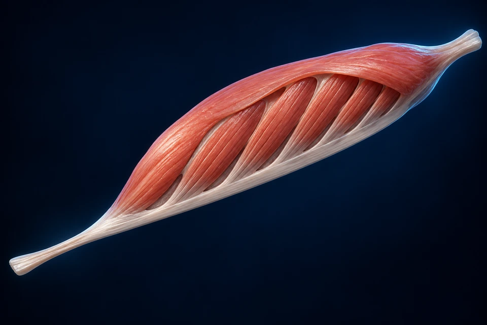
New generated generic pennate architecture, not a named muscle or measured specimen. Fascicles are shown on one side of a connective sheet. Scale, wrapping and angle are illustrative; no numerical force or excursion is inferred. Different architecture from the parallel-fibre organisation exhibit below.
Chapter overview · future video support
No video is available yet. The illustrated reading below provides the current lesson.
Use the source-labelled reference to turn from front to back, select a structure and relate muscle position to nearby bones. Then return to the illustrations for attachment, tissue and force explanations.
Adult male BodyParts3D source reference. The shoulder entry selects the source’s paired supraspinatus meshes; it is not an isolated single shoulder. Source labels and geometry have not received AllHumanBody human anatomical review. This interactive reference supplements the selected teaching plates; it does not complete the missing whole-body or head/neck illustration coverage. The chapter loads no 3D geometry until you open the atlas.
Organ → bundle → cell → contractile machinery
Different levels. One connected force path.
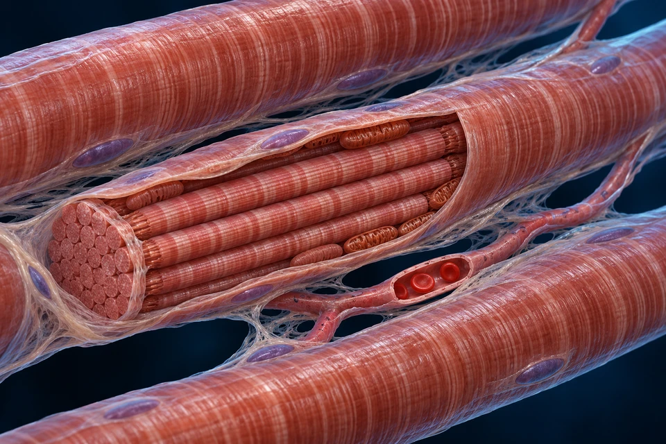
The opened exhibit changes scale as it moves to the right. A fascicle is a bundle of cells; a muscle fibre is one cell. Neither is a motor unit.
New original generated atlas illustration, not genuine microscopy or a named regional muscle. Scale transitions, cell counts, wrapping and fine filament detail are illustrative; do not measure this image. The enlarged right-hand cylinder represents one fibre containing many myofibrils, not another fascicle. Editable points identify the three broad organisational levels; they do not verify microscopic ultrastructure.
Connective wrapping carries force
Epimysium surrounds the organ; perimysium surrounds fascicles; endomysium supports individual fibres outside their membranes. These connective tissues also provide paths for vessels and nerves. Their continuity helps transmit cellular force toward tendon and other attachments.
The fibre has a membrane and cytoplasm
The sarcolemma encloses one long skeletal-muscle cell. Its nuclei usually lie near the periphery. Mitochondria supply metabolic machinery between contractile structures. Connective tissue outside the membrane is not another cell membrane.
Myofibrils repeat sarcomeres
Each myofibril contains repeating units between Z discs. The organised overlap of thin actin and thick myosin filaments produces striation. A myofibril is not one filament, and a fibre contains many myofibrils. Contractile shortening changes overlap rather than simply shortening the filaments themselves.
Skeletal muscle creates force, connective tissues transmit it, joints guide motion and nervous circuits regulate recruitment. This chapter connects regional anatomy to the cellular mechanism rather than treating a red body as a complete explanation.
Muscle
Whole organ Fibres, connective tissue, nerves and vessels work together.
Tendon and bone
Force transmission Attachments translate muscle force into joint torque.
Nervous control
Motor and sensory pathways Recruitment and feedback adjust force for the task.
Three connective levels are not the cell membrane.
Epimysium surrounds the whole muscle, perimysium surrounds fascicles and endomysium surrounds individual fibres. Sarcolemma is the fibre membrane. These tissues provide support, routes for vessels and nerves, and force transmission.
Epimysium
Whole muscle An outer connective envelope.
Perimysium
Fascicle Connective tissue around a bundle of fibres.
Endomysium
Individual fibre Fine connective support outside the sarcolemma.
Rectus suggests a straight course; oblique suggests an angled one. Lateralis and medialis describe position. Biceps and triceps refer to heads, not the number of muscles in an arm. Names help orientation but do not replace attachments and joint relationships.
Position
Tibialis anterior Its name relates a muscle to the tibia and anterior leg.
Shape or parts
Deltoid / biceps Shape or head count offers a clue, not a complete function.
Course
Rectus / oblique Fibre direction can affect how force is transmitted.
Parallel, pennate and convergent arrangements differ. The introductory exhibit shows angled bundles meeting a sheet; it is not a claim that every muscle has the same shape.
A bundle direction is not an activation record
Fascicle arrangement relates cellular shortening to the connective path that carries force. Angle and packing influence the relationship between tissue dimensions and tendon movement; size alone does not predict strength.
From architecture to a joint
Muscular force acts through attachments and moment arms. A muscle can develop force while shortening, remaining nearly the same overall length or lengthening against a load.
Force does not require shortening.
An active muscle may shorten, hold a nearly fixed length or lengthen while controlling a load. Concentric, isometric and eccentric describe these conditions; an organ can develop force in all three.
Shortening
Concentric action The active muscle shortens while moving a load.
Holding
Isometric action Force develops with little overall length change.
Active lengthening
Eccentric action The active muscle lengthens while resisting a load.
Muscles cooperate rather than simply switch roles.
For a particular action, a prime mover and synergists contribute; antagonists can oppose or brake it. Coactivation may stabilise a joint. The roles change with the task, posture and external load.
During elbow flexion, brachialis, biceps and brachioradialis may contribute differently. During extension, triceps contributes; flexors may remain active for control. A static anatomy plate is not an activation recording.
The distal biceps force path passes in front of the elbow. Its attachment to the radius differs from the ulnar attachment of brachialis.
New generated regional illustration, not a patient scan or measured dissection. Proximal heads are outside the crop. The selected distal path is shown; fine articular surfaces, attachment-footprint boundaries and nearby fascial sheets are not a surgical map.
Why the forearm bone matters
The radius changes its relationship to the ulna during pronation and supination. Because the biceps tendon reaches the radius, biceps can contribute to turning the supinated forearm as well as flexing the elbow. Its effect depends on joint position and task, rather than the muscle name alone.
Flexion has several contributors
Brachialis lies deeper than biceps and reaches the ulna. Brachioradialis supplies another contribution from the lateral side. These muscles can cooperate, while extensors may remain active to stabilise or control the movement. The picture is anatomy, not a record of muscle activation.
Compare a posterior triceps path with the anterior biceps path. The tendon attachments reach different forearm bones and produce different joint actions.
New generated cropped regional exhibit. The three proximal heads and their origins are not mapped here. The attachment region is broad anatomical context; exact footprint layers, joint surfaces and neighbouring fascial detail are not measured.
The side of the axis changes the action
A tendon force acting behind the elbow can create an extension moment. An anterior flexor force can create a flexion moment. The external load and its moment arm also matter, so the same muscle can move, hold or brake a segment depending on the task.
Attachment is not the whole muscle
Triceps has long, lateral and medial heads that contribute to a common distal force path. The long head also crosses the shoulder, so shoulder position can affect its behaviour. This local view explains the distal relationship without claiming a complete shoulder or head map.
Gastrocnemius lies superficially over soleus. Their different proximal relationships matter even though both contribute to plantar flexion through the calcaneal tendon.
New generated posterior-lateral layered exhibit, not acquired imaging. The lateral window opens superficial tissue to reveal soleus. Small muscles, vessels and exact insertion facets are not mapped; this is not a complete posterior-leg dissection. Broad force-path relationships, rather than measured fibre twist or foot geometry, are taught.
Similar action, different joint relationships
Gastrocnemius originates above the knee and contributes to knee flexion as well as ankle plantar flexion. Soleus begins below the knee, so it does not cross that joint. Changing knee position therefore changes their mechanical circumstances differently.
A tendon transfers force without becoming a muscle
The calcaneal tendon carries tensile force toward its heel attachment. It can deform elastically as the system is loaded, but it does not generate active contractile force like muscle fibres. During standing or gait, coordinated muscles and external forces determine motion; a still picture cannot reveal their activation.
A muscle attachment can include several continuous tissues. The knee extensor apparatus joins quadriceps, tendon, patella and the patellar ligament in a force-transmitting relationship.
New generated normal regional illustration, not a patient scan. Proximal quadriceps heads are outside the crop. The main extensor tissues and their broad attachment relationship are shown; exact tendon layers, enthesis microstructure, articular thickness and numerical force are not measured.
A tendon above, a ligament below
The quadriceps tendon reaches the patella; the patellar ligament continues from the patella to the tibial tuberosity. The distal band is also commonly called the patellar tendon because it belongs to the muscle’s continuous extensor apparatus. The two names do not mean two separate bands are present below the kneecap.
The kneecap changes the mechanical relationship
The patella helps position the extensor force path relative to the knee. Its alignment and contact with the femur change as the joint moves. The illustration explains tissue continuity, rather than calculating a universal moment arm or predicting an individual’s knee strength.
TRANSVERSE REGIONAL VIEW · MUSCLES AROUND TWO BONES
A cross-section gives location a second dimension
This constructed orientation places anterior at the top and the radial side at the left. It explains broad bone and muscle-group relationships, not a complete named-muscle or neurovascular map.
Generated dimensional transverse teaching exhibit, not genuine histology, MRI or CT. The broad orientation and two-bone context are source-checked. Individual muscle boundaries, small neurovascular identities, tissue counts and cortical proportions are not measured or assigned. Initial thick interosseous slab and shaft-core texture were corrected before integration.
Names refer to groups and actions, not one red sheet
Anterior and posterior forearm tissues include several muscles with different origins and destinations. Finger and wrist muscles can place their bellies in the forearm while their tendons continue toward the hand. A cross-section locates the bellies but cannot show every distal tendon insertion.
Bone relationships change during rotation
The radius and ulna are linked by joints and interosseous connective tissues. Pronation and supination change their spatial relationship; the interosseous membrane is a complex connective system rather than a third bone or a thick solid block. This static section does not show its complete longitudinal bands or a measured rotation.
POSTERIOR RIGHT SHOULDER · A MOBILE JOINT NEEDS CONTROL
A cuff guides the humeral head
Deep to the deltoid, muscles connect the scapula to the proximal humerus. Read the scapular spine first: it separates the supraspinous and infraspinous regions. The smaller teres minor lies along the lateral border below infraspinatus.
Original generated anatomical illustration, not a patient scan or a dissection photograph. Posterior right shoulder; superior is up, medial scapular border is left. Deltoid and surrounding tissues are omitted to expose the cuff. Labels identify broad visible structures, not measured tendon footprints, capsule layers, nerve paths or fibre subdivisions. The anterior subscapularis is not visible.
Four muscles; three visible from behind
Supraspinatus, infraspinatus, teres minor and subscapularis form the rotator cuff. The first three reach the greater tubercle region of the humerus; subscapularis arises on the anterior scapula and reaches the lesser tubercle. A posterior plate cannot reveal its full anterior belly. Teres major is a neighbouring muscle, but is not part of the cuff.
The names give location clues: supra- and infra- refer to above and below the scapular spine. Follow each muscle into paler tendon rather than reading the red tissue as one continuous sheet.
Movement and centring happen together
The humeral head articulates with a relatively shallow glenoid socket. Coordinated cuff force helps maintain its relationship to that socket while larger muscles, including deltoid, move the arm. Infraspinatus and teres minor contribute to external rotation; subscapularis contributes to internal rotation. Supraspinatus contributes to abduction as well as stability.
These are overlapping contributions, not a rule that one muscle works only through one fixed angle. Position, load and recruitment change the contribution of different parts of the cuff. Stabilising a joint is active force control, not simply holding it motionless.
The cuff is not four separate ropes
Near the joint, tendon fibres blend with the capsule and neighbouring tendons. Anatomical studies describe interdigitation, particularly between supraspinatus and infraspinatus. This continuity helps explain the term cuff; the boundaries are more complex than four isolated cords with identical attachment patches.
The scapula also moves on the thorax as the arm rises. Its positioning muscles provide a changing base for the cuff. This local view explains the scapula-to-humerus relationship; it does not depict a complete shoulder-girdle motion or calculate an individual range of movement.
A cutaway in superficial rectus femoris reveals vastus intermedius beneath it. Vastus lateralis and vastus medialis flank this central relationship. Follow the whole group toward the tendon above the patella.
Original generated regional atlas illustration, not genuine MRI or dissection photography. Anterior right thigh, tilted: hip is upper left and knee lower right; the lateral side lies toward the lower-left margin of the thigh. The opened rectus-femoris window is an intentional teaching cutaway, not a normal cavity. Only broad muscle and attachment relationships are labelled; exact proximal heads, insertion layers, retinacula, nerve branches and accessory slips are not mapped.
Depth changes what a surface view can teach
Rectus femoris lies in front of vastus intermedius. The two other main vasti lie laterally and medially, rather than forming four equal strips on the same surface. Here the cutaway reveals a deeper muscle, not another fascicle within rectus femoris. Each component contains its own fibres and connective architecture.
Quadriceps is the conventional name for these four main components. Fine subdivisions and additional slips vary; the visible boundaries are not a complete map of every anatomical variation.
One component crosses an extra joint
Rectus femoris has proximal attachments on the pelvis, including the anterior inferior iliac spine and the region above the acetabulum. It crosses the hip before reaching the knee extensor apparatus, so it can contribute to hip flexion as well as knee extension. The three vasti arise from the femur and do not cross the hip.
Changing hip position therefore changes rectus femoris mechanical circumstances differently from those of the vasti. The resulting force also depends on knee position, activation, tissue properties and load. A still view cannot predict activation or strength from apparent muscle size.
A shared destination has layered connections
The components converge through the quadriceps tendon to the patella, with further force transmission through the patellar ligament toward the tibial tuberosity. The tendon contains interacting layers and fibres; it is not four identical strings simply tied together. Their arrangement varies between people.
The earlier local knee plate gives the distal attachment its own close view. This thigh plate adds proximal origin and depth, rather than repeating the same knee image. For the muscle cells themselves, follow motor-unit recruitment and the separate force–length comparison.
Flexion and extension change the joint angle in the relevant plane. Abduction and adduction move away from or toward a reference midline. Rotation turns a segment; pronation and supination describe the forearm relationship.
Biceps contributes to elbow flexion and forearm supination. Brachialis inserts on the ulna and flexes the elbow; brachioradialis adds a lateral contribution. Triceps on the posterior arm supplies extension through the olecranon.
Tendons transmit force; they do not actively contract.
A tendon connects a muscle force path to a bony attachment. Broad aponeuroses distribute force across sheets. Connective continuity also transmits force within the muscle, alongside the longitudinal path to its tendon.
Biceps
Radial tuberosity context Its principal tendon insertion is on the radius, not the ulna.
Brachialis
Coronoid / ulnar tuberosity context A distinct insertion on the ulna supports elbow flexion.
Triceps
Olecranon of ulna A posterior force path contributes to elbow extension.
The same body includes different lever arrangements. Moment arms and the load position change torque; these diagrams are qualitative and do not predict strength from muscle colour or size.
Most long finger and wrist muscles place their bellies in the forearm, with tendons continuing toward the hand. Pronators, supinators, flexors and extensors are different groups, not one generic red sheet.
Quadriceps comprises rectus femoris and three vasti. All contribute to knee extension; rectus femoris also crosses the hip. Sartorius and gracilis cross two joints, while adductors and posterior-thigh groups have other attachment patterns.
The long head of biceps femoris, semitendinosus and semimembranosus connect hip and knee context; the short head of biceps femoris does not cross the hip. Muscle names alone cannot tell you every movement in every posture.
Quadriceps
Anterior thigh Knee extension is shared; rectus femoris also contributes to hip flexion.
Hamstring context
Posterior thigh Knee flexion and hip extension depend on the muscle and joint position.
Joint geometry
Knee and hip Moment arms and muscle length vary as joints move.
Controlled supporting illustration. All essential steps remain visible; select a step only to highlight it. Not a measured specimen, activation recording or complete cellular reconstruction.
Use Inspect full supporting comparison for the larger diagram. All three explanations remain visible.
First class
Head balance: cervical axis between posterior extensor effort and head-weight context.
Second class
Heel raise: forefoot contact axis, body-load context between it and Achilles effort.
Third class
Elbow flexion: biceps effort between elbow axis and a hand-held load.
NEURAL SIGNAL → MUSCLE MEMBRANE
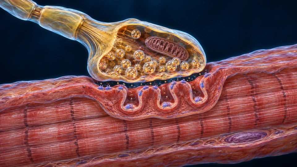
The cells meet across a chemical gap
The nerve ending releases transmitter at a specialised junction. The muscle then generates its own electrical excitation; transmitter does not travel down the myofibrils.
Generated neuromuscular-junction teaching cutaway, not electron microscopy. Vesicle counts, receptor shapes/positions, membrane spacing and fine protein detail are illustrative. No individual receptor/channel type is assigned to the depicted protein-like forms.
A chemical gap, then an electrical wave
Acetylcholine activates endplate receptors. Local depolarisation can initiate a muscle action potential. It is the muscle membrane’s excitation that travels onward; transmitter does not run down the myofibrils.
The signal is cleared
Acetylcholinesterase breaks down acetylcholine in the cleft. Excitation–contraction coupling and calcium recovery within the fibre are distinct from this clearance step.
At the neuromuscular junction, a motor-neuron ending releases acetylcholine. Receptor activation depolarises the endplate, leading to a muscle action potential. The signal travels along sarcolemma and T-tubules.
Controlled supporting illustration. All essential steps remain visible; select a step only to highlight it. Not a measured specimen, activation recording or complete cellular reconstruction.
Use Inspect full supporting comparison for the larger diagram. All three explanations remain visible.
Neuromuscular junction
ACh release activates endplate receptors, leading to a muscle action potential.
Excitation–contraction coupling
Sarcolemma and T-tubules carry excitation; coupling triggers SR calcium release.
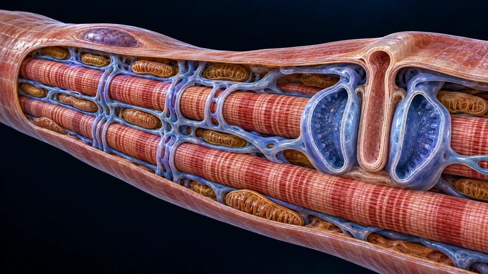
A skeletal fibre is one long cell. Its surface membrane, inward T-tubules and sarcoplasmic reticulum have different roles; a triad is a specific relationship between them.
Generated expanded membrane relationships, not histology or a molecular reconstruction. One T-tubule and two neighbouring SR cisternae form the triad. Fine networks, junctional proteins, counts and compartment widths are illustrative. The red T-tubule and blue SR are separate membrane compartments, not blood vessels.
Excitation reaches an internal store
In skeletal muscle, voltage-sensing machinery links T-tubule excitation to SR calcium release. Calcium then acts on the thin-filament regulatory system. This is not a diagram of calcium simply flowing from a nerve into muscle.
Recovery also uses energy
ATP-dependent pumps return calcium to the SR, reducing thin-filament activation. Skeletal and cardiac coupling differ; this triad should not be used as a universal cardiac-cell map.
Skeletal muscle fibres contain peripheral nuclei, myofibrils, mitochondria and membrane systems. T-tubules carry electrical excitation into the fibre; the sarcoplasmic reticulum stores and releases calcium. A fibre is not a motor unit or a fascicle.
Excitation-contraction coupling triggers calcium release from the sarcoplasmic reticulum. Calcium binds troponin, shifting tropomyosin so myosin can engage actin. Calcium is pumped back into the reticulum as activation falls.
This is the skeletal-muscle teaching mechanism. Smooth muscle uses different regulatory proteins, and cardiac excitation has additional calcium relationships; do not apply this exact sequence universally.
Controlled supporting illustration. All essential steps remain visible; select a step only to highlight it. Not a measured specimen, activation recording or complete cellular reconstruction.
Use Inspect full supporting comparison for the larger diagram. All three explanations remain visible.
Surface
Sarcolemma is the plasma membrane; peripheral nuclei belong to the single skeletal fibre.
Interior
T-tubules carry electrical excitation inward; SR calcium release enables activation.
Repetition
Myofibrils run along the fibre with repeating sarcomeres; mitochondria support metabolism.
CALCIUM REGULATION / FILAMENT INTERACTION
Shorter sarcomeres, unchanged filament lengths
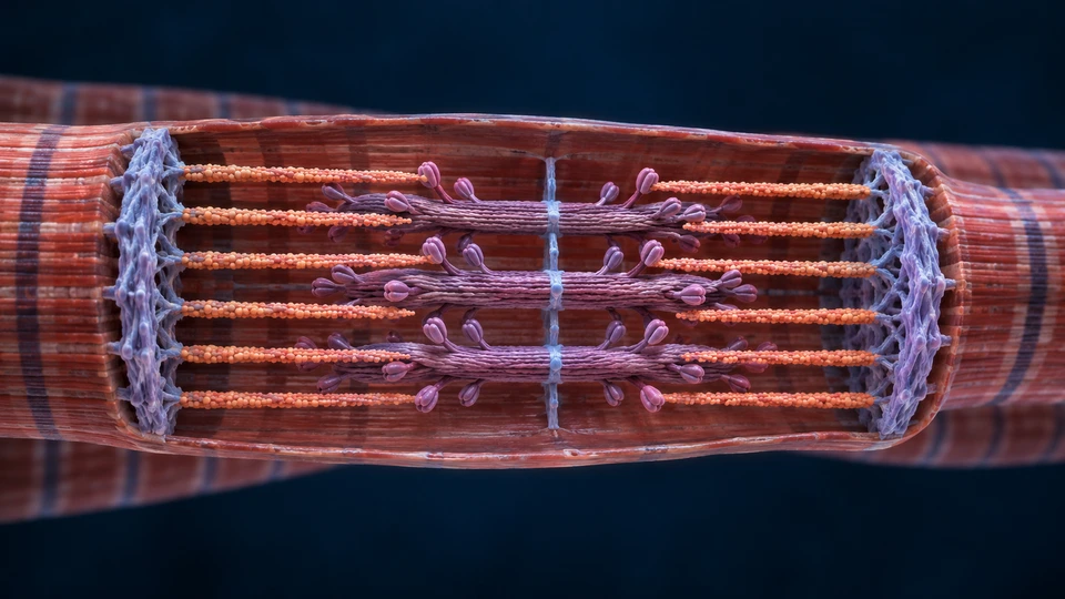
Actin and myosin overlap within repeated units. Calcium-dependent access and ATP-dependent interactions make active force possible; the proteins are not simply cut shorter.
Generated dimensional contractile arrangement, not microscopy or exact molecular geometry. Expanded lattice spacing, filament counts and head shapes are illustrative. The supporting controlled comparison below, rather than this single illustrative arrangement, demonstrates unchanged filament lengths.
Overlap changes
Thin and thick filaments slide relative to one another. During shortening, the Z discs approach and overlap increases; filament length is not the variable being shortened.
Activation and ATP matter
Calcium regulation permits actin–myosin interaction. ATP binding supports detachment; hydrolysis prepares another interaction. ATP is also needed for calcium recovery, not only for shortening.
A sarcomere lies between adjacent Z discs. Thin actin filaments are anchored toward the ends; thick myosin occupies the central region. During shortening, Z discs approach and overlap increases while filament lengths remain essentially unchanged.
ATP binding allows myosin to detach from actin. Hydrolysis primes the head for a subsequent interaction; force-producing steps accompany changes in the bound products. ATP also powers calcium pumping. It is not used only during shortening.
Controlled supporting illustration. All essential steps remain visible; select a step only to highlight it. Not a measured specimen, activation recording or complete cellular reconstruction.
Use Inspect full supporting comparison for the larger diagram. All three explanations remain visible.
Relaxed arrangement
Thin actin extends inward from each Z disc; thick myosin remains central.
Shortened arrangement
Z discs are closer and overlap increases. Actin and myosin lengths are unchanged.
Return / lengthening
External forces and control can restore length. Active lengthening is not the absence of force.
A repeating molecular cycle
Controlled supporting illustration. All essential steps remain visible; select a step only to highlight it. Not a measured specimen, activation recording or complete cellular reconstruction.
Use Inspect full supporting comparison for the larger diagram. All three explanations remain visible.
Detach / prime
ATP binds to detach myosin; hydrolysis primes the head with products bound.
Engage / force
Myosin engages accessible actin; product-release and conformational changes accompany force generation.
Continue / relax
New ATP supports another cycle; ATP also powers calcium pumping. A head does not push actin outward.
RECRUITMENT & RATE CODING
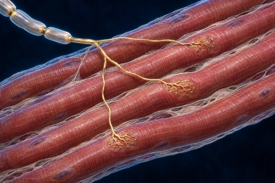
A functional unit crosses connective groupings
Motor-unit fibres are defined by their innervating neuron, not by a fascicle outline. Activating more units and changing discharge frequency provide distinct ways to regulate force.
Generated intramuscular arbor, not microscopy or an activation recording. Only selected branches and contacts are shown; their count, size, exact terminal shapes and spacing are not a measured motor-unit map. Visible contacts remain on distinct fibres connected to one incoming axon. No soma or complete spinal circuit is depicted.
A functional group, not a connective bundle
A motor unit is one motor neuron together with the fibres it innervates. Its fibres may be distributed among fibres belonging to other units. A fascicle is a connective-tissue grouping, so its boundary does not define one motor unit.
Force changes in space and time
Recruitment brings additional units into activity. Rate coding changes how frequently active motor neurons discharge. Both contribute to force control; changing frequency does not turn a fibre into a new unit. Their relative influence depends on the action.
A motor unit comprises one motor neuron and the skeletal fibres it innervates. Units overlap within a muscle rather than being identical to one fascicle. Recruitment and firing frequency help adjust force.
Controlled supporting illustration. All essential steps remain visible; select a step only to highlight it. Not a measured specimen, activation recording or complete cellular reconstruction.
Use Inspect full supporting comparison for the larger diagram. All three explanations remain visible.
Motor neuron
One motor neuron plus all its innervated skeletal fibres forms a motor unit.
Recruitment
Activating more units changes the number of contributing fibres.
Rate
Changing discharge frequency alters force; a unit is not a fascicle or one isolated muscle cell.
LENGTH / FORCE / CENTRAL INTERPRETATION
Different locations provide different feedback
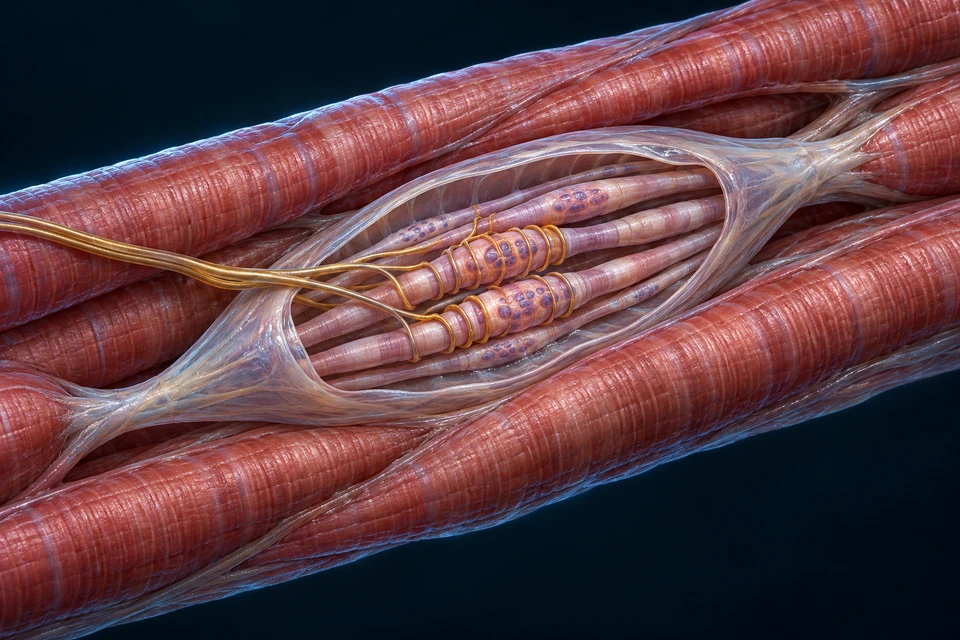
Spindles and tendon organs have distinct anatomical relationships. Their signals are integrated with other sensory and motor information; neither is a complete body-position meter.
Generated expanded spindle cutaway, not microscopy. Fibre counts, nuclear arrangements, neural branching and proportions are illustrative. Only broad capsule, fibre and sensory-region relationships are labelled; no exact Ia/II terminal or gamma axon is identified from an invented shape.
Length is interpreted, not read from a ruler
Spindle discharge supplies information related to muscle length and how it changes. Primary and secondary endings have different response properties; central processing is still needed during active movement.
The receptor also receives motor control
Gamma motor input influences intrafusal fibres and adjusts the receptor’s response to stretch. It is distinct from alpha motor input to the main force-producing fibres. This view does not show a complete efferent circuit.
Generated expanded tendon-organ context, not histology or a measured myotendinous junction. Collagen braids, capsule contour and neural branches are illustrative. Group Ib afferents innervate tendon organs, but this drawing cannot verify an individual axon’s molecular identity or exact termination.
A different placement gives different information
Unlike the spindle’s parallel arrangement, a tendon organ lies in a muscle-to-tendon force path. Its collagen-associated endings contribute force-related feedback rather than simply duplicating a muscle-length signal.
Feedback is broader than an emergency cut-off
Tendon-organ input participates in distributed motor circuits. Its effect varies with task and circuit state; the receptor should not be taught as a switch that always shuts off a muscle when force rises.
Position and movement are inferred from populations of sensory signals, alongside skin, joint, visual and vestibular information. These two local receptors are parts of that system, not a complete map of body awareness.
Muscle spindles lie within muscles and signal length and its changes. Golgi tendon organs relate to musculotendinous force. Skin and joint receptors also contribute; proprioception is a distributed process, not one sensor measuring every state.
Controlled supporting illustration. All essential steps remain visible; select a step only to highlight it. Not a measured specimen, activation recording or complete cellular reconstruction.
Use Inspect full supporting comparison for the larger diagram. All three explanations remain visible.
Muscle spindle
Within muscle, length and length-change information supports control.
Tendon-organ context
Force-related feedback arises at musculotendinous context; it is not a duplicate length sensor.
Integration
Spinal and higher circuits combine this with joint, skin and other sensory information.
SKELETAL / CARDIAC / SMOOTH
Three cellular organisations, different control
Striation, branching, nuclear position and cell shape help compare tissue. Appearance supports understanding, but the source and preparation must be known before interpreting a real specimen.
Skeletal muscle
Muscles attached to the skeleton and selected soft-tissue structures.
Generated cellular cutaway, not histology. Counts, scale, connective texture and fine organelle detail are illustrative.
Motor neurons activate fibres at specialised neuromuscular junctions. Recruitment coordinates force across the organ; a single fibre is still one cell.
Generated tissue arrangement, not an acquired specimen. Junctions are broad context; individual gap junctions or desmosomes cannot be identified from this artwork.
Specialised cardiac cells initiate rhythmic excitation. Autonomic input modulates the activity; it does not provide a separate skeletal-type motor endplate for each beat.
Many hollow-organ and vessel walls, and specialised sites such as the iris.
Generated cellular teaching illustration, not microscopy. Fine filament and anchoring forms are illustrative; no particular dense body or junction is assigned to a drawn dot.
Calcium–calmodulin regulation promotes myosin activation. Neural, hormonal and local inputs vary by tissue; some cells are electrically coupled, while others act more independently.
Skeletal, cardiac and smooth are three tissue types.
Skeletal muscle has long striated fibres. Cardiac muscle has striated branching cells joined at intercalated-disc context. Smooth muscle has spindle-shaped cells without the same sarcomeric striation pattern. Neural and hormonal control differ.
Controlled supporting illustration. All essential steps remain visible; select a step only to highlight it. Not a measured specimen, activation recording or complete cellular reconstruction.
Use Inspect full supporting comparison for the larger diagram. All three explanations remain visible.
Skeletal
Long striated, multinucleated fibres; motor-neuron activation.
Cardiac
Striated branching cells with intercalated-disc connections; cardiac activation differs.
Smooth
Spindle-shaped cells with different contractile organisation and regulation; no skeletal sarcomere pattern.
REAL OBSERVATIONS · SEPARATE FROM GENERATED ATLAS ART
Preparation changes what you can see
These acquired images are not AI recreations. Compare tissue direction and visible features, while retaining each preparation’s source, imaging method and limits.
Skeletal muscle · real histology
University-sourced longitudinal skeletal-muscle section. Species, stain and acquisition magnification are not specified in the source description. Original image is 696 pixels wide; zoom cannot reveal absent detail.
Authentic microscopy. Proportional WebP resizing only; full original field, colours and any embedded scale bar retained. No crop, recolouring, sharpening or AI reconstruction.
A longitudinal view can reveal repeating bands along aligned skeletal fibres. A nucleus must be located relative to a cell boundary before its position can be interpreted; the picture is not a map of every fibre type.
Original source ↗ · Courtesy Department of Histology, Jagiellonian University Medical College; Commons uploader Reytan · CC BY-SA 3.0. Derivatives retain the same licence.
Cardiac muscle · real mammalian specimen
Source identifies mammalian heart, differential interference contrast (Nomarski). It does not identify a human donor or exact mammalian species.
Authentic microscopy. Proportional WebP resizing only; full original field, colours and any embedded scale bar retained. No crop, recolouring, sharpening or AI reconstruction.
The specimen provides a real tissue field to compare with the constructed cardiac atlas. Not every cell boundary or intercalated disc is resolved confidently here, so no drawn line is assigned to an individual junction.
Original source ↗ · Josef Reischig, CSc., archive donated by the heirs · CC BY-SA 3.0. Derivatives retain the same licence.
Intestinal smooth muscle · real section
Source describes a longitudinal muscle section in a transverse intestinal preparation, bright-field microscopy. Species and stain are not specified in that description.
Authentic microscopy. Proportional WebP resizing only; full original field, colours and any embedded scale bar retained. No crop, recolouring, sharpening or AI reconstruction.
Elongated profiles in the muscle region differ from the regular bands of striated muscle. The entire intestinal field also contains other tissue components; every dark dot is not automatically a smooth-muscle nucleus.
Original source ↗ · Josef Reischig, CSc., archive donated by the heirs · CC BY-SA 3.0. Derivatives retain the same licence.
These views are not a matched experiment: imaging methods, section directions, fields and specimen metadata differ. Their pixel sizes do not establish relative cell dimensions. An embedded source scale bar resizes with its own image; a browser zoom level is not the microscope’s magnification. No clinical diagnosis or unverified fine junction identification is offered.
SUPPLY / RECOVERY / MAINTENANCE
Force depends on a living tissue environment
Blood delivery, metabolism, membrane recovery and the surrounding matrix sustain activity. Fatigue and adaptation cannot be reduced to one substance or a universal countdown.
New generated tissue illustration, not acquired microscopy or a measured specimen. The capillary is extracellular; mitochondria and contractile bundles are intracellular. Relative sizes, cell counts, capillary branching and organelle forms are illustrative. No oxygen particles, measured gradients or molecular transporters are drawn.
Delivery and use occur in different places
Blood moves within vessels. Oxygen reaching muscle cells crosses several tissue boundaries on its way from blood to the sites of oxidative metabolism. Capillaries between fibres provide exchange context; they do not become channels inside the myofibrils. The image locates these compartments without claiming a measured transport rate.
ATP is continually regenerated
Phosphocreatine, glycolytic reactions and oxidative metabolism contribute together, with their relative importance changing with demand and conditions. They are not three switches that always turn on in a fixed order. Mitochondria support oxidative metabolism; they are not the sole source of all cellular ATP.
Fatigue is a change in performance
Reduced force can involve neural drive, membrane excitation, calcium handling, contractile responses and metabolites. Phosphate-related changes can affect contractile performance and calcium release. This tissue picture cannot diagnose the dominant cause in a particular task, and fatigue does not mean that every muscle cell has exhausted its ATP.
Recovery is more than stopping motion
After activity, membrane gradients, calcium distribution and energy reserves continue to be regulated. A return of performance and longer-term tissue adaptation are different processes. A still image supplies their anatomical setting, rather than a universal recovery schedule or exercise prescription.
Stored ATP is limited. Phosphocreatine supports rapid regeneration; glycolytic pathways and oxidative metabolism contribute according to task and conditions. Oxygen delivery, mitochondria and available fuels influence sustained work.
Immediate support
ATP / phosphocreatine Rapid ATP regeneration, not an unlimited store.
Glycolytic support
Cell metabolism Contributes without a fixed universal countdown.
Oxidative support
Mitochondria / circulation Uses oxygen-dependent metabolism from several fuels.
A decline in force or task performance can involve central drive, membrane excitation, calcium handling, contractile processes and metabolism. The contribution depends on the task and how it is measured. Lactate is not a universal single explanation.
Recovery and adaptation occur on different timescales. This chapter does not provide exercise prescriptions, injury diagnosis or measured fatigue thresholds.
Fibres, satellite cells, fibroblastic cells, vessels and extracellular matrix have different roles in maintenance. Loading and signalling can change the tissue; connective tissue is more than passive wrapping.
Muscle fibre
Contractile cell Contains the contractile and excitation machinery.
Satellite-cell context
Adjacent to fibre surface Supports skeletal-muscle maintenance and repair.
Matrix / vascular context
Between fibres and bundles Supports force transfer, exchange and tissue organisation.
Original educational explanations connect tissue organisation, skeletal activation, filament interaction, recruitment, feedback and normal muscle types. The existing book inventory guides topics: FB029 muscle tissue, FB031 organisation and FB033 teams/feedback. Motor-unit rate coding and expanded cellular physiology use independent sources; no reference-book prose or figures are reproduced.
The constructed atlas pictures are generated illustrations. The separate microscopy section contains authentic, credited preparations with their own methods and metadata limits. Selected broad structures and their explanatory relationships were source-checked; dimensions, counts, molecular forms and exact ultrastructure are not measured. Where a shape cannot support a precise molecular label, only the supported context is labelled. Human medical review has not been performed; physical-phone and independent live review are separate from narrow-browser testing.
Selected elbow, knee and calf force paths plus a forearm transverse context are included. Full anterior/posterior muscle maps and qualified face/head/neck/trunk layers and complete joint mechanics remain incomplete. The acquired microscopy comparison is source-cleared; it is not a matched three-tissue experiment or a complete microscopy library. No invented specimen, patient scan, measured recording or treatment advice is supplied.
Content version: muscles-atlas-bridge-public-20261015-v9. Original complete muscular knowledge remains preserved in the project; this reading connects completed cellular and force-control teaching with selected regional attachment examples.
 A chemical contact ↗
 Surface to interior ↗
 Overlap produces force ↗
 Sense the movement ↗