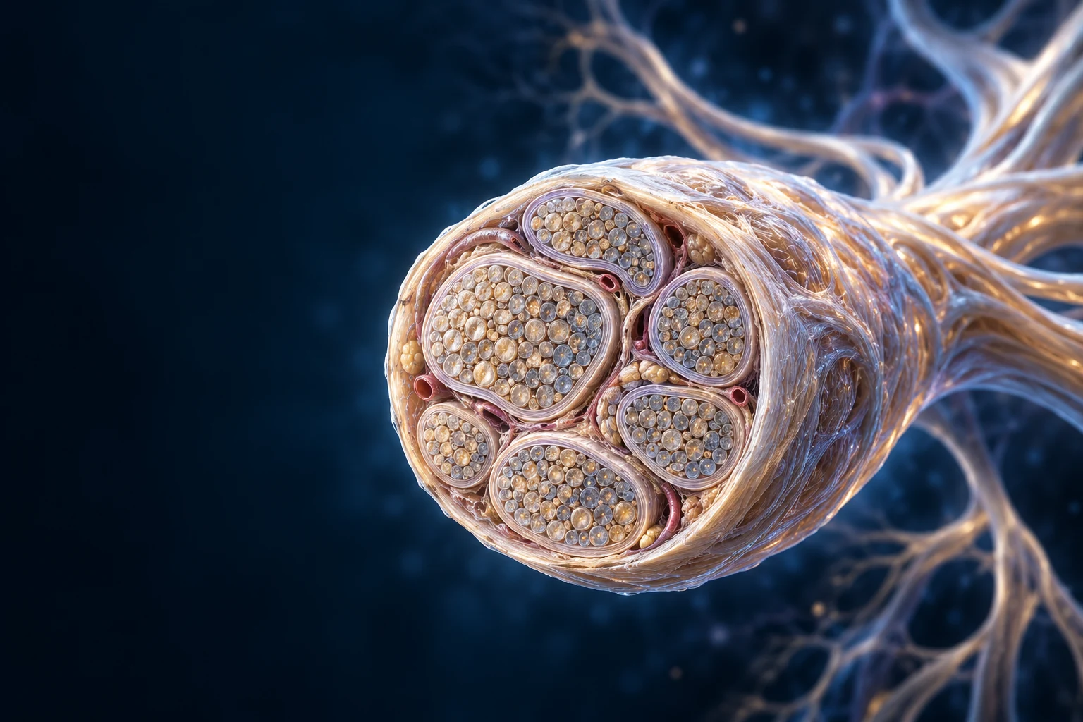
A nerve is more than a cable. Explore its bundles and supporting cells, follow roots into regional pathways, and connect anatomical structure with electrical and chemical communication.
Chapter overview film planned. No playable video is currently available.
Original anatomical exhibit · generated fine detail illustrative
01 / THE TISSUE HIERARCHY
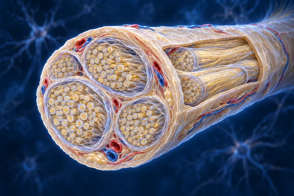
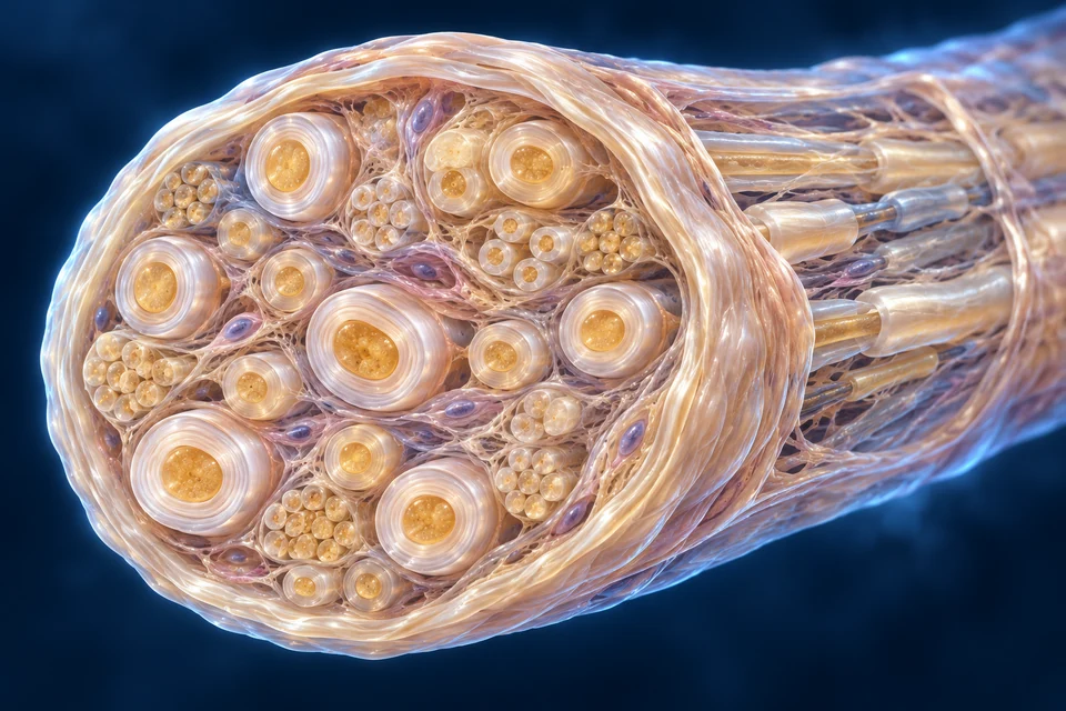
A nerve is organised around bundles.
Epineurium supports the nerve as a whole and between fascicles. Perineurium encloses a fascicle and contributes to a diffusion barrier. Endoneurial tissue supports individual nerve fibres and their local environment. None of these coverings is the axon membrane.
Original source-informed cellular illustration, not microscopy. Visible hierarchy and glial/axon relationships source-verified; fibre counts, lamellar thickness, nuclei, decorative organelles and fine connections illustrative / not independently verified. Human medical review: Not performed.Structure notes · further context
Epineurial support
This is not the axon membrane. Exact collagen weave and connective-tissue thickness are illustrative.
Perineurial boundary
Perineurial cells contribute to a diffusion barrier. The visible pale boundary indicates the relationship, not a counted cell-layer specimen.
Fascicle
A fascicle is not one neuron or one axon. Numbers and packing vary along a real nerve.
Nerve fibre profiles
Myelinated and unmyelinated relationships differ. Generated profiles do not prove measured diameters or identify every fibre.
Between-fascicle tissue
Do not confuse these spaces with the within-fascicle endoneurial environment.
Vascular context
The red vessel lies in connective context, not the central lumen of a nerve fibre. Its detailed course is illustrative.
Between-fascicle tissue: Connective support occupies the spaces between bundles. This connective-tissue region is between bundles, not the pale boundary of one fascicle. No precise point is assigned to an ambiguous generated boundary.
Epineurium
Whole nerve / between bundles
Connective support also accompanies a vascular supply.
Perineurium
Fascicle boundary
A specialised layered boundary, not another myelin sheath.
Endoneurium
Within a fascicle
Local tissue around nerve fibres, not the neuron membrane.
Distinguish central circuits, peripheral routes and the direction of information.
A network spans organs, tissues and cells.
The brain and spinal cord form the central nervous system. Peripheral nerves connect receptors, muscles, glands and viscera with central circuits. A nerve is a tissue containing many axons and supporting cells; it is not one long neuron.
Central
Brain / spinal cord
Integration occurs through many local and long-distance circuits.
Peripheral
Nerves / ganglia
Axons, glial cells and connective tissues link regional targets.
Functional divisions
Somatic / autonomic / enteric
These describe roles and circuits, not separate isolated bodies.
Sensory input and motor output are different directions.
Afferent pathways carry information toward central circuits. Efferent pathways carry commands toward effectors. A mixed peripheral nerve contains different axons serving these roles; one axon does not routinely alternate between being sensory and motor.
Input, integration and output
Start in a tissue, follow an incoming axon, then ask where a response is assembled. Sensory input and motor output travel in different axons.
Original generated anatomical teaching illustration, not microscopy. Representative free endings; colour distinguishes nerve from vessel, not a molecular receptor subtype. Cell counts, layer thickness and terminal density are not measurements.
Cyan: a representative sensory direction away from the skin endings. Not every skin ending detects the same stimulus.
Stimulus → electrical changeMechanical, thermal or chemical conditions can change a sensory ending’s membrane current. A graded receptor potential can bring a spike-generating region to threshold; stimulus information is carried by patterns of action potentials, not by a piece of the stimulus travelling inside the nerve.
One route, a soma beside itThe peripheral and central processes of a typical primary somatic sensory neuron meet at a T junction. Its soma lies in a sensory ganglion. The dorsal-root ganglion is not the central synaptic integration station.
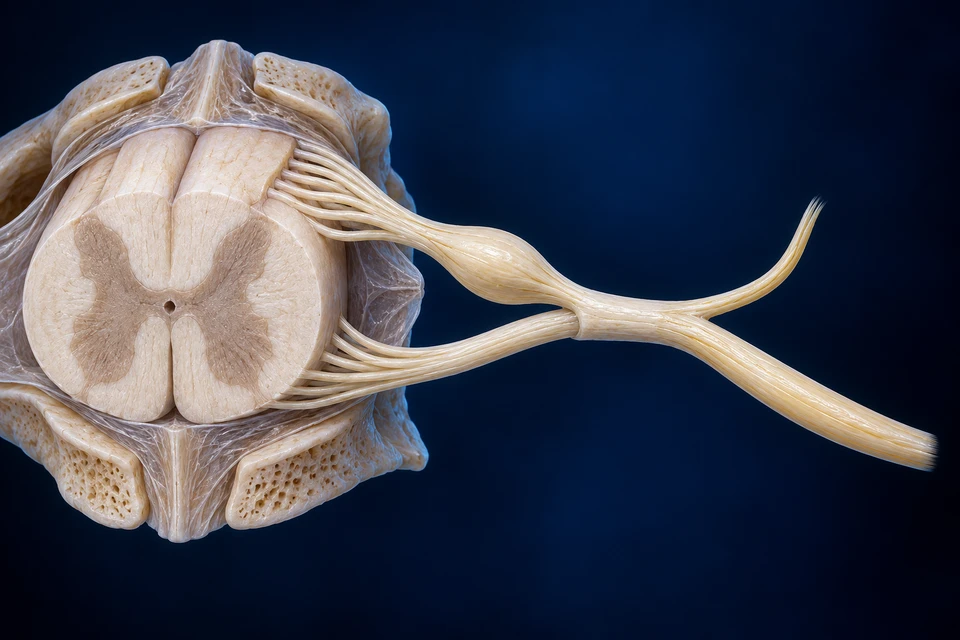
Into the cord. Out toward a target.
Original AI-assisted anatomical illustration, not a specimen or clinical scan. Posterior is above; anterior below. One representative level, not an exact vertebral-level or surgical map. Fine rootlets, horn nuclei, meningeal boundaries and communicating branches are not certified or completely shown.Structure notes · further context
Spinal cord section
The oblique cut gives context for roots. Individual horn nuclei and tracts are not identified by this generated texture.
Posterior root and sensory ganglion
The enlargement belongs to the posterior root before its union with the anterior root. It is not a ganglion of the ramus.
Anterior root
This is not exclusively skeletal motor output: at applicable levels it can also contain preganglionic visceral efferents.
Short mixed spinal nerve
Different sensory and motor axons share the short nerve. The single representative pathway is not a claim that each axon changes its direction of function.
Posterior ramus
Do not confuse this posterior ramus with the sensory posterior root; rami carry both afferent and efferent fibres.
Anterior ramus
Many anterior rami contribute to plexuses. This drawing does not show every connecting or meningeal branch.
Cyan: toward the cord through the posterior root. Gold: outward through the anterior root. Arrows follow broad root corridors, not a verified individual fascicle or intramedullary synapse. Both rami remain mixed.
Integration changes the responseA cutaneous withdrawal response recruits spinal interneurons and motor neurons; it is not a one-synapse skin-to-muscle shortcut. Excitation of selected muscles, inhibition of antagonists and signals to higher centres involve related but distinct circuits. The generated cord section does not locate those individual synapses.
Output meets a different tissueA lower motor neuron’s axon leaves toward skeletal muscle, where a specialised neuromuscular junction uses acetylcholine to initiate endplate depolarisation. This is a different cellular interface from a skin receptor. Reflex action can occur without a conscious decision, while descending circuits can modify the response.
Locate central tissue within its bony protection, then follow selected peripheral routes in real body context. A larger-scale window distinguishes the cord ending from the roots below.
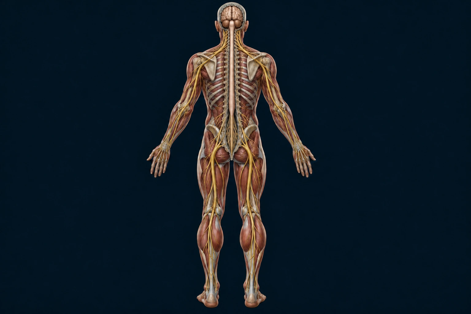
One body. Different neural compartments.
Start with the brain and cord, then find routes travelling into the limbs. The regional nerves lie among real muscles and bones; they are not single neurons drawn across an empty silhouette.
Posterior view. Bone and muscle are selectively opened to show different depths together. This original generated preparation is not a specimen, clinical scan, complete nerve inventory or exact vertebral-level map. Selected gold routes do not resolve every named branch, plexus contribution or skin territory.Structure notes · further context
Brain
Central neural tissue lies within the skull.
Spinal cord
Smooth central neural tissue lies inside the vertebral canal.
Upper-limb route
Peripheral bundles connect with regional arm and hand tissues.
Conus medullaris
The adult cord tapers in the upper lumbar region.
Cauda equina
Separate lower nerve roots continue below the cord.
Posterior-thigh route
A selected peripheral trunk continues through a different tissue compartment.
Locate central tissueCompare the brain with the cord in the opened vertebral canal. The cord is neural tissue; the surrounding vertebral column is bone. Connections within both central structures are more complex than the selected surface view can show.
Look closer · a cord is not the roots below it.
Locate the smooth taper, then the separate fibres within the lower canal. This small window is limited to its original pixel detail; the inspection view retains that limit instead of stretching the anatomy. A genuinely higher-detail local preparation remains to be supplied.
130 × 205 source-pixel window from the same posterior preparation; display size is capped to avoid invented or blurred detail. The smooth cord tapers above separate descending roots. The root bundle is not another solid spinal cord. Exact segment counts, tiny root attachments, meninges and individual exit levels are not resolved by this view.Structure notes · further context
Spinal cord
Smooth central neural tissue lies inside the vertebral canal.
Conus medullaris
The adult cord tapers in the upper lumbar region.
Cauda equina
Separate lower nerve roots continue below the cord.
Central tissue is surrounded by a different supporting skeleton.
The brain and spinal cord form the central nervous system. The skull and vertebral column protect that tissue; they are not themselves nervous structures. The opened posterior view lets you compare the brain, the cord within the canal and nerves travelling outward into the body. Local spinal circuits can organise responses as well as passing information along longer pathways.
A longer vertebral column does not mean a longer spinal cord.
In an adult, the cord usually ends around the first or second lumbar vertebra. Lower nerve roots continue down within the canal before reaching their exits. Those separate roots form the cauda equina: they are peripheral nerve roots, not a solid continuation of the cord. Spinal cord segments and the bones beside them therefore do not keep a one-to-one alignment down the whole back.
The large peripheral trunks are tissue bundles, not single neurons. Supporting cells, connective coverings and blood vessels accompany their axons. Sensory and motor axons can share a mixed nerve while serving different directions. Regional plexuses redistribute axons into routes to a limb; an intersecting line alone does not identify a synapse or a neuronal cell body.
Deeper learning · route, function and scale are different questions
Central versus peripheral describes anatomical organisation. Sensory versus motor describes the direction and role of particular axons. Somatic, autonomic and enteric learning adds further circuit distinctions; the posterior limb view is not a complete map of visceral or cranial pathways.
A lower root can descend inside the vertebral canal before leaving it. A named peripheral nerve can then contain fibres redistributed from several spinal levels. Neither a nerve’s position beside a bone nor the colour used in a teaching illustration establishes its complete root supply.
Directed source and gross-structure checks: 16 October 2026. Original generated illustration, with separate editable names, anchors and interaction. Retained native-source references below preserve their own licensed geometry. Fine anatomy, complete cranial and visceral routes, physical-phone and physician review are not certified.
This source-native overview locates limb routes and spinal sensory ganglia. It deliberately omits brain, cranial nerves and many smaller branches: a selected peripheral reference is not a complete nervous-system atlas.
Retained source-native supporting reference; native topology unchanged. Selected native peripheral routes; pale bones are ghosted for orientation. No independent deformation; incomplete branch coverage and fine source tube/surface detail remains illustrative / not independently verified. BodyParts3D — DBCLS (CC BY-SA 2.1 Japan); Z-Anatomy (CC BY-SA 4.0); FBX: Lluís Vinent Juanico. AllHumanBody render and changes: CC BY-SA 4.0. Full provenance in Sources below.Structure notes · further context
Median nerve, right
Source object: Median_nerver. The visible anchor intersects the selected native nerve mesh. Aggregate roots/ganglia are not an identification of one exact spinal level; tube surfaces are source modelling, not measured individual fascicles.
Ulnar nerve, right
Source object: Ulnar_nerver. The visible anchor intersects the selected native nerve mesh. Aggregate roots/ganglia are not an identification of one exact spinal level; tube surfaces are source modelling, not measured individual fascicles.
Femoral nerve, right
Source object: Femoral_nerver. The visible anchor intersects the selected native nerve mesh. Aggregate roots/ganglia are not an identification of one exact spinal level; tube surfaces are source modelling, not measured individual fascicles.
Tibial nerve, right
Source object: Tibial_nerver. The visible anchor intersects the selected native nerve mesh. Aggregate roots/ganglia are not an identification of one exact spinal level; tube surfaces are source modelling, not measured individual fascicles.
Brachial plexus roots, right
Source object: Roots_of_brachial_plexusr. The visible anchor intersects the selected native nerve mesh. Aggregate roots/ganglia are not an identification of one exact spinal level; tube surfaces are source modelling, not measured individual fascicles.
Spinal sensory ganglia, left
Source object: Spinal_ganglionl. The visible anchor intersects the selected native nerve mesh. Aggregate roots/ganglia are not an identification of one exact spinal level; tube surfaces are source modelling, not measured individual fascicles.
Move from the whole nerve to one fascicle, then distinguish axons from their supporting cells.
One bundle can contain different fibres.
A fascicle contains axons with their Schwann-cell context; a nerve can contain myelinated and unmyelinated fibres. Fascicle number and packing vary by nerve and along its course. The supporting illustration expands spaces to explain organisation.
Original source-informed cellular illustration, not microscopy. Visible hierarchy and glial/axon relationships source-verified; fibre counts, lamellar thickness, nuclei, decorative organelles and fine connections illustrative / not independently verified. Human medical review: Not performed.Structure notes · further context
Fascicle boundary
The opened surface is an educational preparation, not a clinical specimen.
Endoneurial context
Expanded spaces, nuclei and packing are illustrative; no cell identity is inferred from a purple generated shape.
Axon
It is separate from the surrounding glial membrane. This cross-section is not a measured diameter.
Myelin
Lamellar thickness and number are not independently verified; enlarged rings are educational context.
Smaller fibre context
Peripheral nerves include myelinated and unmyelinated fibres. The generated small profiles are not verified individual Remak-bundle reconstructions.
Source-informed educational illustration, not histology. Invented packing, repeated profiles and diameters are not measured anatomy or a universal fascicle count.
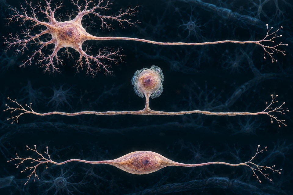
Motor neurons and many central neurons are multipolar. Many primary sensory neurons are pseudounipolar, with a peripheral and central process linked to their cell body. Bipolar cells occur in specialised sensory systems. These are examples, not a complete taxonomy.
Different cell shapes serve different routes
Three separate anatomical examples. Trace a process before naming its direction.
Original generated medical teaching illustration, not microscopy. Broad relationships source-checked; organelle counts, protein shapes and dimensions are not measurements. Labels and mechanism arrows are independently editable. Human medical review not performed.Structure notes · further context
Dendrites · multipolar
Branched input surfaces.
Soma · multipolar
Cell body and nucleus.
Axon · multipolar
One main output route branches distally.
Sensory soma · middle
Cell body beside the through-route.
T junction · middle
Route continues without a soma relay.
Bipolar example · below
Two major processes leave opposite poles.
Multipolar · many inputs, one axonal route
Many central and motor neurons have this arrangement. Inputs combine across dendrites and soma; action potentials commonly begin near the axon initial segment. This is one example, not a universal cell silhouette.
Pseudounipolar · soma beside the route
In a typical primary sensory ganglion neuron, one process leaves the soma and divides. The peripheral and central branches form a continuous route; the soma is not a synaptic relay. Left is peripheral context; right is central context in this example.
Bipolar · processes at opposite poles
Bipolar neurons occur in specialised sensory pathways, including retinal circuits. This shape is not a specific retinal layer map.
Teaching focus: Multipolar example — Dendritic inputs and a separate axonal route; not every cell has the same branching.
Multipolar example
Dendritic inputs and a separate axonal route; not every cell has the same branching.
Pseudounipolar example
Primary sensory soma branches off the continuous peripheral-to-central route; no relay synapse is shown at the soma.
Bipolar example
Distinct processes occur in specialised sensory contexts such as the retina.
Schwann cells support peripheral axons. Oligodendrocytes myelinate central axons. Astrocytes help regulate local tissue conditions; microglia participate in immune surveillance. Ependymal cells line ventricular surfaces. These roles are not interchangeable.
Schwann cells
Peripheral nerve
Myelinating and nonmyelinating relationships differ.
Oligodendrocytes
Central nervous system
One cell can supply internodes on multiple axons.
Other central glia
Astrocytes / microglia / ependyma
Different support, immune and lining roles; no fixed neuron-to-glia ratio.
Similar insulation. Different cellular arrangements.
Compare successive peripheral internodes with the branching support of a central glial cell.
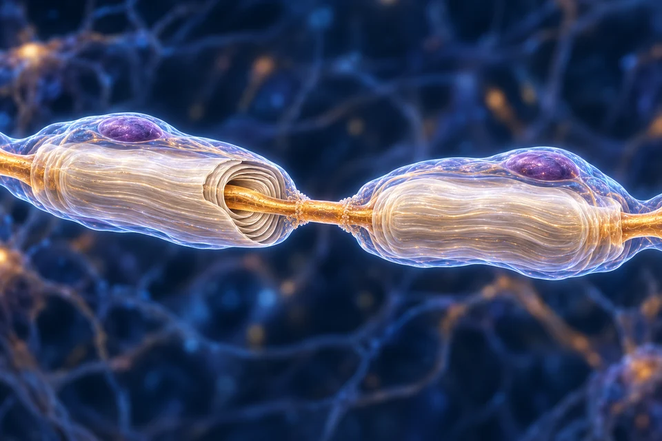
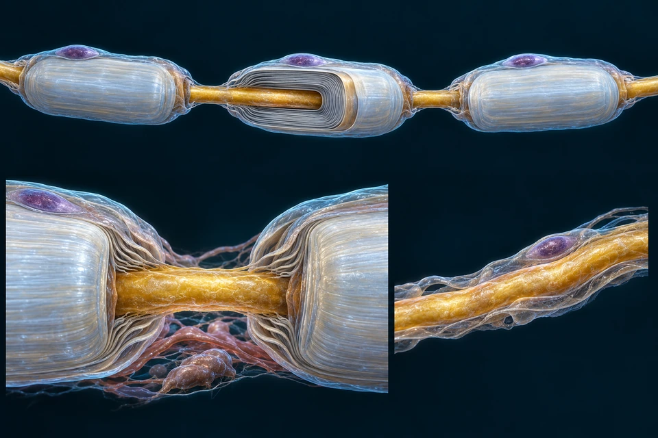
One Schwann cell makes one myelinated internode.
For a myelinated peripheral fibre, an individual Schwann cell wraps one axon segment. Successive cells form successive internodes. Gaps between internodes expose nodal membrane; the axon itself remains continuous.
Original source-informed cellular illustration, not microscopy. Visible hierarchy and glial/axon relationships source-verified; fibre counts, lamellar thickness, nuclei, decorative organelles and fine connections illustrative / not independently verified. Human medical review: Not performed.Structure notes · further context
Continuous axon
There is no severed cable gap. Fine membrane particles are illustrative and not identified channels.
Nodal region
The gap and diameter are enlarged for teaching; this is not a measurement of a node of Ranvier.
Peripheral internode
Successive Schwann cells form successive internodes; one cell does not supply all successive peripheral sheaths.
Membrane wrapping
The opened wedge reveals a relationship, not the exact number or chemistry of layers.
Schwann-cell nucleus
Fine generated cytoplasm and organelles are illustrative; this is not fluorescence microscopy.
The gap is enlarged for teaching. This is not an exposed cable break, measured nodal length or literal electron micrograph.
Central myelin uses a different cellular arrangement.
An oligodendrocyte can extend processes to internodes on different axons. The relationship differs from a myelinating Schwann cell. Exact numbers vary; the illustration does not establish a universal number of axons per cell.
Original source-informed cellular illustration, not microscopy. Visible hierarchy and glial/axon relationships source-verified; fibre counts, lamellar thickness, nuclei, decorative organelles and fine connections illustrative / not independently verified. Human medical review: Not performed.Structure notes · further context
Oligodendrocyte
This differs from one myelinating Schwann cell wrapping one peripheral internode. Exact counts vary.
Glial process
The thinner glial process is separate from the golden axon; it does not become the fibre centre.
Central myelin
Counts, membrane layers and process topology are illustrative / not independently verified as histology.
Central axon
It is distinct from the glial process. Enlarged diameters and visible cutaway details are not literal tissue measurements.
Gradients, channels and junctions carry information.
The fibre remains continuous. Supporting mechanism views explain voltage and transmission in the living tissue.
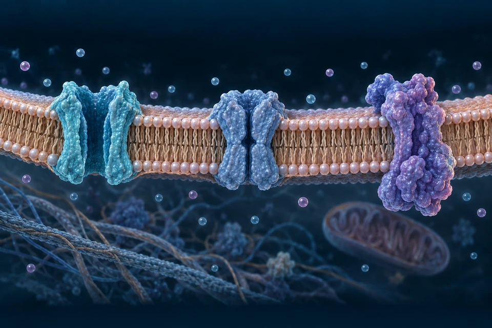
Voltage comes from gradients and selective permeability.
A resting membrane potential reflects ion gradients and membrane permeability. Ion pumps maintain gradients over time. Channels change permeability rapidly during signalling; the sodium–potassium pump does not directly generate each upstroke of an action potential.
Selective permeability changes voltage
Read across a real membrane boundary: a lipid barrier, selective channels and a separate energy-driven pump.
Original generated medical teaching illustration, not microscopy. Broad relationships source-checked; organelle counts, protein shapes and dimensions are not measurements. Labels and mechanism arrows are independently editable. Human medical review not performed.Structure notes · further context
Extracellular fluid
Sodium-rich relative to axoplasm.
Lipid bilayer
Heads face fluid; tails form an inner barrier.
Open channel · example
An aqueous pore permits selective ion flow.
Gated channel · closed example
A gate changes ion permeability.
Ion pump · illustrative
Energy supports gradient maintenance.
Axoplasm
Potassium-rich relative to extracellular fluid.
Gold arrow illustrates inward current through the open channel during a typical sodium-driven depolarising phase. It is not an arrow through the lipid core or a universal direction for every ion.
A selective barrier: lipid tails face inward; heads face the aqueous compartments. Ion distributions and relative permeability, especially resting potassium permeability in many neurons, contribute to membrane voltage.
A rapid change: gates alter permeability. In a typical spike, sodium entry drives the upstroke; sodium-channel inactivation and potassium conductance contribute to recovery. The qualitative waveform remains in the next lesson.
Maintaining the conditions: ATP-dependent pumps sustain gradients over time. They do not directly create each millisecond upstroke. Shown proteins are illustrative classes, not verified molecular structures; ion counts and colours are not measured concentrations.
Teaching focus: Gradients — Different ion distributions are maintained over time; symbols are not measured counts.
Gradients
Different ion distributions are maintained over time; symbols are not measured counts.
Channels
Selective leak and gated channels permit current according to electrochemical conditions.
Maintenance
Pumps sustain gradients; this is not the millisecond mechanism generating every spike upstroke.
Receptor and synaptic potentials can vary with input. Excitatory and inhibitory effects combine across space and time. Whether a cell reaches firing threshold depends on its state and inputs; a stronger stimulus is not simply a taller action potential.
Graded response
Receptor / synaptic regions
Amplitude varies; local changes can weaken with distance.
Summation
Inputs in space and time
Timing and location affect how signals combine.
Spike output
Excitable membrane
Frequency and timing can convey information; all-or-none is a scoped teaching model.
In a typical neuronal action potential, voltage-gated sodium-channel activation drives rapid depolarisation. Sodium-channel inactivation and potassium conductance contribute to repolarisation. Recovery creates refractory behaviour. Channel details vary by cell type.
A qualitative action-potential sequence
Supporting educational diagram; not literal regional anatomy, microscopy or a measured recording. Essential explanations remain visible. Select only to highlight a step.
At narrow widths, scroll the supporting diagram sideways for full-size labels.
Depolarisation
Typical neuronal sodium-channel activation drives a rapid upstroke.
Repolarisation
Sodium inactivation and potassium conductance contribute to voltage recovery.
Recovery
Refractory behaviour changes immediate re-excitability. No universal times or voltages are plotted.
Local current spreads between nodes in a myelinated axon; action potentials are regenerated at nodes. Unmyelinated axons regenerate along their excitable membrane. Saltatory conduction does not mean particles or whole spikes leap across empty axon gaps.
The axon remains continuous
Follow the continuous gold axon. Wrapping stops at nodes; the axon does not.
Original generated medical teaching illustration, not microscopy. Broad relationships source-checked; organelle counts, protein shapes and dimensions are not measurements. Labels and mechanism arrows are independently editable. Human medical review not performed.Structure notes · further context
Schwann-cell nucleus
Outside the compact myelin sheath.
Compact myelin
Many membrane wraps insulate an internode.
Continuous axon
The gold core crosses each nodal gap intact.
Node of Ranvier
Gap in myelin; the axon is not broken.
Gold arrows: local current along the axon, and Na⁺ entry at an exposed node. No particle leaps across a broken fibre.
Closer: where wrapping ends
Same original master, enlarged at a different scale. Myelin ends, axon membrane continues. Fine paranodal folds are illustrative, not a molecular junction model.Structure notes · further context
Node of Ranvier
Exposed excitable axon membrane.
Sheath ends
Compact myelin stops beside the node.
Compare: without compact myelin
Same original master, distinct comparison. Unmyelinated peripheral axons can still have nonmyelinating Schwann support; this is not every Remak-bundle arrangement.Structure notes · further context
Unmyelinated axon
Successive membrane regions regenerate the signal.
Under myelin: insulation reduces current loss and effective membrane capacitance. Local current in the continuous axon helps bring the next node toward threshold.
At nodes: concentrated voltage-gated sodium channels support regeneration of the action potential. Internodes are shortened and nodes enlarged here; no speed or dimension is measured.
Different glia: each myelinating Schwann cell serves one peripheral internode. A CNS oligodendrocyte can myelinate several internodes; existing cell-comparison plates above retain that distinction.
Teaching focus: Internode — Local current spreads under myelin; the axon is not interrupted.
Internode
Local current spreads under myelin; the axon is not interrupted.
Node
Excitable nodal membrane regenerates the action potential.
Sequence
Successive regeneration supports propagation; this is not a particle jumping a gap.
A junction can translate electrical activity into chemistry.
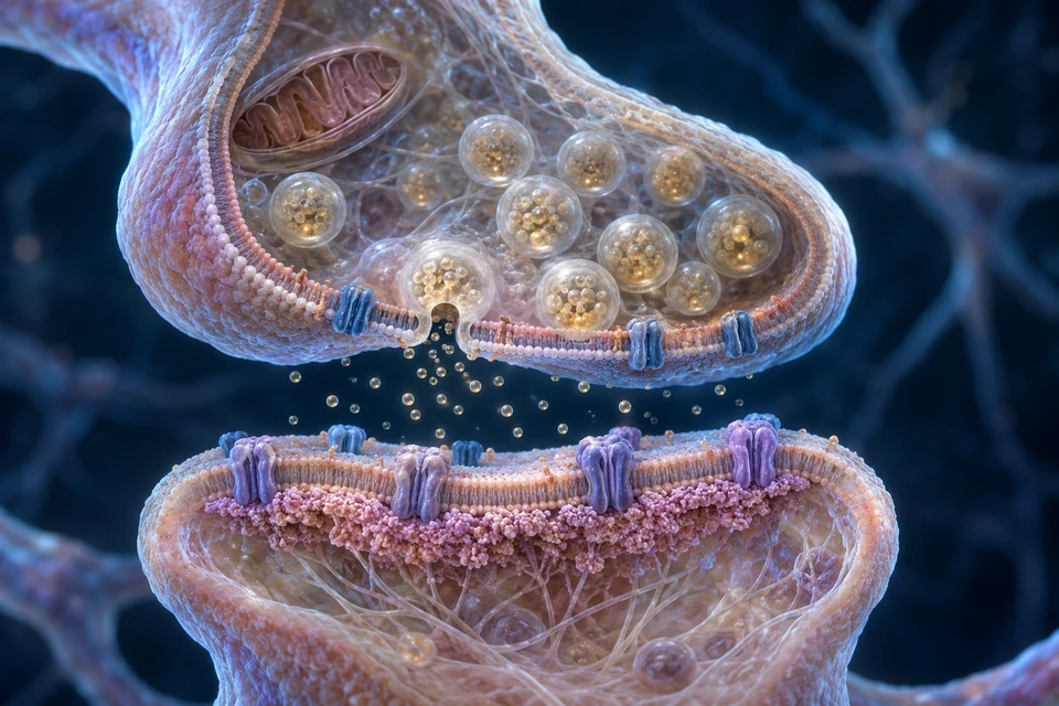
At a chemical synapse, calcium entry into a presynaptic ending supports transmitter release. Receptors change the postsynaptic response. Transmitter is cleared through several mechanisms. Electrical synapses use gap junctions and are a different arrangement.
Electrical arrival, chemical release, receptor response
Read from the upper terminal, across the cleft, to the lower receiving membrane.
Original generated medical teaching illustration, not microscopy. Broad relationships source-checked; organelle counts, protein shapes and dimensions are not measurements. Labels and mechanism arrows are independently editable. Human medical review not performed.Structure notes · further context
Presynaptic terminal
Electrical arrival triggers chemical release.
Synaptic vesicles
Transmitter is stored inside membrane sacs.
Fusion pore
Vesicle membrane joins terminal membrane.
Ca²⁺ channel context
Calcium enters from extracellular fluid.
Synaptic cleft
Released molecules cross extracellular space.
Postsynaptic receptor
A receiving membrane changes its response.
Upward gold arrow: Ca²⁺ entry through presynaptic channel context. Downward arrow: released transmitter into the cleft. Vesicles do not cross the gap.
Arrival → release: terminal depolarisation opens voltage-gated calcium channels. Incoming calcium supports vesicle fusion with the presynaptic membrane and release of its contents.
Release → response: transmitter binds receptors. This illustration scopes an ionotropic receiving junction; other receptors work through intracellular signalling. The postsynaptic effect depends on the receptor and ion gradients, and need not trigger a new spike.
Ending the signal: diffusion, transporter uptake and/or enzyme action clear transmitter according to the synapse. Electrical synapses instead pass current through gap junctions; they are not pictured here.
Locate the sensory ganglion, efferent root and short common nerve. A posterior ramus is not a posterior root.
Roots separate direction before a nerve becomes mixed.
Dorsal roots carry sensory axons into cord context; dorsal-root ganglia hold primary sensory cell bodies. Ventral roots carry efferent axons outward. After roots join, a short spinal nerve is mixed. The rendered source aggregates are regional, not a labelled single spinal level.
The posterior root carries incoming sensory fibres and has a ganglion containing their cell bodies. The separate anterior root carries efferent fibres outward; at applicable levels these include visceral as well as skeletal motor output.
After the roots unite, sensory and motor fibres share a short spinal nerve. Its posterior and anterior rami are both mixed. Their names describe where they go, not a split into purely sensory and purely motor nerves.
The preparation explains joining and branching. It does not establish a surgical exit point, vertebral level, complete meningeal envelope or every communicating branch. The native regional reference below shows a different scale.
Retained source-native supporting reference; native topology unchanged. Selected native peripheral routes; pale bones are ghosted for orientation. No independent deformation; incomplete branch coverage and fine source tube/surface detail remains illustrative / not independently verified. BodyParts3D — DBCLS (CC BY-SA 2.1 Japan); Z-Anatomy (CC BY-SA 4.0); FBX: Lluís Vinent Juanico. AllHumanBody render and changes: CC BY-SA 4.0. Full provenance in Sources below.Structure notes · further context
Posterior sensory roots, right
Source object: Posterior_root_of_spinal_nerver. The visible anchor intersects the selected native nerve mesh. Aggregate roots/ganglia are not an identification of one exact spinal level; tube surfaces are source modelling, not measured individual fascicles.
Anterior motor roots, right
Source object: Anterior_root_of_spinal_nerver. The visible anchor intersects the selected native nerve mesh. Aggregate roots/ganglia are not an identification of one exact spinal level; tube surfaces are source modelling, not measured individual fascicles.
Spinal sensory ganglia, right
Source object: Spinal_ganglionr. The visible anchor intersects the selected native nerve mesh. Aggregate roots/ganglia are not an identification of one exact spinal level; tube surfaces are source modelling, not measured individual fascicles.
Posterior sensory roots, left
Source object: Posterior_root_of_spinal_nervel. The visible anchor intersects the selected native nerve mesh. Aggregate roots/ganglia are not an identification of one exact spinal level; tube surfaces are source modelling, not measured individual fascicles.
After a spinal nerve forms, its dorsal and ventral rami both contain mixed fibres. Dorsal rami supply deep back and associated skin; ventral rami supply anterolateral trunk and limbs. Do not label a dorsal ramus as purely sensory or a ventral ramus as purely motor.
Root direction differs from ramus composition
Supporting educational diagram; not literal regional anatomy, microscopy or a measured recording. Essential explanations remain visible. Select only to highlight a step.
At narrow widths, scroll the supporting diagram sideways for full-size labels.
Dorsal root
Sensory axons enter; primary sensory somata lie in dorsal-root ganglia.
Ventral root
Efferent axons exit toward skeletal or autonomic pathways.
Mixed nerve and rami
After roots join, both dorsal and ventral rami carry mixed fibres. Ganglion is not drawn as a synaptic relay.
The usual arrangement comprises eight cervical, twelve thoracic, five lumbar, five sacral and one coccygeal pair. Cord segments, exiting nerves and vertebrae do not stay at matching heights through the adult column. Descending lower roots form cauda-equina context.
Cervical
8 pairs
C8 exits between C7 and T1 vertebrae.
Thoracic
12 pairs
Many ventral rami follow segmental thoracic routes.
Lower regions
5 lumbar / 5 sacral / 1 coccygeal
Lower roots descend before leaving the vertebral canal.
Licensed native-source references locate selected routes. They omit many surrounding tissues and do not replace a complete anatomical atlas.
A plexus redistributes axons; it is not a new relay.
Anterior rami contribute fibres to limb plexuses. Bundles regroup into trunks, divisions, cords and branches in the brachial plexus. These junctions reorganise axons; they are not ganglia where every fibre synapses.
Trace a contribution into a named nerve.
Original generated expanded nerve preparation, not in-situ anatomy, microscopy or a surgical map. Main connections independently traced against sources; smaller collateral branches are omitted. Spacing and generic muscle/bone context are not measured. Warm/cool nerve colours distinguish routes, not myelin, sensory or motor classes.Structure notes · further context
Musculocutaneous nerve
Arises from the lateral cord.
Axillary nerve
One terminal branch of the posterior cord.
Median nerve
Its lateral and medial roots unite before this route.
Ulnar nerve
Continues from the medial cord.
Radial nerve
A separate terminal branch of the posterior cord.
Musculocutaneous · Lateral cordThe lateral cord supplies a separate musculocutaneous route. It serves major anterior-arm muscles and continues into lateral-forearm sensory context; it is not a branch of the median nerve.
5 anterior ramiC5–T1 contributions
3 trunksUpper, middle and lower unions
6 divisionsEach trunk divides in two
3 cordsDivisions regroup
5 main terminal nervesDifferent routes to targets
A plexus reorganises axons. It does not turn each meeting point into a synapse. Follow the five named branches above, then inspect the two places where fibres regroup.
Closer · the three trunks
Same master, enlarged proximal preparation. These plexus “roots” are anterior rami. No dorsal-root ganglia or relay synapses are being identified.Structure notes · further context
C5 contribution
An anterior ramus, not the cord’s ventral root.
C6 contribution
Joins C5 in the typical superior trunk.
C7 contribution
Continues alone into the middle trunk.
C8 contribution
Joins T1 in the typical inferior trunk.
T1 contribution
The lowest of the five illustrated contributions.
Superior trunk
Typical union of C5 and C6 contributions.
Middle trunk
C7 continues without a paired-root union.
Inferior trunk
Typical union of C8 and T1 contributions.
Closer · divisions into cords
Same master, expanded central connections. Crossings in separate planes are not junctions. Posterior routes are cool-tinted only to aid tracing. The artery-relative cord names require the separate spatial view below.Structure notes · further context
Upper anterior division
Contributes to the lateral cord.
Middle anterior division
Also contributes to the lateral cord.
Lower anterior division
Continues into the medial cord.
Middle posterior division
Joins the other two posterior divisions.
Lateral cord
Receives the upper two anterior divisions.
Posterior cord
Receives all three posterior divisions.
Medial cord
Continues from the lower anterior division.
A root here is an anterior ramus. The separate sensory posterior root and efferent anterior root have already united into a spinal nerve before that nerve branches into rami. Plexus roots should not be confused with the cord’s motor rootlets.
The middle contribution stays distinct. In the typical pattern, C5 and C6 form the superior trunk, C7 continues into the middle trunk, and C8 with T1 form the inferior trunk. Each trunk supplies anterior and posterior divisions.
Recombination is selective. The upper two anterior divisions form the lateral cord; the lower anterior division continues as the medial cord. All three posterior divisions contribute to the posterior cord. The median roots subsequently unite, while axillary and radial branch separately from the posterior cord.
Regional position is a different question. Trunks are generally above the clavicle, divisions pass behind it, and cords occupy the axillary region. The network above is spread out to expose connections; use the retained regional reference and the separate artery view for spatial context.
Why “lateral”, “medial” and “posterior”?
Cord names refer to their typical relationship to the second part of the axillary artery, rather than to the reader’s screen. This separate, expanded tissue window establishes directions; it is not the same magnification or a registered slice of the network above.
Anterior ↑← Lateral · Medial →Posterior ↓
Original generated representative transverse organisation, not CT, MRI, a specimen or a certified exact slice through the artery’s second part. Selected structures only: the axillary vein and other regional structures are not fully shown. Spacing, fascicle counts and fine surrounding tissue are illustrative, not measurements.Structure notes · further context
Axillary artery
The vessel is distinct from the surrounding nerve cords.
Lateral cord
Toward the lateral side in the declared orientation.
Medial cord
Toward the medial side in the declared orientation.
Posterior cord
Behind the artery, not within its wall.
Deeper learning · a named nerve is not one spinal segment
A terminal nerve can contain axons from several spinal levels. A sensory dermatome describes a spinal-root relationship, whereas a named peripheral nerve’s skin territory combines its own branches and root contributions. Neither map is a perfectly sharp boundary in every person. Anatomical variations also change branching and contributions; this typical preparation is not a diagnostic map.
Retained native regional reference · locate the plexus
Retained source-native supporting reference; native topology unchanged. Selected native peripheral routes; pale bones are ghosted for orientation. No independent deformation; incomplete branch coverage and fine source tube/surface detail remains illustrative / not independently verified. BodyParts3D — DBCLS (CC BY-SA 2.1 Japan); Z-Anatomy (CC BY-SA 4.0); FBX: Lluís Vinent Juanico. AllHumanBody render and changes: CC BY-SA 4.0. Full provenance in Sources below.Structure notes · further context
Brachial plexus roots, right
Source object: Roots_of_brachial_plexusr. The visible anchor intersects the selected native nerve mesh. Aggregate roots/ganglia are not an identification of one exact spinal level; tube surfaces are source modelling, not measured individual fascicles.
Superior trunk, right
Source object: Superior_trunk_of_brachial_plexusr. The visible anchor intersects the selected native nerve mesh. Aggregate roots/ganglia are not an identification of one exact spinal level; tube surfaces are source modelling, not measured individual fascicles.
Middle trunk, right
Source object: Middle_trunk_of_brachial_plexusr. The visible anchor intersects the selected native nerve mesh. Aggregate roots/ganglia are not an identification of one exact spinal level; tube surfaces are source modelling, not measured individual fascicles.
Inferior trunk, right
Source object: Inferior_trunk_of_brachial_plexusr. The visible anchor intersects the selected native nerve mesh. Aggregate roots/ganglia are not an identification of one exact spinal level; tube surfaces are source modelling, not measured individual fascicles.
Posterior cord, right
Source object: Posterior_cord_of_brachial_plexusr. The visible anchor intersects the selected native nerve mesh. Aggregate roots/ganglia are not an identification of one exact spinal level; tube surfaces are source modelling, not measured individual fascicles.
Typical major contributions are C5–T1. Source anatomy and individual branching vary; the view does not establish a universal surgical pattern.
C5–C6 contributions usually form the superior trunk, C7 the middle trunk and C8–T1 the inferior trunk. Trunks divide; divisions combine into cords. Cords are named relative to axillary-artery context. Axillary and radial nerves arise from the posterior cord, not one from the other.
Roots
Anterior rami
These plexus roots are not the cord’s separate dorsal/ventral roots.
Trunks → divisions
Trunks above · divisions behind the clavicle
Three trunks each divide into anterior and posterior divisions.
Cords → branches
Axillary regional context
Lateral, medial and posterior cords contribute to terminal branches.
Median, ulnar and radial nerves take different courses and carry different branch combinations. Regional anatomy is more useful than treating the hand as three perfectly separate coloured zones. Sensory territories overlap and vary.
One elbow. Different nerve routes.
Use the bone and muscle context to distinguish a nerve passing behind a prominence from a nerve dividing into superficial and deep branches. These are separate selected views, not two sides of one registered scan.
Medial side · a route behind bone
Original generated selected preparation, not clinical imaging or a surgical map. The opened tissue exposes the posterior groove. Distal muscle heads and the complete cubital-tunnel roof are not individually certified or exhaustively shown.Structure notes · further context
Ulnar nerve
Descends toward the medial elbow.
Medial epicondyle
The nerve passes behind this humeral prominence.
Posterior nerve course
The nerve occupies the groove between the two bony landmarks.
Olecranon
The proximal ulna forms the other bony landmark.
Forearm continuation
The exposed nerve turns toward deeper forearm tissue.
Locate the posterior grooveThe nerve passes behind the medial epicondyle, between that prominence and the olecranon. This bony relationship is different from the radial nerve’s lateral-elbow branching.
Lateral side · one parent, different branches
Original generated teaching anatomy, not a specimen or surgical map. A selected lateral-elbow preparation exposes major branch relationships. Exact muscle attachments, the complete passage through supinator, the arcade of Frohse and a precise posterior-interosseous naming boundary are not identified. The posterior humeral course is outside this view.Structure notes · further context
Radial nerve
The parent nerve approaches the lateral elbow.
Division near the elbow
Two branches continue into different tissue planes.
Superficial branch
Continues beneath brachioradialis toward the distal forearm.
Deep branch
Turns toward the supinator region around the proximal radius.
Exposed deep segment
An opened tissue window reveals part of the deeper route.
Find the splitThe radial nerve has already travelled through the arm. Near the lateral elbow it divides: a common origin does not mean the two branches have the same targets.
Origin is not the same as destination. The ulnar nerve arises from the medial cord; the radial nerve from the posterior cord. Each carries many axons, with branches to different targets. A change in bundle direction does not create a relay synapse. Return to the plexus connections →
Move distally without inventing one common tunnel. At the wrist the median nerve travels beneath the flexor retinaculum in the carpal tunnel. The ulnar nerve uses a separate, more superficial ulnar-side route. A high-detail new wrist window is still in preparation; the elbow views above do not demonstrate those wrist relationships. Continue with existing flexor and pulley anatomy →
Position explains vulnerability, not a diagnosis. A superficial nerve beside a bony prominence and a branch travelling through muscle meet different mechanical surroundings. Symptoms depend on the level, fibres and severity involved; these illustrations cannot locate a lesion in an individual person. Distinguish nerve territory and spinal dermatome →
Deeper learning · what this pair of views cannot show
The radial nerve’s posterior humeral route lies outside the selected elbow view. The complete deep-branch passage through supinator, precise collateral branches, muscle-attachment details and cubital-tunnel roof are not mapped exhaustively. Natural variation also changes branching and territories. The retained licensed native regional reference below gives broader context at a different scale.
Names, short explanations, semantic anchor IDs and focus geometry are independent of the text-free medical images. The same figure data feed body, enlargement and accessible descriptions. English remains the public language.
Retained native regional reference · arm and hand routes
Retained source-native supporting reference; native topology unchanged. Selected native peripheral routes; pale bones are ghosted for orientation. No independent deformation; incomplete branch coverage and fine source tube/surface detail remains illustrative / not independently verified. BodyParts3D — DBCLS (CC BY-SA 2.1 Japan); Z-Anatomy (CC BY-SA 4.0); FBX: Lluís Vinent Juanico. AllHumanBody render and changes: CC BY-SA 4.0. Full provenance in Sources below.Structure notes · further context
Median nerve, right
Source object: Median_nerver. The visible anchor intersects the selected native nerve mesh. Aggregate roots/ganglia are not an identification of one exact spinal level; tube surfaces are source modelling, not measured individual fascicles.
Ulnar nerve, right
Source object: Ulnar_nerver. The visible anchor intersects the selected native nerve mesh. Aggregate roots/ganglia are not an identification of one exact spinal level; tube surfaces are source modelling, not measured individual fascicles.
Radial nerve, right
Source object: Radial_nerver. The visible anchor intersects the selected native nerve mesh. Aggregate roots/ganglia are not an identification of one exact spinal level; tube surfaces are source modelling, not measured individual fascicles.
Deep radial branch, right
Source object: Deep_branch_of_radial_nerver. The visible anchor intersects the selected native nerve mesh. Aggregate roots/ganglia are not an identification of one exact spinal level; tube surfaces are source modelling, not measured individual fascicles.
Common palmar digital branches
Source object: Common_palmar_digital_branches_of_median_nerver. The visible anchor intersects the selected native nerve mesh. Aggregate roots/ganglia are not an identification of one exact spinal level; tube surfaces are source modelling, not measured individual fascicles.
Dorsal ulnar branch
Source object: Dorsal_branch_of_ulnar_nerver. The visible anchor intersects the selected native nerve mesh. Aggregate roots/ganglia are not an identification of one exact spinal level; tube surfaces are source modelling, not measured individual fascicles.
A dermatome is skin associated with a spinal sensory root. A peripheral cutaneous nerve can contain fibres from several roots. A myotome concerns segmental muscle supply. Maps are useful approximations with overlap, not exact boundaries on every person.
Dermatome
Spinal root relationship
Overlapping segmental sensory context.
Peripheral territory
Named nerve relationship
Fibres may derive from multiple root levels.
Myotome
Segmental motor contribution
Muscles commonly receive more than one segmental contribution.
Anterior and medial thigh routes have different roles.
Femoral branches supply major anterior-thigh muscles and sensory routes. Obturator branches contribute to medial-thigh adductors. Their native paths through pelvic and femoral context are retained; surrounding muscles are omitted rather than invented.
Retained source-native supporting reference; native topology unchanged. Selected native peripheral routes; pale bones are ghosted for orientation. No independent deformation; incomplete branch coverage and fine source tube/surface detail remains illustrative / not independently verified. BodyParts3D — DBCLS (CC BY-SA 2.1 Japan); Z-Anatomy (CC BY-SA 4.0); FBX: Lluís Vinent Juanico. AllHumanBody render and changes: CC BY-SA 4.0. Full provenance in Sources below.Structure notes · further context
Femoral nerve, right
Source object: Femoral_nerver. The visible anchor intersects the selected native nerve mesh. Aggregate roots/ganglia are not an identification of one exact spinal level; tube surfaces are source modelling, not measured individual fascicles.
Obturator nerve, right
Source object: Obturator_nerver. The visible anchor intersects the selected native nerve mesh. Aggregate roots/ganglia are not an identification of one exact spinal level; tube surfaces are source modelling, not measured individual fascicles.
Anterior cutaneous femoral branches
Source object: Anterior_cutaneous_branches_of_femoral_nerver. The visible anchor intersects the selected native nerve mesh. Aggregate roots/ganglia are not an identification of one exact spinal level; tube surfaces are source modelling, not measured individual fascicles.
Posterior obturator branch
Source object: Posterior_branch_of_obturator_nerver. The visible anchor intersects the selected native nerve mesh. Aggregate roots/ganglia are not an identification of one exact spinal level; tube surfaces are source modelling, not measured individual fascicles.
The sciatic route contains tibial and common fibular components. Their separation level varies. Tibial branches serve posterior-leg and plantar-foot context; fibular branches serve anterior/lateral-leg and dorsal-foot context. Sensory branches are not synonymous with motor compartments.
Place the route. Then follow its two components.
These posterior right-sided views use different scales. The first locates a proximal route beneath reflected gluteal tissue; the second reveals how two major components continue beyond the thigh.
Hip to thigh · a deep, continuous route
Original generated selected preparation, not a specimen or surgical map. One common pattern is illustrated. The visible sacral side has four posterior openings; the opposite side and exact root-to-foramen mapping are not shown. Fine muscle attachments and every small nerve branch are not certified.Structure notes · further context
Piriformis
The selected sciatic route emerges below this muscle.
Sciatic nerve
A continuous common trunk appears at the muscle’s lower border.
Greater trochanter
The lateral femoral landmark helps orient the route.
Ischial tuberosity
The medial landmark lies beside the descending nerve.
Posterior-thigh continuation
The nerve continues deep toward the hamstring region.
Locate the proximal routeIn the illustrated common arrangement, the sciatic nerve leaves the pelvis below piriformis. Some people have a higher separation or a different muscle relationship; the picture is not an invariant pattern.
Knee to leg · central and lateral continuations
Original generated selected neuro-musculoskeletal view, not clinical imaging or a fixed branching-level standard. Medial is left; lateral is right. Smaller collateral nerves and popliteal artery/vein are omitted. Distal fibular branches and exact muscle attachments are not completely shown.Structure notes · further context
Visible separation
Tibial and common fibular routes become visibly separate here.
Common fibular nerve
The smaller route travels toward the lateral knee.
Tibial nerve
The central route continues toward the posterior leg.
Fibular head
A lateral bony landmark, distinct from the larger tibia.
Around the fibular neck
The nerve curves around the lateral bone before deeper continuation.
Distinguish the componentsA visible separation reorganises the outer bundles; it is not a synaptic relay. Its height varies from the pelvis or thigh to the region above the knee. This image chooses one representative level.
A shared route carries several spinal contributions. The usual sciatic contribution spans L4–S3 through the sacral plexus. Axons travel together without becoming one neuron. A named peripheral nerve and a spinal-root territory therefore answer different questions. Compare nerve territory and dermatome →
Muscle supply is not the same as overlying skin supply. Most hamstring motor fibres are associated with the tibial component; the short head of biceps femoris is a common fibular-component exception. Posterior-thigh skin chiefly uses a separate posterior femoral cutaneous route, omitted from these selected views.
Distal branches support different movements. The tibial route serves posterior-leg and plantar targets. Common fibular branches extend toward anterior-leg dorsiflexors and lateral-leg evertors. This is a functional overview, not a sharp sensory-zone map or a way to diagnose weakness. Connect with calf force and movement →
Deeper learning · variation, sensory overlap and the foot
The level at which the sciatic components visibly separate is variable. High divisions can also have different relationships with piriformis. A single teaching view cannot represent every anatomical variant or establish a compression site in an individual person.
Cutaneous pathways extend beyond a simple anterior-versus-posterior muscle grouping. Sural, saphenous, plantar and fibular branches have different origins and overlapping territories; skin maps are not identical to motor compartments. The separate saphenous route helps explain why the sciatic system is not the nerve supply of every part of the leg.
The images are original generated teaching preparations. Their semantic anchor IDs, names, short meanings, focus geometry and accessible copy remain separate from the text-free anatomy and are reused by the enlargement view. They are not clinical scans or measured dissections.
Retained source-native supporting reference; native topology unchanged. Selected native peripheral routes; pale bones are ghosted for orientation. No independent deformation; incomplete branch coverage and fine source tube/surface detail remains illustrative / not independently verified. BodyParts3D — DBCLS (CC BY-SA 2.1 Japan); Z-Anatomy (CC BY-SA 4.0); FBX: Lluís Vinent Juanico. AllHumanBody render and changes: CC BY-SA 4.0. Full provenance in Sources below.Structure notes · further context
Sciatic nerve, right
Source object: Sciatic_nerver. The visible anchor intersects the selected native nerve mesh. Aggregate roots/ganglia are not an identification of one exact spinal level; tube surfaces are source modelling, not measured individual fascicles.
Tibial nerve, right
Source object: Tibial_nerver. The visible anchor intersects the selected native nerve mesh. Aggregate roots/ganglia are not an identification of one exact spinal level; tube surfaces are source modelling, not measured individual fascicles.
Common fibular nerve, right
Source object: Common_fibular_nerver. The visible anchor intersects the selected native nerve mesh. Aggregate roots/ganglia are not an identification of one exact spinal level; tube surfaces are source modelling, not measured individual fascicles.
Sural nerve, right
Source object: Sural_nerver. The visible anchor intersects the selected native nerve mesh. Aggregate roots/ganglia are not an identification of one exact spinal level; tube surfaces are source modelling, not measured individual fascicles.
Deep fibular nerve, right
Source object: Deep_fibular_nerver. The visible anchor intersects the selected native nerve mesh. Aggregate roots/ganglia are not an identification of one exact spinal level; tube surfaces are source modelling, not measured individual fascicles.
Principal cranial roles and reflex organisation extend the pathways without inventing missing cranial geometry.
Twelve cranial nerves serve different functions.
The directory below names principal learning roles, not every fibre component. The optic pathway is specialised central tissue; cranial nerves are not all identical peripheral cable designs. Current source geometry does not provide a verified complete cranial-nerve plate.
CN I
Olfactory
Smell-related sensory input.
CN II
Optic
Retinal ganglion-cell output in a specialised central visual pathway.
CN III
Oculomotor
Most extraocular movements, eyelid elevation and parasympathetic pupil/accommodation routes.
CN IV
Trochlear
Motor supply to superior oblique.
CN V
Trigeminal
Principal facial sensation and motor supply to mastication muscles.
CN VI
Abducens
Motor supply to lateral rectus.
CN VII
Facial
Facial expression, anterior tongue taste and parasympathetic gland routes.
CN VIII
Vestibulocochlear
Hearing and balance-related sensory pathways.
CN IX
Glossopharyngeal
Posterior tongue sensation/taste, stylopharyngeus and parotid-related parasympathetic routes.
CN X
Vagus
Visceral sensory and parasympathetic routes plus pharyngeal/laryngeal motor roles.
CN XI
Accessory
Motor supply to sternocleidomastoid and trapezius context.
CN XII
Hypoglossal
Most tongue muscle motor supply; palatoglossus is a vagal exception.
A sensory ending, afferent route, central circuit, efferent route and effector form a useful reflex framework. Some circuits include a direct sensory-to-motor synapse; others use interneurons and coordinated inhibition. Reflexes are also modulated by higher circuits.
Internal regulation depends on targets and receptors.
Follow autonomic origins, relays and transmitter exceptions rather than treating the divisions as universal opposite switches.
Visceral regulation is coordinated, not two universal switches.
Autonomic output regulates smooth muscle, cardiac muscle and glands. Sympathetic and parasympathetic pathways have different central origins and ganglion arrangements. Effects depend on target and receptor; not every target receives equal opposing innervation.
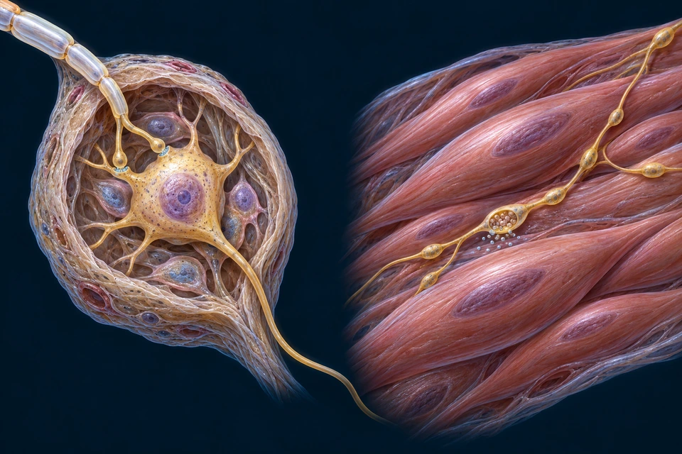
Compare two scoped output arrangements
A typical autonomic output uses two separate neurons. Then inspect how a terminal axon influences visceral tissue through several release sites.
Original generated teaching preparations at different magnifications, not microscopy or a traced continuous dissection. The CNS preganglionic soma is outside the view. Right-side particles are transmitter symbols, not a chemically identified substance. Fine synapses and cell wrapping are illustrative.
Peripheral ganglion · cell-body scale
Original generated teaching preparations at different magnifications, not microscopy or a traced continuous dissection. The CNS preganglionic soma is outside the view. Right-side particles are transmitter symbols, not a chemically identified substance. Fine synapses and cell wrapping are illustrative.
Smooth-muscle contact · closer view
Original generated teaching preparations at different magnifications, not microscopy or a traced continuous dissection. The CNS preganglionic soma is outside the view. Right-side particles are transmitter symbols, not a chemically identified substance. Fine synapses and cell wrapping are illustrative.
A true relay in a ganglionA preganglionic neuron has its soma in the brainstem or spinal cord. Its axon contacts a separate postganglionic neuron in a peripheral ganglion. Acetylcholine acting on neuronal nicotinic receptors supports the typical ganglionic synapse. This differs from the sensory soma beside a through-route.
Release along the targetPostganglionic terminal branches can carry multiple en-passant swellings, or varicosities. Transmitter released near smooth-muscle cells influences them through their receptors. The close-up is a representative diffuse arrangement, not a single folded skeletal-muscle endplate or a complete map of all visceral targets.
Somatic motor → skeletal muscle
One lower motor neuron connects the CNS to a skeletal-muscle fibre through its axon and specialised motor endplate. Acetylcholine evokes endplate depolarisation; muscle membrane excitation and internal calcium handling follow.
Most sympathetic postganglionic neurons use norepinephrine at targets; many parasympathetic targets use acetylcholine. Sweat-gland sympathetic cholinergic fibres are an important exception. A target’s receptor and intrinsic activity determine its response; divisions are not universal opposite switches.
Keep the exceptions: the adrenal medulla receives preganglionic input and releases catecholamines into blood, rather than supplying a conventional postganglionic axon to each target. Enteric networks include local circuits. Neither is forced into the illustrated two-neuron arrangement.
Sympathetic ganglia lie in paravertebral chains beside the column; other relay sites are prevertebral. Preganglionic fibres can relay at different levels or continue to other ganglia. A chain is not the spinal cord and not a single continuous neuron.
Beside the spine. Connected across levels.
Locate the paired chain, distinguish a ganglion from the connecting cord, then follow how an outflow can reach a different level or another relay. The spinal cord lies deeper, within the vertebral canal; it is not either of these gold chains.
Original generated regional teaching model, not a scan or specimen. The spinal cord and microscopic synapses are outside this view. This cropped thoracic segment is not a count or exact-level map of the entire chain. Fine branching and joint surfaces require separate verification.Structure notes · further context
Sympathetic trunk
The paired chains lie outside the vertebral canal.
Chain ganglion
A local swelling contains postganglionic neuronal cell bodies.
Between ganglia
Axons connect neighbouring ganglia along the chain.
Ramus communicans
A visible short side connection joins chain and local nerve.
Thoracic nerve
Selected mixed-nerve segment emerges from deeper context.
Rib
Posterior chest-wall bone provides spatial orientation.
Vertebral body
The chains lie beside this central bony column.
Locate the chainThe chain lies outside the vertebral canal. A ganglion contains neuronal cell bodies; the slimmer connections carry axons between ganglia. Neither structure is the spinal cord. The opposite chain supplies another regional route, rather than forming a single cord down the midline.
Look closer at a short connection.
The neighbouring nerve has its own deeper course. A communicating branch joins it from the chain; the chain does not create the whole nerve. Compare this local connection with the larger paired arrangement above.
A local view of the same teaching model, not another specimen. One short connection is visible; white versus gray fibre composition cannot be identified. The complete distal intercostal route and costal groove continuity are not verified by this preparation.Structure notes · further context
Chain ganglion
A local swelling contains postganglionic neuronal cell bodies.
Ramus communicans
A visible short side connection joins chain and local nerve.
Thoracic nerve
Selected mixed-nerve segment emerges from deeper context.
Outflow levels differ from distribution levels.
Preganglionic sympathetic cell bodies lie in thoracic and upper lumbar spinal gray matter, usually described as T1–L2. White rami enter the chain at those outflow levels; gray rami connect with spinal nerves throughout the column. Axons travelling within the chain help reach regions above or below the central origin.
A route can contain a relay, or pass toward another one.
A synapse transfers influence to another neuron; a branch or crossing alone is not a synapse. Thoracic splanchnic pathways can carry preganglionic axons beyond the chain to a prevertebral relay. The adrenal medulla is a further exception, explained with the existing transmitter section. Do not treat every splanchnic nerve everywhere as the same fibre type.
Returning to a spinal nerve reaches a different set of targets.
Postganglionic axons distributed through spinal nerves can reach cutaneous blood vessels, sweat glands and arrector pili muscles. Motor axons to skeletal muscle remain a different neuronal pathway. Visceral afferent routes can accompany autonomic nerves, but their sensory cell bodies are not the sympathetic chain’s postganglionic relay cells.
“Preganglionic” and “postganglionic” describe axons relative to a sympathetic relay. White rami include myelinated preganglionic axons; gray rami predominantly carry unmyelinated postganglionic axons. The gold surfaces in this regional image do not resolve those microscopic properties. The local short connection is therefore not individually certified as white or gray.
Axons can enter a ganglion, continue within the trunk, or pass onward toward another relay. Thoracic splanchnic and direct visceral branches are omitted from this regional preparation. Their destinations must not be inferred from a missing line or from a nearby rib. The complete intercostal nerve course and finer communicating-branch topology still need a separate anatomical view.
Regional sensory ganglia and sympathetic chain ganglia are also different structures. Compare their positions and cell-body roles before treating every swelling beside the spine as the same kind of relay.
Directed source and gross-structure checks: 16 October 2026. Original generated illustration with independent editable names and positions; not microscopy, a surgical map, physician review or a count of all ganglia. The same raster and semantic terms supply the full view, local window and enlarged views.
Retained source-native supporting reference; native topology unchanged. Selected native peripheral routes; pale bones are ghosted for orientation. No independent deformation; incomplete branch coverage and fine source tube/surface detail remains illustrative / not independently verified. BodyParts3D — DBCLS (CC BY-SA 2.1 Japan); Z-Anatomy (CC BY-SA 4.0); FBX: Lluís Vinent Juanico. AllHumanBody render and changes: CC BY-SA 4.0. Full provenance in Sources below.Structure notes · further context
Sympathetic trunk, right
Source object: Sympathetic_trunkr. The visible anchor intersects the selected native nerve mesh. Aggregate roots/ganglia are not an identification of one exact spinal level; tube surfaces are source modelling, not measured individual fascicles.
Sympathetic ganglia, left
Source object: Ganglia_of_sympathetic_trunkl. The visible anchor intersects the selected native nerve mesh. Aggregate roots/ganglia are not an identification of one exact spinal level; tube surfaces are source modelling, not measured individual fascicles.
Bone context is ghosted to reveal source paths. This selected chain view does not show every visceral target or establish exact ganglion counts.
Cranial and sacral parasympathetic pathways use ganglia near or in target tissues. The vagus supplies important thoracic and abdominal routes; sacral pathways supply other pelvic contexts. Their complete courses are not rendered in this selected peripheral atlas.
Cranial outflow
III / VII / IX / X contexts
Different pathways reach eye, glands and visceral targets.
Sacral outflow
Pelvic contexts
Distinct routes contribute to lower visceral regulation.
Transmitters and receptors explain important exceptions.
Both divisions use acetylcholine at autonomic ganglia. Most sympathetic postganglionic fibres use noradrenaline; many sweat-gland fibres use acetylcholine. Parasympathetic postganglionic fibres use acetylcholine. The adrenal medulla releases catecholamines into blood, a neuroendocrine exception.
Ganglion
ACh / nicotinic receptors
The shared preganglionic transmitter does not imply identical target effects.
Effector
Receptor-dependent response
Muscarinic and adrenergic receptor contexts differ.
Adrenal medulla
Circulating signal
Not another ordinary long postganglionic nerve ending.
Cardiac pacemaking has intrinsic mechanisms modulated by nerves. Gut circuits organise local activity with autonomic influence. Pupillary and glandular responses use specialised routes. Visceral control operates together with hormonal and local tissue signals.
Heart
Rate / conduction context
Autonomic modulation is not the sole source of each heartbeat.
Gut
Enteric circuits
Local networks interact with sympathetic and parasympathetic inputs.
Glands / vessels
Target-specific receptors
Responses are not uniformly opposite in every tissue.
Different representations answer different questions. Established related chapters extend the learning without duplicating accepted anatomy.
An anatomy mesh and microscopy answer different questions.
A render shows source-recorded regional paths. Histology reveals fascicles and connective boundaries; electron microscopy resolves axon and myelin detail; labelled fluorescence identifies molecular distribution. None of the controlled illustrations is a patient scan or measured specimen.
Regional source render
Spatial anatomy
Native source mesh, limited branch coverage.
Histology / EM
Tissue and ultrastructure
Actual licensed high-resolution peripheral specimens remain a gap.
Physiology
Electrical recording
Not inferred from colour, diameter or a still rendering.
Explore the established brain chapter for central structures, spinal section, sleep and cognition. Muscle, skin, eyes and hand/foot pages extend the peripheral routes into targets and sensory contexts without duplicating their accepted main anatomy.
Normal-function depth with explicit source limits.
This chapter adds peripheral organisation, tissue hierarchy, myelin, signalling, roots, plexuses, limb routes, cranial roles and visceral regulation. Clinical disorders, diagnostic maps and treatment belong to a later phase. Coverage is not a fabricated completeness percentage.
Source-supported educational relationships; native source surfaces and fine illustrative tissue detail are not independently validated histology. Human medical review: not performed. Real physical-phone acceptance: not performed. No complete cranial/enteric/visceral-target geometry or measured conduction data.
Peripheral organisation, tissue hierarchy, myelin, signalling, roots, plexuses, limb routes, cranial roles and visceral regulation. Existing normal-function knowledge remains intact. Independent references support broad relationships; no source illustration or private-book prose is reproduced.
Representation limits
AI-assisted original teaching. Generated tissue and root preparations are not acquired histology, patient scans or surgical maps. Fine membranes, nuclei, channels, counts and tiny branches remain illustrative. Native regional renders are supporting references, not complete premium anatomy. Verified complete cranial geometry and autonomic routes remain gaps. Human medical review: not performed.
Regional source attribution
BodyParts3D — The Database Center for Life Science — CC BY-SA 2.1 Japan; Z-Anatomy — The open source atlas of anatomy — CC BY-SA 4.0; FBX export by Lluís Vinent Juanico. AllHumanBody selected native peripheral and skeletal objects, changed visibility, camera, lighting and materials, and exported proportional rasters. Regional derivatives: CC BY-SA 4.0. Mixed-licence third-party brain, cranial, ear and kidney meshes were excluded. Application code is not asserted to be licensed as source artwork.
Revision: nervous-system-english-2026-10-02-deep-v1. Stable local key: ahb_ch_nervous_system_english_v1. No new CMS entity, publication-state change or fabricated model mapping. Text-free masters and editable labels remain separate. Fine generated anatomy, human medical review and physical-phone acceptance are not certified. Educational only; not diagnostic or surgical guidance.