Nervous / VISUAL EXPLAINER
Which Cells Line the Fluid Spaces Inside Your Brain?
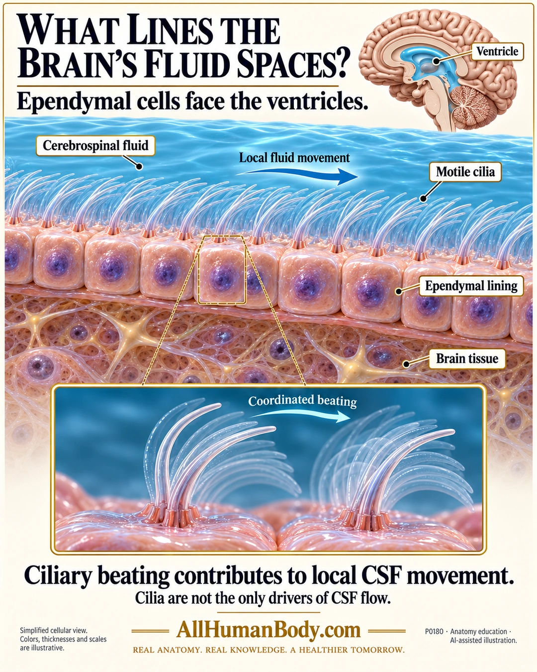
Multiciliated ependymal cells line brain ventricles, with cilia projecting from the fluid-facing surface. Coordinated beating contributes to local cerebrospinal-fluid movement, rather than acting as the only driver of its circulation.
READ THE IMAGE
What the illustration shows
Ivory-and-gold poster with a small sagittal brain locator indicating a blue ventricular region. Main enlarged wall has blue cerebrospinal fluid above one row of pink ependymal cells with purple nuclei, and neural tissue below. Fine pale cilia project from the fluid-facing surfaces, with an arrow indicating local fluid movement. Labels identify CSF, motile cilia, ependymal lining and brain tissue. Lower inset enlarges cilia with lighter curved positions suggesting coordinated beating. Text states cilia contribute to local CSF movement and are not its only drivers. Gold AllHumanBody.com and complete slogan appear at the bottom. Colors, thicknesses, locator and motion sequence are schematic.
How to interpret this poster
- Many mature ependymal cells are multiciliated; the poster does not establish that every ependymal cell is identical.
- Locator is a simplified ventricular region, not a full sagittal CSF map. Ciliary overlays do not measure beat mechanics, human flow speed or direction throughout the brain.
- Ordinary ventricular lining is not shown as the sole CSF source or a completely sealed blood–brain barrier. No hydrocephalus diagnosis or prevention claim.
- AI-assisted educational illustration; not physician certification, microscopy, a diagnosis or individualized medical advice.
- Fine annotations need enlargement; the full description provides an accessible text alternative.
- Display the full uncropped illustration; do not trim titles, legends or boundary annotations.
AI-assisted educational illustration, adapted into web-sized images without changing its composition. This is general education, not an individual diagnosis or treatment recommendation.
EVIDENCE / CONTEXT
Sources and what they support
- Planar Organization of Multiciliated Ependymal (E1) Cells in the Brain Ventricular Epithelium ↗
Multiciliated ventricular lining and coordinated ciliary contribution to CSF flow; review abstract, not full-article verification.
Evidence correspondence is limited to the abstract.