Hormones connect distant tissues, but circulation alone does not choose a target. A response depends on the message, the receiving cell and the changing needs of the body.
Chapter overview film planned. No playable video is currently available.
A source → a message → a responsive target · fine generated detail illustrative
30-SECOND ORIENTATION · THREE CONNECTED SCALES
Sources, delivery and reception are different jobs.
Use the pictures to distinguish where a message originates, how it reaches the circulation and why a receiving cell responds. Then follow the deeper anatomy and mechanisms below.
Locate a source · distinguish delivery · identify reception
1 · Where is the source?
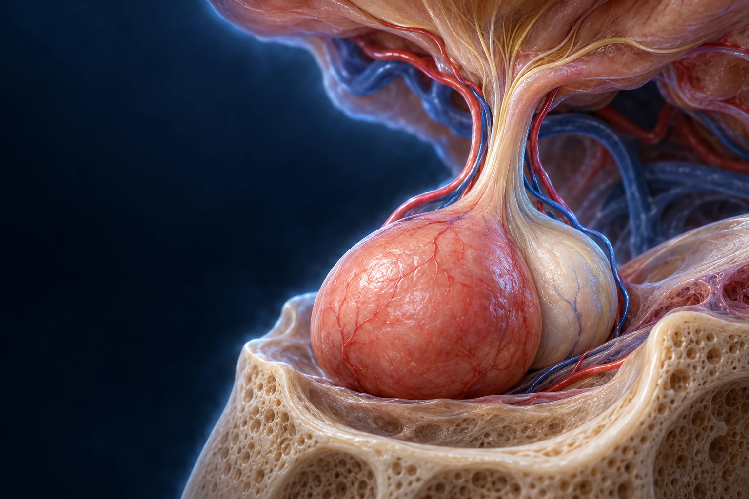
Brain section · pituitary beneath the hypothalamic region
Retained original teaching illustration. One regional example, not a claim that every endocrine source is controlled by this route.Teaching notes
Hypothalamic region
Neural information helps regulate selected endocrine outputs.
Pituitary
A small gland below the brain; anterior and posterior outputs use different routes.
A source can be a gland or a cell population within another organ. Begin by locating the tissue, not by assuming one master gland.
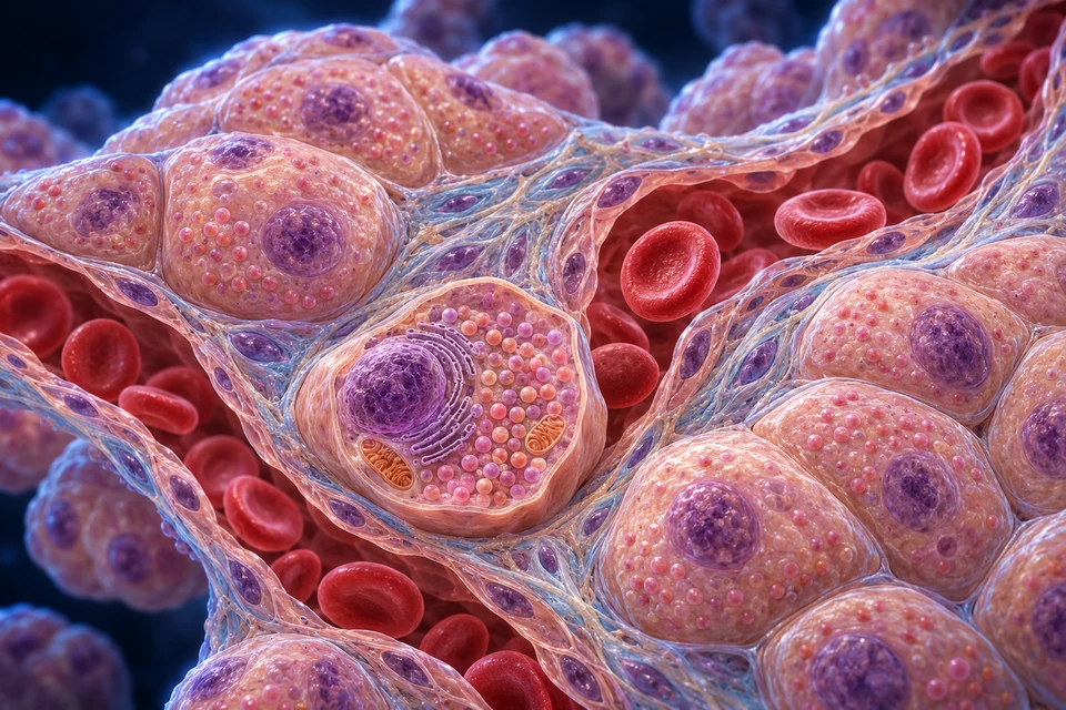
Tissue scale · source cells beside a separate vessel
Representative tissue illustration, not microscopy or one identified gland. Released-product symbols do not depict whole secretory vesicles entering blood.Teaching notes
Endocrine cell group
A source releases a chemical message into its surroundings.
Tissue space
The extracellular interval is separate from both the cell and the vessel lumen.
Blood space
Circulation can carry a released hormone beyond its source.
Endocrine delivery uses surrounding fluid and circulation. The blood channel is not a gland duct, and red cells are not the hormonal message.
Selected receptor contexts, not measured molecular structures. The intracellular example concerns steroid access; thyroid-hormone entry requires transport mechanisms.Teaching notes
Surface receptor
Many peptide signals are recognised at the cell boundary.
Intracellular receptor
Selected hormone classes act through receptors within the cell.
Receptors, cellular machinery and local handling shape the response. One circulating hormone can have different effects in different tissues.
These preparations connect three learning scales. They are not one traced vessel, a hormone animation or a universal three-gland chain. Mineral changes and neural drive can regulate outputs without copying the same pituitary axis.
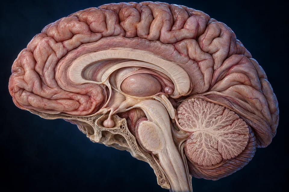
The brain contains several different signalling roles.
Hypothalamic regulation, pituitary output and pineal timing occupy different regions. The pituitary is important, but neither the entire brain nor every endocrine tissue follows one master-gland pathway.
New Golden-family text-free teaching illustration. Broad midline orientation, front is image left. Regional teaching only: fine folds, nuclei, stalk fibres, vessels and hidden junctions remain illustrative / not independently verified. The image does not locate a microscopic clock nucleus or draw its full autonomic route. Human medical review: Not performed.Structure notes · further context
Hypothalamic region
Releasing signals reach anterior-pituitary blood pathways; other neurons send axons toward the posterior pituitary. Exact nuclei are not identified by this generated surface.
Pituitary
Anterior and posterior mechanisms differ. Its important role does not mean it directly controls every endocrine tissue.
Pineal context
Human light–dark information arrives through neural regulation; the gland is not depicted as directly seeing light through the skull.
Thalamic orientation
This context landmark is not a claim that the entire thalamus is an endocrine gland.
Cerebral context
Cortical folds are illustrative. Endocrine timing and behaviour involve networks, not one isolated control centre.
Anterior lobe · instructions arrive in blood
Hypothalamic neurons release regulating signals into local blood. Portal vessels deliver those signals to anterior-pituitary endocrine cells. The receiving cells make and release their own products, such as growth hormone or thyroid-stimulating hormone (TSH), into circulation.
Posterior lobe · supply arrives along axons
Antidiuretic hormone (ADH, or vasopressin) and oxytocin are made in hypothalamic neurons and carried along their axons. Terminals in the posterior pituitary release them into blood. Release location therefore differs from synthesis location; ADH can then act on responsive kidney cells.
Mechanism source: Anterior and posterior pituitary routes. The broad brain illustration locates regions; it does not trace these tiny vessels or individual axons.
The endocrine network extends beyond a list of glands.
Selected locations below connect to distinct realistic organ lessons. Hormone-producing cells also occur in organs whose main role is pumping, filtering, digesting or storing energy.
Nearby organs can have different outputs.
Anterior regional preparation · patient right is image left
Original teaching illustration. Adrenals are separate glands, not kidney collecting structures. Individual contours and fine vessels are not an operative map.Teaching notes
Right adrenal
A separate gland lies near the upper right kidney.
Left adrenal
The opposite gland lies near the upper medial left kidney.
Right kidney
Renal tissue filters and regulates substances, and also has endocrine roles.
Left kidney
A circulating adrenal hormone can act on renal tissue without a direct gland-to-urine duct.
The adrenal cortex produces steroids, whereas the medulla provides a distinct neuroendocrine output. Use the control comparison below to distinguish the neural trigger from a hormonal axis.
Selected source-labelled organ locations · supporting reference
New controlled unchanged BodyParts3D geometry render; source CC BY 4.0, The Database Center for Life Science. This selected regional preparation is not patient imaging or a complete endocrine atlas. Thyroid/parathyroid/gonadal geometry is absent, not invented. Surface meshes do not identify hormone-producing cell subtypes. Fine source contours/material detail illustrative; Human medical review: Not performed.Structure notes · further context
Right adrenal
The gland is a separate source, not renal tissue or a urine route. Both cortex and medulla have their own source-supported chapter.
Left adrenal
Typical gland contour differs from the right, and venous drainage varies. The model is one source preparation, not all individual anatomy.
Pancreas
An organ-surface anchor cannot identify an individual islet, alpha or beta cell. The linked tissue plate supplies the separate scale.
Right kidney
Renin-related input, EPO support of marrow production and vitamin-D activation occupy different cell processes, not one surface point.
Left kidney
Kidney identity is source-tracked. No exact ADH, aldosterone or PTH target cell is inferred from this external organ surface.
Heart context
This native landmark is ventricular wall for organ orientation, not an atrial ANP secretion site. Specific source cells are not marked here.
Hypothalamus & pituitary
Below the central brain
Neural integration, releasing signals and several distinct pituitary outputs.
Adipose tissue also releases signals such as leptin. Gonads and pregnancy-related tissues belong in the broader network; their detailed anatomy and developmental regulation remain separately staged, not represented by a substituted male or female body.
The source-tracked regional reference supplies selected thoracoabdominal context, not a complete gland atlas. The directory adds further locations in words; thyroid, parathyroid and gonadal surfaces are not identified in this preparation. Reproductive and developmental anatomy remain separately staged.
Many endocrine cells form cords or clusters beside capillaries. A released product passes through local tissue space before vascular delivery. A blood channel is not a secretory duct.
New Golden-family text-free teaching illustration. Representative tissue illustration, not a specific gland specimen or measured H&E/EM. Broad cell–interstitium–blood separation is taught; exact cells, granules, fenestrae, dimensions and red-cell count remain illustrative / not independently verified. Not all endocrine tissues share this architecture. Human medical review: Not performed. Released-product symbols do not depict intact vesicles entering blood.Structure notes · further context
Endocrine cell group
No hormone subtype is established by colour or the appearance of an individual generated cell. Thyroid follicles are a different arrangement.
Blood space
Red cells remain inside the vascular compartment. Circulating hormone is not represented by the red cells themselves.
Vascular boundary
The plate does not independently resolve real membrane thickness or pores. Endocrine release passes through tissue space before vascular entry.
Interstitial support
Pericapillary spaces and connective support are distinct from the blood channel and cell interiors.
Cellular context
A nucleus does not reveal the specific hormone, receptor or measured activity. Appropriate specimen evidence is needed.
Released products are symbolic; intact storage vesicles are not shown entering blood.
This representative arrangement is not universal: thyroid follicles use an extracellular colloid store, and hypothalamic neurons provide another cellular route. Tissue architecture and product chemistry should be taught together.
Blood transport is not identical to cellular entry.
Many steroid and thyroid hormones circulate largely bound to proteins, in equilibrium with a smaller free fraction. Binding, distribution and metabolism influence availability.
1
Enter circulation
Vascular delivery carries a released product beyond its source.
2
Exchange with carriers
Free and bound pools can exchange; circulating total amount is not the same as an immediately available fraction.
3
Reach and enter tissue
Local access and transport matter. Thyroid cellular entry involves carriers, despite lipid-soluble hormone chemistry.
4
Convert or remove
Tissue metabolism can change the active form or remove a signal. A measured concentration is not simply a gland’s output rate.
Supporting transport sequence, not exact pharmacokinetics or clinical assay interpretation.
Receptors and cellular machinery select the effect.
A circulating hormone does not affect every cell equally. Receptor expression, signal pathways and local handling shape the response.
Original educational cell illustration. These are selected surface and intracellular receptor contexts, not every hormone pathway or measured protein structures. Thyroid cellular entry involves transporters; it is not represented by the steroid-access example. Human medical review not performed.Structure notes · further context
Extracellular signal
The blue signal is approaching, not visibly bound; the picture does not prove receptor activation.
Cell-surface receptor
This is not a pore for the whole hormone. Its shape does not establish a particular receptor family.
Intracellular machinery
These representative proteins are not a measured cAMP cascade. Surface receptors use different downstream pathways.
Intracellular receptor
The small amber signal and protein are explanatory symbols, not an atomic reconstruction or a specified hormone.
Nuclear compartment
The static image locates the nucleus; it does not demonstrate completed gene expression or show a universal receptor movement sequence.
Plasma membrane
The right illustration is a steroid-access example. Thyroid hormones require cellular transport mechanisms; they are not depicted here.
Surface reception
Many peptide signals bind membrane receptors and change intracellular processes without entering as a whole hormone–receptor pair.
Intracellular reception
Major steroid and thyroid responses involve intracellular receptors and gene regulation. This is not the only action of those classes.
Amplification
One recognition event can recruit a downstream pathway. The receptor is not itself the entire physiological response.
Adaptation
Receptor numbers and signalling responsiveness can change. Neither receptor colour nor organ location predicts an exact effect by itself.
Local communication is not always endocrine circulation.
Compare who sends the message, where it travels and which cell receives it. A supporting map clarifies delivery after the realistic tissue plate establishes the compartments.
Endocrine
Cell → interstitial space → blood → responsive distant tissue
Circulation delivers the message broadly. Receptors and cellular machinery determine where an effect occurs.
Paracrine
Cell → nearby extracellular space → neighbouring cell
A local signal need not complete a systemic blood journey. Distance and breakdown shape its reach.
Autocrine
Cell → released local signal → the same cell
A cell can respond to a signal it releases. This is a relationship, not a separate universal organ.
Neural information is converted into an endocrine output. Posterior-pituitary release is one connected example.
Supporting communication map, not literal vessel or nerve anatomy. All four routes are visible before clicking. A signalling molecule can participate in more than one context.
Hormones, minerals and neural drive supply different inputs.
Feedback can help regulate output, but a copied three-gland chain would misrepresent every other endocrine mechanism.
Which tissue receives TSH?
Anterior neck · patient right is image left
Original retained teaching illustration, not a patient scan. Thyroid tissue surrounds a separate airway; exact vessels and proportions are not an operative map.Teaching notes
Thyroid lobe
Follicular tissue responds to pituitary TSH; it does not make TSH.
Isthmus
A bridge of gland tissue crosses the front of the trachea.
Separate airway
Air passes through the trachea. Thyroid output enters blood, not this lumen.
Hypothalamic thyrotropin-releasing hormone (TRH) promotes anterior-pituitary thyroid-stimulating hormone (TSH) release. TSH acts on thyroid tissue, which releases T3/T4. These thyroid hormones restrain upstream hypothalamic and pituitary output; the airway is a separate path.
Hormonal output can be episodic or rhythmically coordinated. Not all hormones are simply slow, and not every physiological signal follows the same clock.
Pulses within a longer pattern
ACTH/GH-related secretion illustrates episodic output. Pulse timing and amount differ by hormone and physiological context.
Melatonin normally conveys biological-night information. This is not a direct light beam from the eye to the pineal gland or a universal switch that instantly causes sleep.
The shape of a blood concentration curve does not reveal secretion alone. No clock times, hormone values or individual rhythm measurements are assigned here.
Supporting timing illustration, not clinical data or a diagnostic graph. Labels remain HTML and legible at narrow widths.
Hormone interactions connect filtered substances, regulated tubular recovery and endocrine output. Different signals describe different aspects of balance.
Selected normal coordination examples
Signal/context
Connected response
Keep distinct
ADH
Responsive renal water permeability
Not a substitute for aldosterone’s ion effects
Aldosterone / RAAS
Responsive sodium/potassium handling
Renin is an upstream enzyme signal, not aldosterone
Cardiac natriuretic signals
Salt/volume-related renal and vascular effects
The heart does more than pump blood
PTH / calcitriol
Bone–kidney–gut mineral coordination
Renal reabsorption differs from intestinal absorption
EPO
Supports bone-marrow red-cell production
Not a hormone that makes red cells inside the kidney
The gut sends signals as well as absorbing nutrients.
Enteroendocrine populations communicate with digestive organs and distant targets. A classic one-letter/one-hormone cell list is useful orientation, but not a complete modern identity map.
Gut → pancreas
GIP and GLP-1 contribute to glucose-dependent insulin secretion. GIP’s historic name should not be read as an instruction that it inhibits insulin.
Nutrient exposure → gut signalling → responsive islet output
Gut → coordinated digestion
CCK and secretin help connect luminal information with digestive secretion and movement. Local, neural and circulating messages cooperate rather than form one isolated straight line.
Adipose leptin also conveys energy-state information to responsive tissues. It is not a universal instant fullness switch or a treatment recommendation.
Growth depends on coordinated messages, not one gland.
Thyroid, GH-related, metabolic and reproductive signals interact with nutrition, neural input and tissue responsiveness. The complete developmental and reproductive atlas is separately staged.
Growth hormone is a signal, not the whole growth response.
Growth hormone (GH) is released by anterior-pituitary cells. It acts through its own receptors and also promotes insulin-like growth factor 1 (IGF-1) production in the liver and other tissues. Circulating and locally produced IGF-1 contribute to tissue responses; growth cannot be reduced to a liver-only relay.
These pathways support growth in childhood and remain metabolically important later. Nutrition, other hormones and the responding tissue affect the outcome.
One pituitary signal. More than one route to a tissue response.
Follow GH from a secretory cell, distinguish liver-derived circulating IGF-1 from local IGF-1, and keep direct GH actions in view.
Keep both branches: GH → responsive tissue directly; GH → IGF-1 production → responsive tissue. Local IGF-1 adds a nearby route. The liver is a major circulating source, not a compulsory relay for every GH action.
New original Golden-family text-free teaching preparation. Representative tissue illustration, not H&E microscopy, a subtype stain or a measured cell model. Broad endocrine-cell/capillary relationships are taught; fine generated granules, membranes and cell identities remain illustrative. Human medical review: Not performed.Structure notes · further context
Endocrine-cell cords
The cell types have different hormone products. This unstained illustration cannot independently identify a somatotroph, lactotroph or other subtype by color.
Cell nucleus
Hormone synthesis depends on the cell’s molecular machinery. Nuclear size and chromatin appearance here are decorative, not a diagnostic observation.
Granule context
The exposed dots illustrate secretory storage, not an electron-microscope count, exact composition or universal granule geometry.
Capillary channel
Anterior-pituitary hormones enter capillary blood after secretion into the surrounding tissue interface. This lumen is not a gland duct.
Red blood cells
Their generated number, diameter and spacing are not physiological measurements or evidence of flow speed.
Vessel boundary
Fenestrated pituitary capillaries support exchange. Individual pores, basement membrane and molecular passage are not resolved or independently verified in this artwork.
Gold locates broad tissue and blood-facing contexts on unchanged anatomy. It does not identify a GH-specific cell, trace hormone molecules or map a receptor. These are different magnifications, not one continuous specimen.
Pituitary · release · Where does the message travel?
Somatotrophs in the anterior pituitary make GH. They release it across the surrounding tissue interface into blood, rather than through a duct to the liver. The image shows representative endocrine cords; colour alone cannot identify a GH-producing cell.
A neural input controls a different endocrine cell.
Hypothalamic neurons and anterior-pituitary somatotrophs are different cells. Releasing and inhibiting messages arrive through portal vessels; the somatotroph then produces its own GH. The hypothalamic message is not a parcel of GH passed along an axon to the anterior lobe.
GHRH, somatostatin and ghrelin-related inputs participate in a changing release pattern. IGF-1 feeds back on the system. Negative feedback changes output; it does not mean the pituitary and hypothalamus become anatomically disconnected.
GH and IGF-1 cooperate through different receptors.
The GH receptor belongs to the cytokine-receptor family and works with an associated kinase, JAK2. It is not itself an IGF-1 receptor. GHR signalling includes STAT5-dependent gene regulation and other pathways with context-dependent effects.
The IGF-1 receptor is a receptor tyrosine kinase. Its downstream networks include PI3K–AKT and MAPK pathways. This comparison identifies different signalling machinery, not two interchangeable keys or a measured molecular structure in the illustration.
Blood delivery and local production can act together.
Liver-derived IGF-1 contributes to an endocrine pool, while many tissues produce IGF-1 locally. A local signal may act on the producing cell or neighbouring cells. A blood sample therefore cannot describe every local IGF interaction.
The growth plate receives endocrine inputs while its own cells and surrounding tissues contribute local regulation. No universal percentage of a tissue response is assigned to liver-derived versus local IGF-1. These contributions vary with the tissue, developmental stage and experimental conditions.
A circulating store is not the same as immediate access.
IGF-binding proteins bind IGFs and influence their distribution, persistence and access to receptors. Many circulating complexes contain IGF, IGFBP-3 or IGFBP-5, and the acid-labile subunit. These are protein complexes in plasma, not blood cells or bile droplets.
Local binding, release, proteolysis and tissue interactions affect availability. Binding proteins do more than simply block a signal, and some have IGF-independent actions. The drawing locates a blood compartment but does not resolve or count these molecular complexes.
Activation does not require inventing a new receptor pair.
Experimental work supports GH receptor subunits being associated before ligand binding. GH can change their arrangement and the activity of associated JAK2. A drawing that requires two isolated monomers to collide would make one structural model seem more certain than the evidence warrants.
Biochemical experiments and molecular simulations address receptor activation at a different scale from a tissue illustration. No exact receptor conformation or molecule number is inferred from the cell surface shown here.
Within an active growth plate, chondrocyte proliferation, enlargement, matrix changes and subsequent replacement with bone contribute to longitudinal growth. IGF-1 participates with local regulators; it is not the sole switch for each cartilage zone.
GH and IGF actions on the developing skeleton are related but not identical. The gross bone plate locates the cartilage and neighbouring bone. Its texture cannot identify an individual receptor, measure cell division or establish a child’s growth stage.
Growth signals also matter after height growth slows.
GH contributes to metabolism, including lipid mobilisation and context-dependent protein and carbohydrate handling. Its physiological role does not end when longitudinal bone growth stops. Effects differ with tissue, nutrition and other signals; “growth hormone” does not mean every response is enlargement.
Thyroid hormones, insulin, sex steroids, local factors and adequate substrate support coordinated development. Increasing one signal cannot replace all these conditions. This normal physiology lesson supplies no height prediction, laboratory interpretation, supplementation or anti-aging advice.
An experiment can separate routes without erasing a human organ.
In liver-specific Igf1-deletion mouse experiments, circulating IGF-1 fell markedly while substantial postnatal growth persisted. This challenged a model in which all growth must pass through a hepatic IGF-1 relay.
The experiment does not show that circulating IGF-1 or the liver is unimportant in people. Compensation, tissue-specific production, species and timing all matter. The study helps distinguish mechanisms; it cannot predict the consequence of an individual person’s hormone result.
Does GH become IGF-1 while travelling through the liver?
No. They are different proteins. GH signalling can promote IGF1 expression and production in responsive cells.
Must every tissue IGF-1 signal first come from the liver?
No. Liver-derived circulating IGF-1 and locally produced IGF-1 contribute through different delivery relationships.
Does more total circulating IGF-1 specify a larger local response?
Not by itself. Binding, tissue access, receptors, downstream machinery and physiological context all matter.
Does the visible liver-cell channel deliver IGF-1 as bile?
No. The blood-facing exchange route and the bile-facing canalicular route are separate.
Normal physiological coordination, not a hormone-treatment or height-prediction guide. Sources checked 21 October 2026. Original generated anatomy is not acquired microscopy; fine generated histology, physician review and physical-phone acceptance are not certified.
Connected scales
A circulating signal can change cellular activity, which affects tissue and organ function. Growth-factor production and local response need not share the same anatomical source.
Gonadal, placental, pregnancy, puberty and aging anatomy cannot be represented by a generic reused torso. This foundation introduces their place in the network; it does not declare their teaching complete.
A routine section can reveal cell groups, blood spaces and ducts. Specific hormone identity and activity often need additional markers and physiological evidence.
Human tissue record
Loyola’s DMS142 record identifies human pancreas H&E, vascularized islet cords and no islet duct. Its older embryonic-origin uncertainty is not adopted as a current factual claim.
Generated tissue detail is representative, not a stained specimen or receptor measurement. Clinical imaging, diagnosis and disease comparisons remain later-phase work.
Selected brain/tissue orientation, delivery compartments, chemical classes, receptor specificity, control/timing and connected normal organ roles. Linked deep chapters supply their own distinct organ anatomy.
Explicit limits
AI-assisted visual/content production. Fine generated neural, vascular, cellular and membrane details remain illustrative / not independently verified. Human medical review: not performed. Educational only; not diagnostic or surgical material.
Revision: endocrine-system-english-2026-10-01-foundation-v1. Stable local key: ahb_ch_endocrine_system_english_v1. No new canonical CMS/atlas entity or 3D dataset. Migration physical-phone acceptance: Pending, non-blocking. New controlled thoracoabdominal context uses unchanged source geometry; thyroid/parathyroid/gonadal source surfaces are not invented. Complete body-wide/reproductive/developmental atlas remains separate scope. Text-free illustration masters and editable English labels are separate. Target-cell receptor shapes are explanatory, not molecular reconstructions. BodyParts3D source, The Database Center for Life Science, CC BY 4.0; controlled materials/camera/export by AllHumanBody. New representative cell/brain illustration is not actual microscopy. No authentic specimen images were redrawn or copied.