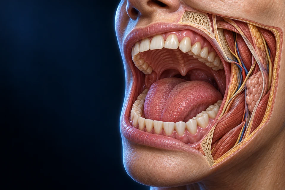
Digestion begins with coordinated tissue, secretion and movement. The mouth handles food; the throat coordinates a shared crossing; the esophagus carries a prepared bolus toward the stomach.
▷
Watch chapter overviewFilm planned · not yet available
Oral entrance preparation · fine generated detail illustrative
The mouth receives material, teeth divide it, and tongue and cheeks reposition it. Saliva moistens the mixture. A prepared bolus passes through the pharynx into the esophagus, while air uses the larynx and trachea.
Blue: resting air-route orientation · Gold: posterior food-tube direction
New original Golden-family text-free teaching preparation. Resting sagittal-region teaching preparation, not a timed swallowing frame or a literal single-plane specimen. Anterior is image left. Fine generated teeth, cartilage, muscle and hidden lumen connections remain illustrative. Human medical review: Not performed.Structure notes · further context
Hard palate
It provides a firm surface against which the tongue can handle and propel material.
Soft palate
Elevation during the pharyngeal swallow helps close the nasopharyngeal route. The hanging uvula is part of this region, not the sole closing mechanism.
Tongue
Intrinsic and extrinsic muscle groups cooperate with cheeks and jaw. Surface texture is illustrative, not a map of individual taste buds.
Pharynx
Oropharyngeal and laryngopharyngeal regions participate in both food and air passage at different times. The nasopharynx is not a normal food route.
Epiglottis
Its inversion works with laryngeal movement and closure at other levels. It is not an independently operated single trapdoor guaranteeing airway protection.
Trachea
Cartilage supports the airway. The resting anatomy plate does not show the changing vocal-fold or laryngeal-vestibule state during swallowing.
Esophagus
It is usually not a wide open tube at rest. The preparation introduces the posterior relationship; exact sphincter roots and individual lumen geometry are not independently verified.
Tongue, teeth and saliva work in one oral space
The tongue and cheeks reposition material; teeth change its size; saliva moistens it. Preparation is not yet intestinal absorption.
These are orientation cues within a resting preparation, not a measured airflow trace or a continuous food-and-air animation. Continue to the separate protective configuration below.
Roof, floor and movable boundaries
The hard palate gives the oral roof a bony support; the posterior soft palate is mobile. Tongue muscle changes shape as well as position. These are interacting surfaces rather than a rigid tube through which everything drops.
One throat, coordinated destinations
Only part of the pharynx is a shared digestive/respiratory route. The nasopharynx normally carries air. The resting preparation locates structures; the later process guide explains changing protection without pretending this static image is a swallow recording.
02 / GLAND LOCATIONS
Secretion reaches the mouth through ducts.
Three pairs of major glands occupy different regions. Many smaller glands also contribute to oral moisture and protection. They are exocrine glands: their products enter ducts and surface spaces, not directly into blood.
Three gland regions, one oral destination
3
Sublingual gland
A smaller paired gland occupies the floor of the mouth.
More about sublingual gland
Its secretion is predominantly mucous. Multiple small ducts contribute; exact outlet number and variants are not asserted.
5
Mandible
The lower jaw supplies structural context for gland positions.
More about mandible
The local opening exposes sublingual context; it is not an intact clinical view or a dental alignment model.
2
Submandibular gland
A paired gland lies around the lower mandibular region.
More about submandibular gland
It has mixed secretory tissue with a strong serous contribution. The deep part and full duct route are not surgically mapped here.
12345
4
Parotid duct
A duct carries saliva across the masseter region toward the cheek.
More about parotid duct
Its normal oral opening is near the upper second molar. That hidden exit is described from sources, not verified from this generated image.
1
Parotid gland
A paired gland lies in front of and below the ear.
More about parotid gland
Its secretion is predominantly serous. The exposed view shows one side; it does not imply a single unpaired gland.
Three gland regions, one oral destination · names and basic functions are visible
New original Golden-family text-free teaching preparation. Expanded lateral preparation with a local mandibular cutaway. Broad gland positions are source-supported. Fine duct exits, nerves, vessels and generated lobules remain illustrative; hidden connections are not inferred. Human medical review: Not performed. Leader lines or matching visible key numbers identify broad teaching locations, not measured borders or a surgical map.
Different secretory mixtures
Parotid tissue is predominantly serous, submandibular tissue has a strong serous contribution with mucous elements, and sublingual tissue is predominantly mucous. These broad tendencies do not establish a universal exact ratio or the identity of every generated lobule.
Routes beneath the surface
The parotid duct crosses the masseter region and opens near an upper second molar. Submandibular and sublingual outlets serve the floor of the mouth. Hidden openings are taught from independent anatomy, not guessed from visible AI-generated lines.
03 / ORGAN TO SECRETORY UNIT
Small end pieces produce a flowing secretion.
Acinar cells surround initial luminal spaces. Small ducts join a branching duct system, while tissue blood supply remains outside that secretory route. Connective tissue organizes the gland into supported groups.
New original Golden-family text-free teaching preparation. Source-supported acinar-lumen and duct relationships at enlarged teaching scale. Individual generated cells, nuclei, granules, basket-like fibers and branch proportions remain illustrative / not independently verified. Human medical review: Not performed.Structure notes · further context
Acinar cells
Acinar secretion brings fluid and proteins into the glandular space. No particular generated cell is independently identified as serous or mucous.
Acinar lumen
This lumen belongs to the exocrine route. It is separate from tissue blood vessels.
Small collecting duct
Intercalated ducts connect secretory units with subsequent duct segments. The exact generated branch tree is illustrative.
Larger duct lumen
Duct cells alter ion composition. This image does not independently prove basal striations, every membrane transporter or a precise duct subtype.
Supporting tissue
It also carries vessels and nerves. Fine fibers in the master do not establish a validated myoepithelial-cell basket.
Separate blood supply
Blood does not empty directly into saliva ducts. Individual capillary geometry and flow direction are not mapped.
Begin in the secretory end piece
Ion movement supports osmotic water entry and protein secretion contributes to the initial luminal fluid. The blood supply stays outside this exocrine route.
Gold outlines locate a tissue context. They do not identify a molecular transporter, prove a cell subtype or measure secretion.
Why does a duct change more than the amount of water?
Saliva is first produced and then modified. Relatively low duct water permeability means ion handling cannot be described as simply reabsorbing a matching volume of water. Flow and gland contribution affect the final mixture; this is not a fixed universal recipe.
Secretory granules and membrane ion transport support different parts of saliva production. Water follows osmotic conditions established by ion movement. A cell-level transporter map cannot be established solely from visible granule-like texture.
Histological appearance is not living geometry
Processing can change how gland cells appear. Serous crescent profiles in some prepared slides should not be treated as a universal living three-dimensional cap. This plate makes no precise demilune or generated-cell identity claim.
04 / SECRETION AND FUNCTION
More than water around a mouthful.
Saliva supports lubrication, buffering, oral defense and initial chemical processing. Dissolving food components also helps them reach taste receptors. Its mixture changes with stimulation, flow and contributions from different glands.
NORMAL PHYSIOLOGY / FROM GLAND TO ORAL ENVIRONMENT
Produce fluid. Reshape the mixture. Protect a surface.
Follow the tissue first: secretory end piece, modifying duct and onward secretion. Then separate the amount collected from its composition and its jobs.
New original Golden-family text-free teaching preparation. Source-supported acinar-lumen and duct relationships at enlarged teaching scale. Individual generated cells, nuclei, granules, basket-like fibers and branch proportions remain illustrative / not independently verified. Human medical review: Not performed. Selected relationship: Start in the acinar lumen Acinar cells contribute initial fluid and proteins to their luminal space. Ion gradients and osmotic water movement support fluid production; blood remains outside this exocrine route.Structure notes · further context
Acinar cells
Acinar secretion brings fluid and proteins into the glandular space. No particular generated cell is independently identified as serous or mucous.
Acinar lumen
This lumen belongs to the exocrine route. It is separate from tissue blood vessels.
Small collecting duct
Intercalated ducts connect secretory units with subsequent duct segments. The exact generated branch tree is illustrative.
Larger duct lumen
Duct cells alter ion composition. This image does not independently prove basal striations, every membrane transporter or a precise duct subtype.
Supporting tissue
It also carries vessels and nerves. Fine fibers in the master do not establish a validated myoepithelial-cell basket.
Separate blood supply
Blood does not empty directly into saliva ducts. Individual capillary geometry and flow direction are not mapped.
Start in the acinar lumen
Acinar cells contribute initial fluid and proteins to their luminal space. Ion gradients and osmotic water movement support fluid production; blood remains outside this exocrine route.
Retained original text-free teaching image and six anatomical endpoints. Gold outlines locate tissue context; they do not identify a molecular transporter, measure secretion or certify generated fine-cell structure.
ProduceAcinar fluid and protein secretion enter the starting lumen.
ModifyDuct epithelia handle ions with relatively little matching water movement.
UseThe oral mixture supports surface films, chemical buffering and material movement.
The secretory end piece moves more than a packet of water.
An acinar cell has a tissue-facing side and a luminal side; transport and protein release do different jobs.
Ion gradients provide the conditions for fluid secretion. Chloride movement toward the acinar lumen, with accompanying ions and osmotic water movement, contributes to the initial fluid. Cellular energy maintains the gradients; water is not lifted through a molecular water pump. The initial fluid is broadly near-isotonic, before ductal modification changes the mixture.
AQP5 is one water-channel route associated with salivary acinar membranes. A knockout experiment in mice reduced stimulated fluid secretion without simply eliminating the separate protein-secretory task. That experiment supports a water-permeability role; it is not a human clinical test or a direct prediction for every gland.
Secretory proteins are made and packaged within gland cells, then released to the luminal route. Fluid transport and exocytosis cooperate, but a granule-like texture in the illustration does not identify a transporter, a protein species or a particular living cell subtype.
Why keep blood outside the lumen?
Tissue vessels supply gland cells from their surrounding side. The acinar and duct lumen is the exocrine output route; secretion is not blood pouring directly into the mouth. The original picture shows that gross separation without tracing every capillary or nerve.
Does every signal produce the same mixture?
No. Cholinergic and adrenergic stimulation can both evoke salivary secretion. Their effects on fluid and protein output differ with gland and conditions; sympathetic activity is not simply an off switch.
The duct changes ions without matching every ion movement with water.
The first fluid and the final secretion are related stages, not identical solutions.
As secretion passes through the duct system, epithelial handling removes sodium and chloride from the luminal mixture and adds potassium and bicarbonate. Different duct regions and transport mechanisms contribute; the gross preparation does not resolve every channel or membrane face.
Compared with the acinar secretory pathway, the duct epithelium has relatively low water permeability. Ion modification can therefore occur without equivalent bulk water reabsorption. Final saliva is generally hypotonic relative to plasma under ordinary conditions.
This is a functional two-stage account, not a claim that every gland cell has one fixed rate or that the ducts are absolutely watertight. Gland contribution, stimulation and regulated epithelial activity all matter.
Why not copy the pancreatic duct explanation?
Both are exocrine systems, but similar-looking ducts have different functional roles and transport conditions. Do not replace salivary modification with a pancreatic bicarbonate-and-water secretion model.
Does faster flow alone determine the answer?
Residence time is a useful component of a simplified model. It does not by itself specify the mixture in every stimulated human whole-mouth collection. Compare what was measured and which glands contributed before predicting an ion concentration.
Three different quantities answer three different questions.
A watery sample, a concentrated sample and a large amount delivered per minute are not synonyms.
Concentration × volume per time = amount per time
Hypothetical solute concentration
2 µmol/mL
Collected volume per time
1 mL/min
Amount delivered per time
2 µmol/min
Two quantities define the output
2 micromoles in each millilitre, carried by 1 millilitre each minute, gives 2 micromoles per minute.
Invented arithmetic examples for one hypothetical solute. These values are not human measurements, normal ranges, a bicarbonate/pH model or a prediction of stimulation.
Flow rate reports fluid volume collected per time. Concentration reports solute amount within a volume. Their product gives the amount carried out per time for that solute. A larger volume can deliver more material even when its concentration is unchanged.
Conversely, greater flow and a lower concentration can yield the same amount-per-time output. This is an arithmetic identity, not proof that a particular gland must behave in that way. The examples below are invented teaching cases and are not patient measurements or reference ranges.
Osmolality concerns the combined concentration of dissolved particles, rather than the concentration of one selected ion. Neither a single ion nor a flow reading alone establishes the complete mixture.
How should a collected sample be described?
Name the collection: a particular gland or whole-mouth mixture, the stimulus, duration and handling. Keep concentration separate from the amount delivered during the collection. These distinctions prevent an equation from being mistaken for an individual physiological law.
A human response is not one universal flow-only curve.
Stimulus, gland contribution and epithelial regulation interact.
A study of 102 adults compared unstimulated oral saliva with chewing-stimulated collections. Individual changes in flow did not track changes in every measured ion in the same way. That observation cautions against predicting the full mixture from flow alone.
The sample was whole-mouth saliva. It cannot isolate each gland's contribution or directly measure each acinar and ductal transport process. The authors' regulatory interpretation is informative, but one study does not erase all earlier single-gland results.
How can a model and a study appear to disagree?
Check population, gland versus oral collection, stimulus and whether the comparison concerns group levels or within-person changes. Those are different observations. A useful model retains its conditions rather than becoming a rule for every reader.
A pH reading is not a measure of the whole buffering reserve.
Buffers limit a chemical change; they do not make every oral surface permanently neutral.
H⁺ + HCO₃⁻ ⇌ CO₂ + H₂O
Bicarbonate participates in reversible chemistry with hydrogen ions, carbon dioxide and water. When added hydrogen ions are taken up through this system, the balance shifts toward carbon dioxide and water. Phosphate and proteins also contribute to buffering.
pH describes the current hydrogen-ion activity. Buffer capacity describes how strongly a sample resists a pH change when acid or base is added under specified conditions. Two samples can share a pH while differing in composition and buffering behaviour.
A primary human study collected saliva while limiting carbon-dioxide loss, then measured buffering by titration. This matters because exposure and gas exchange can change the measured chemistry. A sample reading is not automatically the exact condition at every tooth or mucosal site.
Is bicarbonate the whole explanation?
No. The contributions of bicarbonate, phosphate and proteins depend on the sample and chemical conditions. Fluid renewal and removal of substances are separate physical processes; neither should be collapsed into one universal pH value.
Why no instant neutralization animation?
It could imply that a finite secretion immediately eliminates all acid. The displayed reaction is an equilibrium relationship, not a measured rate, a prediction of dental outcome or a treatment instruction.
A surface film does a different job from a bulk stream.
Proteins, glycoproteins and fluid together support the oral environment.
Resist a change in acidity
Bicarbonate, phosphate and protein systems contribute under different chemical conditions. This is not the same as fluid volume or an unchanging pH.
Hydrogen ions + bicarbonate ⇌ carbon dioxide + water
A functional comparison beside the anatomical lesson, not a simulated acid challenge, surface-force recording or clinical outcome.
Mucins and other salivary components contribute to moist surface films and lubrication. The fluid surrounding food is not simply pure water, and the material left near a surface need not have the same behaviour as a freely collected sample.
Experiments with adsorbed human-saliva films demonstrate lubrication on controlled substrates. They do not reproduce every living mouth surface, chewing load or biological condition. The lesson uses them to distinguish a film role from the amount of fluid collected.
Normal protection also includes dilution, movement and clearance of material, buffering, and antimicrobial components. None makes the mouth sterile. Minor glands and residual saliva matter locally even when they are not the dominant source of a large collected volume.
Why not judge protection by volume alone?
Chemical composition, surface interactions, local renewal and clearance answer different questions. A generous flow reading does not on its own characterize every one of these functions.
Does this course recommend a product or a test?
No. The comparisons explain normal structure and function. They do not diagnose dry mouth, predict disease or prescribe a rinse, medicine or stimulation programme.
Does a larger collected volume prove a higher concentration?
No. Flow, concentration and amount-per-time output are different quantities.
Why is ductal modification not simply water recovery?
Relatively low duct water permeability permits ion handling without equivalent bulk water reabsorption.
Does the mouse AQP5 experiment certify a human salivary test?
No. It is controlled animal evidence about water-channel function, with a different scope.
Can one pH reading establish buffer capacity?
No. Buffering must be described under defined chemical conditions, not inferred from a pH value alone.
Do the images identify every transporter?
No. They locate gross acinar, luminal, ductal and surrounding tissue relations; molecular positions are not resolved.
Sources and measurement boundaries
Directed normal-physiology checks:22 October2026. Existing FB114 coverage and qualified gland/unit artwork are reused. No book image or prose is reproduced and no new salivary bitmap is generated.
NIDCR · Normal saliva rolesOfficial readable normal lubrication, digestion and oral-protection context; no diagnostic or treatment guidance adopted.
Proctor · Physiology of salivary secretionOfficial indexed review: autonomic stimulation, acinar fluid/protein production and ductal modification. Direct fresh PubMed read returned a minimal page; do not claim full methods.
Salivary duct epithelial ion transportReadable official review abstract and retained project source: duct electrolyte handling. A gross tissue picture does not locate every transporter.
AQP5-deficient mice · secretion experimentOfficial primary abstract: AQP5 knockout changed stimulated fluid secretion in mice. Animal intervention, not human diagnostic evidence or a patient response prediction.
Salivary gland function · physiological reviewOfficial indexed functional framework for polarized acinar cells, osmotic water and duct handling. No copied figure or universal molecular map.
Human oral saliva · chewing and ion compositionReadable human primary study,102 adults, unstimulated versus chewing collections. Changes in flow and individual ions were not interchangeable; not a universal single-gland rule.
Human whole-saliva buffer systemsReadable primary abstract,20 volunteers, CO2-preserving collection and titration. pH, composition and buffer capacity are distinct observations.
Human salivary films · ex-vivo lubricationOfficial indexed primary surface-force study using adsorbed human-saliva films on smooth experimental substrates. Supports surface-film teaching, not direct living-mouth force or treatment claims.
Residual saliva and minor-gland secretionsOfficial indexed primary study of30 healthy individuals: residual oral film and minor-gland/protein relations. No fixed contribution percentage inferred.
Original generated anatomy is distinct from acquired microscopy. The figure locates gross tissue relationships; hidden molecular positions, granule identities and fine neural/vascular routes are not certified. Mouse interventions, ex-vivo films and human collection studies have different limits. This AI-assisted source check is not physician review, a clinical salivary test, a physical-phone test or a new WebGL acceptance.
NERVE SIGNAL / CELLULAR RESPONSE / RECOVERY
How does a nerve signal make your mouth water?
Start at a gland cell after a signal arrives. The cell connects that input to ion-and-water movement and release of stored proteins. Follow separate membranes and destinations rather than treating everything as one secretion channel.
What the evidence means: this lesson combines human tissue observations with animal gland and cell experiments. The detailed image is an explanatory serous-cell model, not acquired microscopy or an identical molecular map of every human gland. Human medical review has not been performed.
Receive and mobilizeA receptor triggers messenger production and calcium release from the ER.
Sustain different outputsEntry, gradients and channels support fluid; granules provide a separate protein route.
Recover working conditionsInput eases while calcium clearance and store refilling continue.
Terms used in this lesson
Acinus
A secretory end piece whose cells face a shared starting lumen.
Apical
Facing the acinar lumen in this epithelial cell.
Basolateral
The basal and lateral cell surfaces, distinguished from the apical domain.
Cytosol
The fluid compartment inside the plasma membrane but outside membrane-bound organelles.
Endoplasmic reticulum (ER)
A membrane-bound internal network involved in protein production and calcium storage.
Muscarinic receptor
A cell-surface receptor through which acetylcholine can regulate gland function.
Gq/11
G proteins that link selected activated receptors to PLC signaling.
Phospholipase C (PLC)
An enzyme that cleaves the membrane lipid PIP2 to form signaling messengers.
IP3
A messenger that can activate calcium-release channels in the ER membrane.
IP3 receptor
An ER calcium-release channel, not the outside acetylcholine receptor.
STIM
An ER calcium-sensing protein involved in activating store-operated entry.
ORAI
A plasma-membrane calcium-channel family involved in store-operated entry.
SERCA
An ATP-dependent pump moving cytosolic calcium into the ER.
PMCA
An ATP-dependent pump moving cytosolic calcium across the plasma membrane to the outside.
NKCC1
A coupled sodium/potassium/chloride transporter supplying a selected chloride-loading route.
ANO1 / TMEM16A
Two names for a calcium-activated chloride channel important in the selected apical secretory route.
AQP5
A water-channel protein that increases membrane water permeability.
cAMP
A second messenger that can activate PKA and other signaling machinery.
PKA
A protein kinase regulated by cAMP; it changes target-protein activity through phosphorylation.
Exocytosis
Release of packaged material when a granule membrane fuses with the cell surface.
Electrochemical gradient
The combined influence of concentration differences and electrical potential on ion movement.
Paracellular
Passing between adjacent epithelial cells rather than through their interiors.
Which side of a salivary cell faces the saliva?
The apical surface faces the acinar lumen. The ER has a separate internal space, and the basal/lateral surfaces connect the cell with surrounding tissue and neighboring cells.
Original AI-assisted explanatory serous-cell model, not acquired microscopy or an identical representation of every salivary gland. ER labels target lower ribosome-studded cisternae, not the smooth central stacked profiles. Fine apical projections and luminal droplets are illustrative, not proof of a particular molecule, a fusion pore or intact extracellular granules. Molecular locations and arrows belong to the separately keyed mechanism diagrams.Structure notes · further context
Acinar lumen
An extracellular space receiving secretion. It is not the ER interior, cytosol or a blood vessel.
Apical cell surface
The upper cell surface faces the shared lumen. Fine projections are illustrative and do not certify a measured microvillar structure.
Membrane-bounded granules
Intact intracellular granules are biased toward the lumen-facing pole. Their internal texture does not identify individual proteins.
Rough ER cisternae
Ribosome-studded membrane profiles occupy the lower cell. Their enclosed spaces are separate from cytosol and acinar lumen. Named release sites are taught in the supporting diagram, not inferred from these pixels.
Basal-central nucleus
A nucleus helps orient this selected secretory-cell model. Organelle size and position are illustrative, not measurements.
Mitochondrial profile
A membrane-bounded profile with internal folds provides organelle context; no exact metabolic output is measured here.
Neighboring-cell interface
Adjacent cell contours remain separate. This endpoint does not identify a particular junctional protein.
Basal tissue-facing boundary
The cell boundary separates intracellular material from the connective environment below.
Extracellular tissue context
Support lies outside the basal cell contour. It is a different destination from the acinar lumen above.
The close-up enlarges an unchanged window of the same original preparation. Named channels and receptors below are explanatory glyphs on selected membranes, not identities assigned to specks in this image. Return to the retained acinus-and-duct view →
Follow the same compartments through changing emphasis.
Separate release, entry, refill and extrusion.
The cell maintains different compartments and ion gradients. Low resting cytosolic calcium does not mean zero calcium or an empty ER.
Solid arrows show possible material-movement directions; dashed arrows show regulation. Highlights emphasize a teaching step, not measured activity or a claim that every depicted channel is open. Molecular glyphs are explanatory, not identified from the anatomical bitmap. PMCA is shown at a selected apical site, not an exclusive universal location.
Release
ER interior → cytosol, through IP3R.
Entry
Outside fluid → cytosol, through plasma-membrane entry pathways including ORAI.
Refill
Cytosol → ER interior, through ATP-dependent SERCA.
Extrusion
Cytosol → outside the cell, through ATP-dependent PMCA.
The apical surface faces the acinar lumen. The ER has a separate internal space, and the basal/lateral surfaces connect the cell with surrounding tissue and neighboring cells.
The apical surface faces the small acinar lumen. The basal and lateral surfaces face the supporting tissue or neighboring cells. Junctions near the apical boundary help keep these membrane domains distinct. This polarity lets the cell move material in a useful direction rather than merely exchange it equally on every side.
The cytosol is the fluid compartment inside the outer cell membrane. The ER has its own membrane and internal space, which stores calcium. Secretory granules are another compartment: they hold material that can be released by membrane fusion. The acinar lumen is outside the cell, even though it lies inside the gland.
Read the new cutaway as a selected secretory-cell model, not a microscope image or a census of every salivary cell. Use the retained tissue view to locate this smaller-scale lesson within the acinus and duct.
Deeper question: Is the ER lumen the same as the acinar lumen?
Answer: No. One is inside a membrane-bound organelle; the other receives the gland’s exocrine secretion. Crossing between them would require crossing several separate boundaries.
A surface receptor activates messenger production. IP3 acts on a channel in the ER membrane, releasing calcium into the cytosol; acetylcholine does not enter through the receptor.
Acetylcholine can activate muscarinic receptors at the tissue-facing cell surface. M3 is an important receptor in this response; M1 can contribute too. A receptor is not a hole through which acetylcholine pours into the cell.
Through Gq/11 proteins, receptor activation engages phospholipase C. This enzyme acts on the membrane lipid PIP2, generating IP3 and another messenger, DAG. IP3 can reach receptors in the ER membrane. Those receptors are calcium-release channels: opening them allows calcium to move from the ER interior into the cytosol.
Calcium changes channel activity and other cellular processes. A localized rise can matter greatly without calcium becoming high everywhere. Experiments in living mice found strongly apical signals during secretion, so a whole-cell calcium flood is not an obligatory step.
Deeper question: Does the calcium signal have to travel as one wave across the entire cell?
Answer: No. Its location and timing matter. Isolated-cell experiments and living-gland measurements need not show the same pattern, and a drawing should not turn one pattern into a universal rule.
How can secretion continue without emptying the calcium store?
Calcium entry from outside the cell and uptake back into the ER support the response. Entry across the plasma membrane is not release across the ER membrane.
ER calcium release and calcium entry are different movements. Release crosses the ER membrane into the cytosol. Entry crosses the plasma membrane from the fluid outside the cell.
As ER calcium falls, STIM proteins sense the change and help activate store-operated calcium entry. ORAI channels form one important entry pathway in the plasma membrane. STIM is a sensor, not the entry pore; ORAI is not a hole in the ER.
Incoming calcium can support ongoing signaling and can be taken into the ER by SERCA pumps. Refilling therefore happens during activity as well as after it. Entry, uptake and release cooperate; they are not three isolated turns of a conveyor belt.
Deeper evidence note: A recent experiment in isolated mouse salivary cells found that calcium entry coupled with ER uptake and release supported larger chloride currents than entry alone. The paper also studied other cell models and sweating. It does not establish a complete human salivary-cell map.
Ion gradients and channel activity create conditions for osmotic water movement. AQP5 provides a water-permeable route; it does not pump water.
Calcium signaling helps activate the apical chloride channel ANO1, also called TMEM16A. Chloride then moves from the cell toward the acinar lumen under its electrochemical driving force. Opening a channel and maintaining that driving force are different jobs.
On the tissue-facing side, NKCC1 brings sodium, potassium and chloride into the cell. The sodium–potassium pump uses ATP to move sodium out and potassium in, helping sustain the conditions for coupled transport. Potassium-channel activity helps maintain a membrane voltage favorable for continued chloride exit. The map shows selected sites, not a claim that every potassium channel or sodium–potassium pump exists only on the basal side.
Sodium can move between cells toward the lumen, and water follows the resulting osmotic conditions. AQP5 provides an important water-permeable route. Neither AQP5 nor an ion channel is a water pump. The initial acinar fluid then enters the existing duct-modification pathway.
Deeper question: Does ANO1 explain every possible salivary response?
Answer: No. Adult-mouse experiments establish its importance for the tested muscarinic, calcium-dependent route, while a beta-adrenergic fluid response remained. A selected pathway is not an exhaustive atlas.
No. Granules release packaged material by apical membrane fusion, while channels and gradients support fluid secretion. The regulatory branches can interact.
A secretory granule carries packaged material to the apical region. When its membrane fuses with the apical cell membrane, its contents gain access to the lumen. This is exocytosis. It is not chloride moving through a channel, and it does not mean the whole cell opens or dissolves.
Beta-adrenergic signaling can increase cAMP and engage protein kinase A and other cAMP-sensitive machinery that supports protein secretion. Calcium-dependent signals can also participate, depending on gland, cell type and conditions. A simple protein branch is useful for orientation, but it should not be drawn as the only route to every salivary protein.
The branches interact. In isolated human parotid cells, increasing cAMP modified calcium responses. Therefore, “parasympathetic makes only water; sympathetic makes only protein” is too rigid, just as “parasympathetic on; sympathetic off” is wrong. Fluid output, protein output and protein concentration remain different quantities.
Deeper question: Does more protein in a sample prove more protein was released per minute?
Answer: Not by itself. A concentration also depends on fluid volume. Use the existing flow × concentration comparison to distinguish the two.
Easing receptor input reduces messenger-driven release. SERCA refills the ER and PMCA extrudes calcium; these continuing processes do not instantly replace every released protein.
As receptor stimulation eases, less new IP3 is produced while existing IP3 is metabolized. This reduces the drive for further calcium release from the ER. This qualitative explanation is supported by rat-cell work, not a measured human time course. Calcium clearance and store refilling are separate, continuing parts of recovery. SERCA moves calcium from the cytosol back into the ER. PMCA moves calcium from the cytosol across the plasma membrane to the outside. Both are ATP-dependent pumps, but their destinations differ.
Refilling the ER reduces the store-depletion signal that maintains entry. With cytosolic calcium returning toward its low resting level, calcium-dependent channel activation subsides. Pumps, buffering and local feedback also work during an active response; recovery is not an emergency process that begins only after everything else stops.
A quiet calcium signal does not prove that every released protein has been replaced. Protein synthesis, intracellular packaging and membrane handling are separate parts of maintaining the secretory cell. There is no single recovery time for every gland or stimulus. Readiness means restored working conditions, not a cell with zero calcium or an instantly replenished granule inventory.
Deeper question: If cytosolic calcium is low, must the ER already be full?
Answer: No. Removing calcium from the cytosol and restoring the store are related but distinct processes. A single cytosolic reading cannot establish every compartment’s state.
From the ER interior into the cytosol, not from a granule of saliva.
If extracellular entry pauses, must the first ER-release signal vanish immediately?
Not necessarily. A filled internal store can still release calcium, but maintaining repeated or sustained activity is a different requirement.
Is ORAI an ER calcium-release channel?
No. ORAI supports calcium entry through the plasma membrane; IP3 receptors release calcium through the ER membrane.
Does an active chloride channel alone guarantee sustained fluid output?
No. A driving force, ion loading, water permeability and coordinated transport are also needed.
Where should sodium move in the simplified paracellular path?
Between cells toward the lumen, not through AQP5.
Can secretion occur without calcium becoming high throughout the cell?
Yes. Living-mouse experiments observed strongly apical signaling during secretion.
Does the selected basal K-channel drawing mean no apical K channels exist?
No. It is a selected map; mouse evidence supports apical contributions too.
Does beta-adrenergic signaling necessarily stop fluid production?
No. It can stimulate secretion and interact with calcium signaling.
What makes protein release different from chloride transport?
Granule contents leave through membrane fusion; chloride crosses a channel in a membrane.
Which pump refills the ER, and which sends calcium outside the cell?
SERCA refills the ER; PMCA extrudes calcium across the plasma membrane.
Does low cytosolic calcium prove the ER and all granules are fully replenished?
No. Cytosolic clearance, store filling and protein supply are different variables.
Does an animal knockout provide a normal human recovery time?
No. Its species, intervention and measured endpoint must remain attached to the finding.
Sources, experimental models and actual access
Source checks dated 23 October 2026. Primary mouse/rat/rabbit experiments, human tissue studies and selected other-cell models answer different questions. The HSG line is excluded as normal human acinar evidence because of documented misidentification. No concrete misuse was established in the retained course. Access limits and targeted integrity checks are stated, not presented as comprehensive publication clearance.
Model: Muscarinic receptor single- and double-knockout mice; agonist-evoked salivation.
Actual access: Official PubMed indexed abstract and bibliographic data read through search. Direct open returned a one-line footer; full text not read.
Supports: M1 and M3 contributions depend on stimulation; removing both abolished the tested muscarinic response while a beta-adrenergic response remained.
Limits: Not a universal human receptor proportion, not a claim that all secretion requires M3 alone. No dose or treatment advice.
Integrity: Exact-title correction/retraction search found no relevant notice in returned results; this is not an exhaustive integrity guarantee.
Model: Rat sublingual gland membranes/acinar signaling experiments.
Actual access: Direct PubMed abstract read; publisher PDF search extract includes the abstract and relevant receptor-G-protein discussion. Whole PDF was not reviewed.
Supports: Muscarinic receptors couple to Gq/11 and phosphoinositide hydrolysis.
Limits: Does not determine all human gland receptor stoichiometry or a universal PLC isoform.
Integrity: Important transcription discrepancy: PubMed abstract begins M2 and M3, whereas its title, conclusion and publisher PDF abstract say M1 and M3. Do not propagate the M2 phrase. This is an observed indexing discrepancy, not a verified published correction.
Actual access: Official indexed abstract and publisher PDF excerpt read; full-text direct read not claimed. Additional official publisher-indexed page 264 text was checked for qualitative turnover; direct full PDF retrieval was unavailable.
Supports: Receptor activation drives inositol trisphosphate generation; alpha1-adrenergic stimulation can also engage this pathway. Receptor-blockade observations and discussion of continuing IP3 formation and breakdown support a qualitative decline of messenger drive when stimulation eases.
Limits: Old experimental receptor conditions do not justify declaring beta/cAMP signaling isolated from Ca2+ signaling. Do not copy the source wording as a universal no-beta-cross-talk rule.
Integrity: No notice surfaced in the inspected source excerpts; comprehensive correction history not established.
Model: IP3R2/IP3R3 double-knockout mice, salivary and pancreatic acinar cells.
Actual access: Official indexed abstract read through search. Direct PubMed open returned footer only; methods/full text not read.
Supports: ER calcium-release channels connect intracellular signaling with exocrine secretion; double deletion severely impaired the tested calcium signal and secretion.
Limits: Animal causal evidence, not a human knockout study. Avoid saying only these receptor isoforms exist.
Integrity: No source notice surfaced in accessible metadata; no exhaustive clearance claim.
Limits: Use only normal physiological mechanism and experimental perturbation. No disease inference, therapy or diagnostic content. Does not by itself experimentally establish every ORAI localization.
Integrity: Use final journal article, not PMID 38260625 preprint, whose record has an UpdateIn link. Final record lists a Calcimedica cofounder/consultant conflict for S. Feske. A displayed figure-caption P<0.5 appears suspect; no statistics or figure values are adopted.
Model: Intravital imaging and secretion measurements in mice; separately identified mathematical model.
Actual access: Publisher full text opened and relevant abstract, results, discussion, methods/model assumptions and source links read.
Supports: Apically concentrated signals can support secretion without an obligatory global Ca2+ wave; apical and basolateral transport distributions require nuance.
Limits: Observed mouse signals are not a human recording. Model channel/pump distributions and proportions are not measured universal constants.
Integrity: No Correction match on the inspected publisher page; targeted title search found no relevant correction/retraction. Model assumptions remain explicitly labeled.
Model: Adult mice with acinar-specific Tmem16A ablation; secretory and electrophysiological tests.
Actual access: PubMed indexed metadata and official PMC indexed abstract/relevant results read. PMC direct open hit CAPTCHA; full text not claimed.
Supports: ANO1/TMEM16A is the apical Ca2+-activated Cl pathway for muscarinic Ca2+-dependent secretion in this model.
Limits: Beta-adrenergic fluid secretion remained, so do not label ANO1 the only route for every kind of saliva. Do not extend proposed alternative-channel identity into a complete human atlas.
Integrity: No notice surfaced in accessed records; comprehensive correction history not established.
Model: Aqp5 knockout mice and stimulated saliva collections.
Actual access: Official indexed primary abstract read; direct PubMed open returned only footer. Existing course already uses this source.
Supports: AQP5 contributes water permeability and fluid secretion; fluid and protein outputs are not identical processes.
Limits: No universal human percentage, no automatic AQP5 insertion/gating animation, no claim all water crosses only AQP5.
Integrity: The abstract calls amylase/protein secretion functions of mucous cells; do not reproduce that overbroad cell-type wording. Retained course already avoids it.
Model: Mouse parotid acini; AKAP5 deletion and signaling manipulations.
Actual access: Official indexed PubMed abstract and PMC source excerpt read. Direct PMC full text hit CAPTCHA.
Supports: Beta-adrenergic/cAMP protein secretion includes PKA-related signaling and a PKA-independent Epac contribution.
Limits: Illustrate cAMP/PKA as a major branch, not the sole mechanism or a universal terminal fusion switch. No need to expand the lesson into AKAP/Rap1 molecular detail.
Integrity: No notice surfaced in the accessed excerpts; full correction history not established.
Actual access: Official PMC indexed abstract and relevant source excerpts read. Direct PMC hit CAPTCHA and direct PubMed yielded empty content; no full-text claim.
Supports: Large secretory granules fuse with the apical surface under beta-adrenergic stimulation; granule fusion is distinct from channel transport.
Limits: No universal human fusion timing, compound-exocytosis sequence or obligatory same response in every isolated preparation.
Integrity: No notice surfaced in inspected excerpts; comprehensive correction history not established.
Model: Acutely isolated human parotid acinar cells from non-tumor tissue remaining after surgical gland resections; additional transfected HEK293 experiments.
Actual access: Official indexed PubMed abstract read; publisher search extract includes relevant methods and discussion. Direct publisher request returned 403; direct PubMed minimal footer. No full-text-access claim.
Supports: cAMP/PKA can modify Ca2+ responses in human parotid acinar preparations; fluid/protein signaling branches interact.
Limits: Not healthy-volunteer whole-mouth flow, not every gland, and not direct proof of an exact in-vivo human autonomic response. HEK293 findings are separate.
Integrity: No notice surfaced in inspected source excerpts; comprehensive correction history not established.
Model: Human parotid/submandibular tissue; RT-PCR and immunohistochemistry.
Actual access: Direct PubMed abstract read.
Supports: Human tissue expresses SERCA/PMCA machinery; PMCA2 apical/canalicular localization was reported.
Limits: Expression/localization supports a proposed transport model, not measured human recovery flux. Do not replace modern SOCE with the paper’s proposed CaT-Like-only influx route.
Integrity: No notice visible in accessed record; comprehensive correction review not claimed.
Model: Primary submandibular acinar cells from adult mice; distinct HeLa architecture, NCL sweat-cell experiments and mouse paw sweating.
Actual access: Direct PMC full text read in relevant results, discussion and methods. DOI/publisher route failed; not retried.
Supports: Entry, SERCA uptake and IP3R release can cooperate during ongoing signaling; ER refill is not solely an after-secretion event.
Limits: The engineered-inhibitor in-vivo endpoint was sweating, not salivation. Spatial measurements mainly from HeLa are not a normal human acinar map. Tunneling is a bounded deeper note, not a new required mini-course.
Integrity: No Correction match on the readable full text; targeted title search found no relevant notice. Mixed models are explicitly separated. Article CC BY 4.0 does not license copying third-party BioRender art indiscriminately; no figures copied.
Model: Authentication of HSG samples from three laboratories by STR analysis.
Actual access: Official PubMed indexed abstract read; direct PubMed minimal footer. Cellosaurus and official cell-bank indexed descriptions corroborate the identity warning.
Supports: Evidence exclusion only: HSG cannot be treated as authenticated normal human salivary acinar tissue.
Limits: Not a finding that the existing retained saliva course contains an HSG error. No blanket old-course review.
Integrity: Material model-identity warning. Keep this exclusion when selecting new sources; do not silently count HSG studies as human physiological replication.
One original AI-assisted cellular primary and editable supporting mechanisms. No exact human molecular positions, salivary composition, quantitative simulation or universal recovery time is claimed. No diagnosis, drug, product or dry-mouth treatment advice. Existing oral anatomy, six saliva-flow units, amylase/film teaching and posters remain intact.
Starting starch processing
Salivary amylase begins splitting susceptible starch bonds; it does not complete all carbohydrate digestion in the mouth. Mechanical preparation and enzyme action are different processes. Later pancreatic and intestinal steps continue the chemical work.
Mucins help lubrication, buffers moderate chemical conditions, and antimicrobial components contribute to oral defense. This does not make the mouth sterile or imply a treatment claim. No fixed daily volume or salivary pH is used as a universal rule.
05 / MECHANICAL PREPARATION
Crown shapes change the contact with food.
Incisors, canines, premolars and molars bring different crown shapes to biting and chewing. Tongue and cheeks guide material between contacts. Dental structures are not simply miniature bones.
Different crowns distribute different contacts.
Original generated crown-form comparison, not extracted specimens or an individual dental chart. These examples teach contact surfaces, not a measured scale, exact cusp count for every tooth, or a universal root-number rule. Root anatomy and tooth presence vary. Human medical review: Not performed.Structure notes · further context
Shape works with position and movement
The tongue and cheeks reposition food between the teeth while the jaw brings opposing surfaces into contact. A tooth is not an independent cutting machine.
Crown form is not a full tooth atlas
These selected examples do not identify side, tooth number, occlusal relationships, eruption stage or a treatment need. Use the following tooth/socket preparation for enamel, dentin, pulp and supporting-tissue relationships.
The original crown-role meanings are retained as default-visible editable labels. The realistic tooth/socket plate below provides tissue relationships. Root number, eruption and tooth presence vary; this is not an individual dental chart.
The usual primary dentition has twenty teeth; the usual complete permanent pattern has thirty-two including third molars. Tooth presence and eruption vary, and this overview is not an individual dental chart or an age-based diagnostic schedule.
Preparation is not absorption
Chewing creates smaller pieces and mixes material with secretion. It prepares a bolus and exposes surfaces; it does not move nutrients across the intestinal wall. That later epithelial step has its own transport mechanisms.
Enamel covers the crown; dentin surrounds the pulp; cementum covers the root. A periodontal ligament joins root and socket bone. The pulp contains a vascular and neural supply reaching through root-end openings.
A hard crown, living pulp and a flexible attachment
1
Enamel
A highly mineralized outer layer protects the crown.
More about enamel
Mature enamel is acellular. It is not bone, and it is distinct from root cementum.
2
Dentin
The main mineralized tissue surrounds the pulp space.
More about dentin
Odontoblast cell bodies lie near the pulp-dentin boundary; their processes extend into tubules. The generated fine radial texture is not a verified tubule specimen.
3
Pulp
Living connective tissue contains vessels and nerves.
More about pulp
The pulp chamber continues into root canals. Exact neural branches and capillary patterns are illustrative.
4
Cementum
A thin mineralized layer coats the root.
More about cementum
It provides attachment for periodontal-ligament fibers. The pale root coat should not be read as enamel extending to the apex.
12367A
7
Gingiva
Gum tissue surrounds the cervical tooth region.
More about gingiva
It belongs to the supporting oral tissues, rather than being a hard layer within the crown.
5
Periodontal ligament
A fibrous attachment links the root coat to socket bone.
More about periodontal ligament
It distributes load and contributes sensory information. The tooth is not directly fused to its alveolus.
6
Alveolar bone
The jaw socket supports the tooth through its attachment.
More about alveolar bone
Bone and ligament remodel. Exact trabecular geometry in this generated preparation is not patient evidence.
A · Enlarged local detail
The outlined region is enlarged from the same artwork. Matching keys keep close teaching locations separate; no new anatomical detail is invented.
A hard crown, living pulp and a flexible attachment · names and basic functions are visible
New original Golden-family text-free teaching preparation. Educational molar section, not universal root anatomy or a dental scan. Enamel covers the crown; the thin root coat represents cementum. Fine tubules, nerves, vessels, fibers and tissue thicknesses remain illustrative. Human medical review: Not performed. Leader lines or matching visible key numbers identify broad teaching locations, not measured borders or a surgical map.
Different renewal abilities
Mature enamel has no living cell population rebuilding the crown surface. Dentin can be maintained and added by odontoblasts at its pulpal boundary. This distinction does not justify a clinical treatment or a promise of whole-tooth regeneration.
Attachment and sensation
Ligament fibers transmit and distribute forces between root cementum and alveolar bone. Supporting tissues also contribute sensory information. Fine fiber orientations and nerves in the master remain illustrative rather than independently validated microanatomy.
07 / SELECTED CELL ROLES
Secretion, protection and mineralized support.
Different populations answer different mechanical and chemical demands. These selected roles are broader than an individual generated cell and are not a complete oral-tissue census.
Acinar cells
Initial secretory fluid and proteins
Secretory end pieces
Ion movement drives osmotic water entry; protein secretion and fluid secretion contribute differently. Individual generated cell subtypes are not identified.
Mucus-rich secretory cells
Mucins for lubrication
Selected gland end pieces and minor glands
Mucins organize the hydrated protective secretion. Not every gland has the same mix or secretion ratio.
Duct cells
Ion modification and passage
Ductal tree
They reabsorb sodium/chloride and secrete potassium/bicarbonate through regulated pathways. Relatively low duct water permeability is important.
Myoepithelial cells
Contractile support
Around secretory units and small ducts
Their contraction assists secretion movement. Basket-like fibers in the artwork are not independently verified cell boundaries.
Squamous epithelial cells
Protective layered barrier
Oral and esophageal surfaces
Region-specific differentiation protects against friction. Oral lining and gingival/masticatory surfaces are not all identical.
Odontoblasts
Dentin production and maintenance
Pulp-dentin boundary
Cell bodies remain by the pulp; processes enter dentinal tubules. Mature enamel has no equivalent living cell layer renewing its surface.
Oral mucosa varies by region: masticatory gingiva/hard-palate surfaces differ from more flexible lining mucosa. A detailed tongue-papilla, enamel-prism and taste-bud microscopic atlas remains separate visual debt.
08 / A COORDINATED PROCESS
A sequence with overlap, not four isolated boxes.
Preparation and oral propulsion introduce the movement; pharyngeal coordination protects routes; esophageal activity continues transfer. These are useful learning phases, not one fixed clock that every food texture follows.
Original generated anatomy, not a patient scan or swallowing recording. One representative protective configuration is compared with the earlier resting view. The bolus is behind the airway. Nasal separation and the folded epiglottic region are visible; deeper vocal-fold closure, precise upper-esophageal opening and three-dimensional lateral bolus routes are explained from sources rather than certified from this cutaway. Human medical review: Not performed.Structure notes · further context
Protection has several cooperating parts
Soft-palate elevation separates the nasal region. Laryngeal movement, vestibular and vocal-fold closure, and epiglottic inversion help protect the airway. A single leaf-shaped cartilage does not guarantee safe swallowing.
An opening is a coordinated event
Relaxation of upper-esophageal muscle, traction associated with hyolaryngeal movement and bolus forces contribute to opening. The illustrated bolus position does not measure sphincter pressure or certify a precise opening state.
Configuration is not a fixed timeline
Liquids and solid-food eating differ. Preparation and propulsion overlap in ordinary eating. These focus controls identify cooperating regions within one static illustration, not consecutive frames or instructions for a swallowing manoeuvre.
An elevated soft palate separates the upper passage
The raised mobile palate meets the posterior region; the lower pharyngeal food route remains distinct from the nasal cavity.
Separate the nasal route
The raised mobile palate meets the posterior region; the lower pharyngeal food route remains distinct from the nasal cavity.
Protect the airway
The folded epiglottic region is visible above the larynx. Laryngeal movement and closure at other levels also participate; the generated cutaway does not certify hidden vocal-fold contacts.
Continue into the food tube
The posterior pharyngoesophageal route receives the illustrated bolus. Upper-esophageal opening depends on coordinated relaxation, traction and bolus forces, not a one-way flap.
Focus changes the explanation on one static anatomy, not the swallowing phase. The four-phase guide below describes overlapping events.
Why are four learning phases not a fixed swallowing clock?
Liquids and solid food have different preparatory patterns. During ordinary eating, chewing, bolus preparation and transfer overlap. A static cutaway cannot supply a universal duration or a safe-swallowing instruction.
Airway protection and upper-esophageal opening cooperate with brief respiratory interruption. The epiglottis is one contributor; it is not the entire protective mechanism. Continue from protection to esophageal transfer and neural coordination.
Targeted facts and broad figure relationships checked 18 October 2026. AI-assisted review; human medical review not performed.
Preparation, transfer and protection cooperate.
Teeth + tongue + saliva
Jaw and tongue redistribute solid food and mix it with secretions. Liquids and solids do not always follow the same preparatory pattern.
Airway generally available before the pharyngeal swallow
Tongue contact → posterior transfer
Tongue-palate contact moves material toward the pharynx. Chewing, bolus preparation and transfer may overlap in ordinary solid-food eating.
Preparation and propulsion are not rigid sealed steps
Protected laryngeal route + open upper food entrance
Soft palate elevation, upward/forward laryngeal movement, closure at laryngeal levels and upper-esophageal opening cooperate. Breathing briefly pauses.
Multiple protective mechanisms, not one epiglottic lid
Peristaltic coordination → stomach
Relaxation ahead and contraction behind help transport the bolus. The distal junction relaxes for passage and then resumes its barrier role.
Airway reopens after pharyngeal passage; timing varies
Focused phase: Prepare
Supporting process guide, not a timed anatomy animation. Oral preparation and propulsion are separated for learning; solids and liquids differ, and ordinary feeding includes overlapping events. The resting main plate is not transformed into unverified moving anatomy.
09 / ROUTE PROTECTION
Several protective events work together.
The soft palate separates the nasal route during pharyngeal transfer. Laryngeal movement and closure at laryngeal levels cooperate with epiglottic inversion. Upper-esophageal opening permits a different onward path.
Nasal separation
Soft-palate elevation contributes to closing the nasopharyngeal route. The normal swallowed bolus is not directed upward into the nasal cavity.
Laryngeal protection
Vocal-fold closure and laryngeal-vestibule closure work with upward/forward movement and epiglottic inversion. The epiglottis alone is not the complete mechanism.
Food-route opening
Upper-esophageal relaxation, traction from surrounding movement and bolus forces cooperate. A simple hinged valve does not represent the full upper entrance.
Breathing briefly pauses
Respiratory and swallowing networks coordinate, so pharyngeal swallowing briefly interrupts breathing. The protective process is neural as well as mechanical; no precise timing or respiratory-pattern instruction is prescribed here.
Real bolus routes around the laryngeal inlet are three-dimensional. A sagittal teaching image cannot prove every lateral recess, closure contact or hidden passage. This chapter does not present a patient swallow study, safety instruction or aspiration diagnosis.
10 / A WALL FOR TRANSFER
Protection differs from intestinal absorption.
The esophageal lining is layered non-keratinized squamous epithelium. Supporting tissue and mucus glands lie beneath it; deeper muscle organizes transfer. It is not a small intestine with absorptive villi.
A protective lining over a moving wall
1
Protective epithelium
Many cell layers form a non-keratinized squamous lining.
More about protective epithelium
Superficial cells flatten toward the lumen. This is not the absorptive villous surface of the small bowel.
3
Gland duct
A small channel connects secretion with the luminal surface.
More about gland duct
It is an exocrine path, not a blood vessel. The precise generated duct shape is not independently verified.
4
Submucosa
Supporting tissue lies beneath the mucosal compartment.
More about submucosa
It contains glandular and vascular elements. The much thinner muscularis mucosae is distinct from the major movement coat.
123456
2
Mucus glands
Submucosal secretory tissue helps lubricate the food route.
More about mucus glands
These glands are distinct from gastric acid glands and from salivary glands in the head.
6
Outer supporting tissue
Connective tissue links much of the tube with its surroundings.
More about outer supporting tissue
Much of the esophagus has adventitia; a universal complete shiny serosal covering is not claimed.
5
Muscle coat
Coordinated muscle activity propagates a swallowed bolus.
More about muscle coat
Inner circular and outer longitudinal arrangements cooperate; skeletal-to-mixed-to-smooth composition changes along the esophagus without rigid equal-third boundaries.
A protective lining over a moving wall · names and basic functions are visible
New original Golden-family text-free teaching preparation. Source-supported protective lining, submucosal glands and muscle relationships. Scale is intentionally expanded; individual generated cells, ducts, fiber directions and layer thicknesses remain illustrative, not diagnostic histology. Human medical review: Not performed. Leader lines or matching visible key numbers identify broad teaching locations, not measured borders or a surgical map.
Changing muscle composition
The proximal region is more skeletal-muscle dominated, the distal region smooth-muscle dominated, with a mixed transition. Their boundaries vary; equal upper/middle/lower thirds are a teaching simplification, not precise human compartment measurements.
Layered movement
Circular and longitudinal muscle cooperate through neural coordination. A skeletal-muscle component does not mean the whole food tube is voluntarily controlled. The thin muscularis mucosae is different from this larger movement coat.
11 / ONWARD PROPULSION
Peristalsis continues the journey.
Esophageal transfer combines accommodation and propulsion. Gravity can help in an upright position, but it does not replace coordinated muscle activity. The distal junction works with surrounding support to manage entry into the stomach.
Opening and propulsion are coordinated.
Resting barrier and narrow passage
The upper entrance normally maintains tone; the distal gastroesophageal barrier limits reverse movement. The lumen is not a permanently rigid open pipe.
Relaxation ahead + contraction behind
Swallow-related neural coordination opens the upper entrance and propagates esophageal activity toward the stomach. The lower junction relaxes for entry.
Return toward resting coordination
Sphincter tone and surrounding support resume their barrier roles. Secondary peristalsis can respond to esophageal distension; not every clearing wave starts with a new oral swallow.
Focused state: rest
Source-supported functional comparison, not a measured pressure plot or a diagnosis. Sphincter regions are functional zones; the lower junction is not drawn as a separate anatomical one-way flap.
Feeding combines planned actions with sensory feedback and automatic coordination. Cranial pathways and brainstem networks link secretion, oral movement, swallowing and respiratory activity.
Secretion responds to both autonomic branches
Parasympathetic signaling strongly supports fluid secretion; sympathetic signaling can also stimulate glandular secretion and alter its character. “Parasympathetic on, sympathetic off” is an inadequate switch model. Stimuli and flow affect the mixture.
Muscle type is not a voluntary-control map
Oral initiation permits voluntary participation, while subsequent patterning requires coordinated reflex and central mechanisms. Skeletal muscle in pharyngeal and proximal esophageal regions can participate in automatically organized movements.
The oral region links digestive preparation with breathing, taste, speech, skeletal load and local defense. Each connection has a different role; similar location does not make every tissue part of one digestive gland.
Sense, secretion and protection
Food components reaching sensory receptors influence experience and secretion. Mucosal and immune functions protect tissue in a populated oral environment. A full taste/smell and mucosal-immunity lesson remains a linked foundation, not silently claimed here.
From preparation to absorption
Bolus transfer leads to gastric processing, pancreatic/biliary coordination and selective intestinal uptake. Saliva handling is not pancreatic secretion; swallowing is not gastric emptying; chewing is not intestinal transport.
Institutional human sections show cellular patterns and tissue boundaries. A generated cutaway is useful for orientation but cannot independently establish stained cell identity, measured layer thickness or a clinical finding.
Human gland specimen
Histology Guide MH 095 shows submandibular secretory tissue, stroma and ducts. Fixation and staining affect the appearance; specimen proportions are not a universal gland ratio.
External tissue reference only. No specimen pixels copied or browser-redistribution rights assumed.
Human esophageal specimen
MH 109 shows layered squamous mucosa, supporting tissues, mucus glands and muscle. This particular middle-region sample does not establish the muscle composition of the entire human tube.
Clinical swallow imaging, disease comparison, diagnosis and treatment remain later-phase work. No clinical pixels or patient material acquired.
EDITORIAL RECORD
Sources & Editorial Status
Source-supported educational scope
Broad oral/throat/gland/tooth/wall anatomy, selected tissue roles, secretion and normal feeding/swallowing/transfer relationships. Independent sources support those claims, not every generated pixel.
Illustration remains qualified
AI-assisted visual/content production. Fine generated cells, organelles, fibers, vessels, ducts, cartilage, exact root geometry and hidden contacts remain illustrative / not independently verified. Human medical review: not performed. Educational purpose only; not a complete oral/dental or clinical guide.
English revision: mouth-swallowing-english-2026-10-23-cell-coupling-v1. Chapter identity: ahb_ch_mouth_swallowing_english_v1. No canonical oral CMS entity is asserted. Native-source learning links provide related context; they do not certify a chapter-specific anatomical model or swallowing simulation. Migration physical-phone acceptance: Pending, non-blocking. Six retained Golden-family text-free masters and31 original independent labels remain separate. Two new text-free swallowing and crown-form preparations use independent semantic anatomy anchors and editable English copy. Supporting process maps retain editable English code. Fine generated teeth/ducts/cells/layer thickness and hidden contacts remain illustrative / not independently verified.