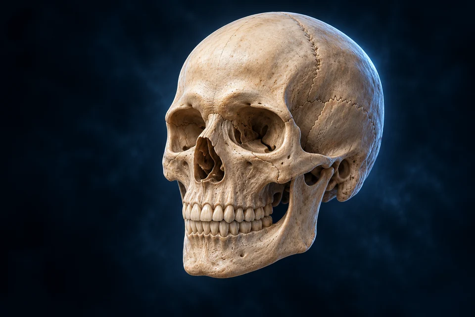
The skull surrounds the brain and supports the face. Explore its outer landmarks, hidden floor and neighbouring cavities, then see how living bone builds the framework.
▷
Watch chapter overviewFilm planned · not yet available
Original adult skull preparation · fine detail illustrative
02 / LATERAL
A cranial enclosure continues into facial support.
Brain-case bones and facial bones meet around the orbit, nose and jaws. The zygomatic arch spans the side above the moving mandible.
New original Golden-family text-free teaching preparation. Anterior is image left; representative adult left lateral orientation, not a measured patient, sex classification or complete skull-bone count. Major visible bone/space relationships are source-supported. Fine generated sutures, foramina, dental roots, pores and grooves remain illustrative / not independently verified. No small opening is assigned an exact nerve route. Human medical review: Not performed. Presentation crops only empty surrounding background, never anatomy; the labelled detail retains this viewport and the full-resolution image entry opens the uncropped original master.Structure notes · further context
Frontal region
The frontal bone contributes to both the cranial enclosure and orbital roof. Exact sutural boundaries in this generated view are illustrative.
Parietal region
Right and left parietals meet superiorly; only the broad left-side region is identified here.
Occipital region
Its lower surface participates in the skull–neck junction. The lateral image does not expose every basal opening.
Temporal region
Different portions include squamous and petrous regions. Fine generated canals are not a verified ear atlas.
Zygomatic arch
Zygomatic and temporal contributions form the arch. Nearby muscles and the jaw are explained in linked oral teaching.
Maxillary region
Paired maxillae also contribute to palate and orbital context. Tooth number and each generated root are not independently verified.
Mandible
Movement combines rotation and translation. This does not imply every other bone in the head, including ear ossicles, is immobile.
In this left lateral view, the face points toward image left. The preserved background-only viewport enlarges the skull; it does not move any anatomical label to a different source point.
Conventional adult teaching counts eight brain-case and fourteen facial bones, excluding ear ossicles and hyoid. Individual sutural bones and developmental differences make this a classification convention rather than a diagnostic count for every person.
One structure looks different from different directions.
A lateral skull explains the face-to-vault relationship. Looking down into the cranial floor reveals internal tiers; a facial cutaway reveals spaces hidden by the outer surface.
Left lateral · face toward image left
New original Golden-family text-free teaching preparation. Anterior is image left; representative adult left lateral orientation, not a measured patient, sex classification or complete skull-bone count. Major visible bone/space relationships are source-supported. Fine generated sutures, foramina, dental roots, pores and grooves remain illustrative / not independently verified. No small opening is assigned an exact nerve route. Human medical review: Not performed. Presentation crops only empty surrounding background, never anatomy; the labelled detail retains this viewport and the full-resolution image entry opens the uncropped original master.Structure notes · further context
Frontal region
The frontal bone contributes to both the cranial enclosure and orbital roof. Exact sutural boundaries in this generated view are illustrative.
Parietal region
Right and left parietals meet superiorly; only the broad left-side region is identified here.
Occipital region
Its lower surface participates in the skull–neck junction. The lateral image does not expose every basal opening.
Temporal region
Different portions include squamous and petrous regions. Fine generated canals are not a verified ear atlas.
Zygomatic arch
Zygomatic and temporal contributions form the arch. Nearby muscles and the jaw are explained in linked oral teaching.
Maxillary region
Paired maxillae also contribute to palate and orbital context. Tooth number and each generated root are not independently verified.
Mandible
Movement combines rotation and translation. This does not imply every other bone in the head, including ear ossicles, is immobile.
The vault, cheek bridge and upper/lower jaws share an exterior view. It cannot also expose all of the internal cranial floor.
Lateral exterior
Anterior left → posterior right
Vault, cheek bridge and jaws share one visible region.
Superior interior
Anterior top → posterior bottom
Removing the roof reveals the connected floor tiers.
Anterior cutaway
Face toward viewer → exposed spaces
A layered educational cut is not an individual CT plane.
These relationship steps supplement the regional anatomy. All remain readable before highlighting; they are not exact hidden geometry, measured airflow or a universal movement sequence. The full craniocervical and hyoid atlas remains a separate lesson.
View controls support orientation. The four primary plates below add new relationships instead of repeating the same image.
Six named groups contribute eight conventional cranial bones.
Paired parietal and temporal bones join four unpaired groups. Their contributions overlap the roof, side and base rather than making six isolated boxes.
Frontal
Anterior vault and orbital-roof context
Links forehead support with the anterior cranial floor.
Broad position; small boundaries are not all resolved in one view.
Parietal pair
Upper and lateral vault
Forms much of the roof/side enclosure.
The two sides meet at the sagittal seam context.
Temporal pair
Lower lateral skull and petrous context
Relates to ear spaces, jaw articulation and cranial floor.
Tiny ear anatomy is staged, not inferred from pores.
Occipital
Posterior skull and basal context
Relates to posterior support and the large skull–cord opening.
The opening alone does not show its full contents.
Sphenoid
Central cranial-base and lateral contributions
Connects several floor, orbital and facial relationships.
Not a disconnected butterfly floating inside the head.
Ethmoid
Anterior midline / nasal–orbital interface
Contributes to nasal shelves, septal and olfactory-roof context.
Fine air cells and nerve pores are not individually validated.
A suture is a connective joint, not a drawn crack.
Fibrous tissue joins skull bones at sutures. Their developmental and adult condition varies; a generated jagged line cannot establish an individual fusion stage.
Coronal context
Frontal ↔ paired parietal bones
A broad anterior-to-posterior vault boundary.
Exact fine serrations are illustrative.
Sagittal context
Right parietal ↔ left parietal
A superior midline relationship.
Not fully visible in a left lateral plate.
Posterior / lateral seams
Lambdoid and squamous relationships
Link occipital or temporal regions with adjoining vault bones.
Not every tiny generated seam receives a precise name.
The cranial base relates to different brain regions.
Anterior, middle and posterior fossae form connected tiers. Ridges, depressions and openings organise support and passage rather than divide the head into sealed compartments.
New original Golden-family text-free teaching preparation. Roof removed; anterior is image top and posterior image bottom. Broad cranial-fossa tiers, central sphenoid context, petrous ridges and foramen-magnum relationship are source-supported. Fine generated small foramina, vascular grooves, cribriform pores and exact sella boundaries remain illustrative / not independently verified, not a surgical or complete nerve-route map. Human medical review: Not performed.Structure notes · further context
Anterior fossa context
Frontal and selected ethmoid/sphenoid contributions form this region. It is not an isolated sealed box.
Middle fossa context
Sphenoid and temporal contributions shape the floor. Not every visible hole is an independently verified canal.
Posterior fossa context
The broad region is shown; dura, vessels and neural structures are not included as a complete reconstruction.
Central sphenoid context
The pituitary sits in the sella region in life. Fine generated seat boundaries and openings are not independently verified here.
Petrous ridge context
The petrous temporal region contains inner-ear context. This plate does not resolve its tiny internal compartments.
Foramen magnum
Brainstem–cord transition and other structures traverse this region. A hole alone does not show every passing tissue.
Frontal-brain support lies toward the front
The shallow anterior cranial fossa supports frontal-brain context. Its surface joins the rest of the cranial floor; the fossa is not a separate sealed chamber.
Gold emphasis selects an anatomical context. It does not measure its boundary, reconstruct hidden structures or certify the fine generated detail.
The central sella region relates to the pituitary; petrous temporal regions also contain ear structures. Detailed foraminal routes require more precise views than this broad regional plate.
How does a floor shape relate to the tissues it supports?
The fossae are connected depressions at different depths. In life, meningeal tissues, vessels and neural structures intervene between a bony surface and a functional system. A regional view gives orientation; it cannot certify every small passage or hidden nerve route.
The orbit, nose and sinuses are neighbouring spaces.
The orbit houses the eye; the nasal cavity is an airway; paranasal sinuses extend into neighbouring bones. Their proximity does not make them one open cavity.
New original Golden-family text-free teaching preparation. Anterior educational layered cutaway, not an individual CT plane or precise sinus-drainage reconstruction. Broad orbital/nasal/frontal/maxillary/ethmoid relationships are source-supported. Fine generated mucosa, conchae, ostia, tooth roots and bony boundaries remain illustrative / not independently verified. Sinus shapes and symmetry vary between people. Human medical review: Not performed.Structure notes · further context
Frontal sinus context
Development, size and side-to-side form vary. This is not a universal volume or fixed pair of identical cavities.
Orbital socket
The eye is absent to reveal surrounding bone. An orbit is not a paranasal sinus.
Maxillary sinus context
Upper dental roots are nearby, but each generated root–sinus distance is not a measured human relationship.
Ethmoid air-cell context
This is a broad neighbourhood, not a verified count or exact labyrinth of individual cells.
Nasal septal context
Real septa include cartilage and bony contributions and may not be perfectly straight. Exact fine material boundaries are illustrative.
Conchal region
Superior and middle conchae belong to ethmoid; the inferior concha is an independent bone. This view does not verify every tiny shelf or meatus.
An orbit houses a sensory organ and its supporting tissues
The paired sockets sit above the maxillary sinuses and beside the nasal region. Eye muscles, fat, vessels and nerves occupy an orbit in life; an empty socket is not a sinus or the eyeball itself.
Gold emphasis selects an anatomical context. It does not measure its boundary, reconstruct hidden structures or certify the fine generated detail.
The hard palate separates the nasal and oral compartments below. The central septum includes bone and cartilage; real septa and sinus shapes vary.
Upper and middle conchae are ethmoid contributions, while inferior conchae are independent bones. Conchae and their mucosal surroundings shape the airway interface.
Sinus groups are named by their bony surroundings.
Frontal, maxillary, ethmoid and sphenoid groups have different locations and drainage context. Size, symmetry and development vary; the sphenoid group is not directly exposed in this anterior cutaway.
Frontal / maxillary
Above orbital level / beneath orbit in upper jaw
Distinct neighbouring bone cavities, not eye sockets.
Shown for broad position, not universal volumes.
Ethmoid
Small-cell context near upper nose and medial orbit
A complex collection of air spaces.
Not a fixed identical set of bubbles.
Sphenoid
Deeper central skull-base context
A posterior sinus relationship requires another view.
Introduced here; no invisible cavity is labelled as exposed.
Several proposed sinus functions remain debated. The chapter teaches location and mucosal connection without declaring every suggested acoustic, impact-buffering or climatic role proven.
Living lining adds functions that bone alone cannot perform.
Respiratory mucosa, mucus and ciliary activity help condition and clear the nasal/sinus environment. The olfactory region is a specialised sensory interface, not the same lining everywhere.
Bone locates the airway; living lining conditions the air
New original Golden-family text-free teaching preparation. Anterior educational layered cutaway, not an individual CT plane or precise sinus-drainage reconstruction. Broad orbital/nasal/frontal/maxillary/ethmoid relationships are source-supported. Fine generated mucosa, conchae, ostia, tooth roots and bony boundaries remain illustrative / not independently verified. Sinus shapes and symmetry vary between people. Human medical review: Not performed.Structure notes · further context
Frontal sinus context
Development, size and side-to-side form vary. This is not a universal volume or fixed pair of identical cavities.
Orbital socket
The eye is absent to reveal surrounding bone. An orbit is not a paranasal sinus.
Maxillary sinus context
Upper dental roots are nearby, but each generated root–sinus distance is not a measured human relationship.
Ethmoid air-cell context
This is a broad neighbourhood, not a verified count or exact labyrinth of individual cells.
Nasal septal context
Real septa include cartilage and bony contributions and may not be perfectly straight. Exact fine material boundaries are illustrative.
Conchal region
Superior and middle conchae belong to ethmoid; the inferior concha is an independent bone. This view does not verify every tiny shelf or meatus.
Conchal shelves and the nasal partition frame the passage. The vascular respiratory lining helps warm and humidify inspired air; this gross cutaway does not resolve its microscopic cells.
Air interface
Incoming air → living nasal surface
Mucosa helps warm and humidify the environment.
Clearance context
Mucus / ciliary activity → normal transport
A supporting concept, not a measured exact drainage route.
Sensory context
Selected olfactory interface → neural information
Smell requires specialised tissue; not all nasal lining is olfactory.
These relationship steps supplement the regional anatomy. All remain readable before highlighting; they are not exact hidden geometry, measured airflow or a universal movement sequence. The full craniocervical and hyoid atlas remains a separate lesson.
Supporting interface map, not an exact sinus drainage model. No surgical ostium route, measured airflow or smell-detection threshold is claimed.
Why does the same bony passage need different living surfaces?
Respiratory lining conditions inspired air and supports mucus clearance. A specialised olfactory region supports smell. Their cellular structures and tasks differ; a visible concha is a bony shelf with a covering, not a giant cilium or an exposed sensory nerve.
A bony partition supports separate oral and nasal spaces.
Maxillary and palatine contributions form the hard palate. Teeth sit in alveolar sockets, while tongue, soft palate and pharyngeal structures add movement and swallowing relationships.
Hard palate
Oral roof / nasal floor context
Separates neighbouring compartments.
The soft palate is a different moving tissue structure.
Dental support
Roots → periodontal interface → alveolar bone
A tooth is supported by specialised tissues.
Not directly welded into solid jaw bone.
Posterior continuation
Nasal / oral passages → pharyngeal context
Shared downstream relationships support breathing and swallowing.
The routes are not identical at every stage of swallowing.
Many adult vault regions combine dense cortical tables with intervening trabecular diploe. The configuration varies by region, age and individual; it is not one uniform shell thickness.
New original Golden-family text-free teaching preparation. Enlarged regional adult calvarial context, not every skull site or an acquired specimen. Broad cortical tables, diploe and outer covering are source-supported. Fine generated pores, marrow, vessels, cellular identity and exact layer thickness remain illustrative / not independently verified. The inner table is not a second guarantee against impact. Human medical review: Not performed.Structure notes · further context
Outer table
Thickness and microarchitecture vary across skull regions and people. The shown curve is a teaching model.
Diploe
Spaces contain living marrow and vascular context. Not all cranial sites have identical layered proportions.
Inner table
The brain is separated from bone by meningeal context in life; the plate does not include a complete meninges reconstruction.
Outer connective covering
A lifted edge reveals the underlying table. It is not scalp or a detached piece of cranial bone.
Marrow / vascular context
The red detail is illustrative; it does not identify each diploic vein, blood cell or exact vascular connection.
Connective covering lies outside the outer bony table
The lifted periosteal context reveals the external bone surface. It is separate from the scalp and from the meningeal tissues that lie internally in life.
Gold emphasis selects an anatomical context. It does not measure its boundary, reconstruct hidden structures or certify the fine generated detail.
Vascular and marrow context occupies internal spaces. Fine generated red texture is not a named complete diploic venous map or a measured blood-cell population.
Meningeal tissues lie between bone and brain in life. Their layers and CSF relationships are taught in the linked Brain chapter rather than duplicated as another shallow overview.
What does a layered skull preparation reveal—and what is missing?
Dense tables and intervening trabeculae reveal a material arrangement. Living cells, marrow, vessels and coverings maintain that framework. Meninges and fluid spaces belong to a different internal layer of protection; an enlarged bone section does not show all of them.
Formation, sensing and resorption participate in maintenance. Cortical and trabecular materials, blood and connective interfaces coexist at different scales.
Matrix / cells
Collagen and mineral with living cell context
Bone is maintained rather than remaining inert after growth.
Exact turnover is not measured in these pictures.
Marrow / vessels
Internal diploic and other regional spaces
Support living tissue and wider circulation.
No complete venous pathway inferred from texture.
Connective interface
Outer covering, sutures and meningeal relationships
Neighbouring tissues communicate mechanically and biologically.
A covering alone is not a guarantee against injury.
A developing skull is not a scaled-down adult skull.
Fontanelles and sutures provide developmental interfaces while the brain and face grow. Bone formation and remodelling change shape and tissue organisation over time.
Vault growth
Connective seams and broader tissue interactions
Growth involves linked bony and meningeal contexts.
No single universal closure timeline or pathological classification.
Facial development
Changing teeth, jaws and neighbouring spaces
Proportions and sinus development change across life stages.
An adult image cannot prove fetal or infant anatomy.
Variation
Individuals differ in shape, seams and cavities
Normal variation matters when learning from an example.
A generated skull cannot classify sex, ancestry or personal identity.
The head is supported by both joints and soft tissues.
Occipital condyles articulate with the atlas at the skull–neck junction. The jaw has paired temporomandibular joints; the hyoid is supported by muscular and ligamentous attachments rather than directly articulating with another bone.
Skull–neck
Occipital condyles ↔ atlas context
Bony support works with surrounding joints and soft tissues.
Jaw articulation
Mandible ↔ paired temporomandibular joints
Rotation and translation contribute to oral movement.
Hyoid support
Muscles / ligaments → suspended bone context
No direct joint with another bone is asserted.
These relationship steps supplement the regional anatomy. All remain readable before highlighting; they are not exact hidden geometry, measured airflow or a universal movement sequence. The full craniocervical and hyoid atlas remains a separate lesson.
Supporting relationship comparison; a full craniocervical/atlas-axis/hyoid primary atlas remains staged. No exact movement range or patient positioning advice.
Protection and sensory function share the same bony neighbourhood.
Cranial spaces relate to brain, eyes, ears, airway and oral function. Shared boundaries explain why a regional bone chapter needs links to functional chapters instead of repeating their main images.
Brain
Enclosure, openings and meningeal relationships
The skull supplies support; neural activity is another level.
No one-bone/one-behaviour claim.
Eyes / ears
Orbits and temporal internal spaces
Bone surrounds sensory tissues with different structures.
Tiny ear canals and ossicles are not resolved by these pores.
Airway / oral function
Nasal passage, palate and moving jaw
Bone and living soft tissues cooperate.
An isolated skull is not a complete breathing model.
Views and preparations answer different questions.
A dry skull shows bony surfaces; a section shows tissue arrangement; imaging can show living spatial context. None of those is identical to a source-informed generated teaching plate.
A regional foundation, not every opening and attachment.
Visible gross anatomy, cranial tiers, facial spaces, bone layers and selected normal functions now connect. Detailed nerve routes, ear microanatomy, spine regions and clinical topics remain distinct roadmap jobs.
Now taught
Gross region → space → tissue → function → related system
Four primary views add different angles or scales.
No invented complete-anatomy percentage.
Still staged
Exact foraminal/vascular maps, full spine/ear atlas and clinical branches
These need precise views, independent sources and their own acceptance.
Broad adult skull regions, cranial-floor tiers, facial spaces and calvarial tissue layers; selected normal connective, developmental, airway, jaw and cross-system relationships. Authoritative references are used critically rather than repeating every simplified or historical claim.
Explicit limits
AI-assisted visual/content production. Fine generated sutural, vascular, foraminal, dental and matrix details remain illustrative / not independently verified. Human medical review: not performed. Educational only; not diagnostic, clinical or surgical material.
Revision: skull-spaces-english-2026-10-19-eyes-foundation-v1. Stable local key: ahb_ch_skull_spaces_english_v1. No invented canonical CMS/atlas entity or 3D dataset. Five original Golden-family text-free masters and twenty-four editable English labels remain separate. Fine generated small foramina, dental roots, sutural serrations and matrix texture are not independently verified; no exact nerve routes or measured layer thickness are inferred. Migration physical-phone acceptance: Pending, non-blocking.