A Visual Human Body Encyclopedia
Small structures.
A connected body.
Follow anatomy from the compartments of a cell to the tissues and networks of the brain. Original illustrations make broad relationships visible; authentic scientific images show a different kind of evidence.
Explore the chapters →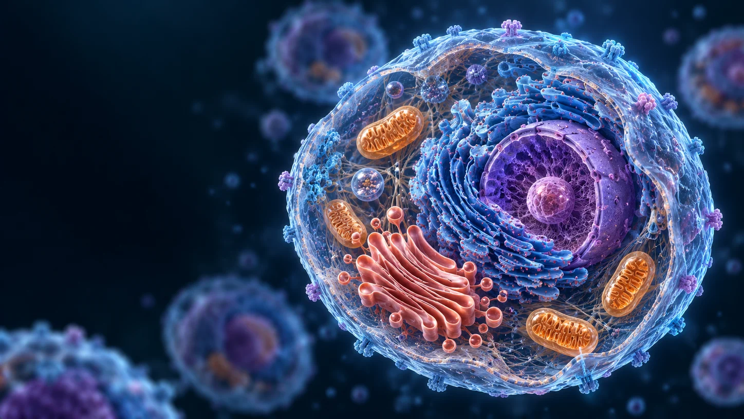
Published chapter selection
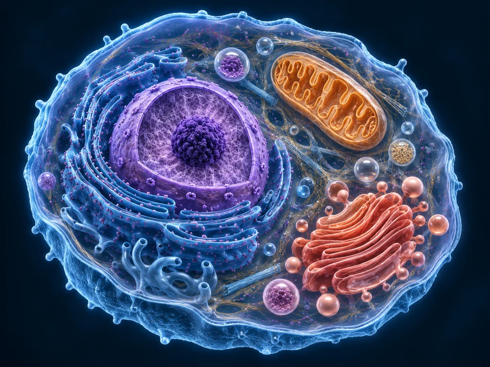
Cells & Tissues
Cell boundaries, organelles, exchange and the four tissue families.
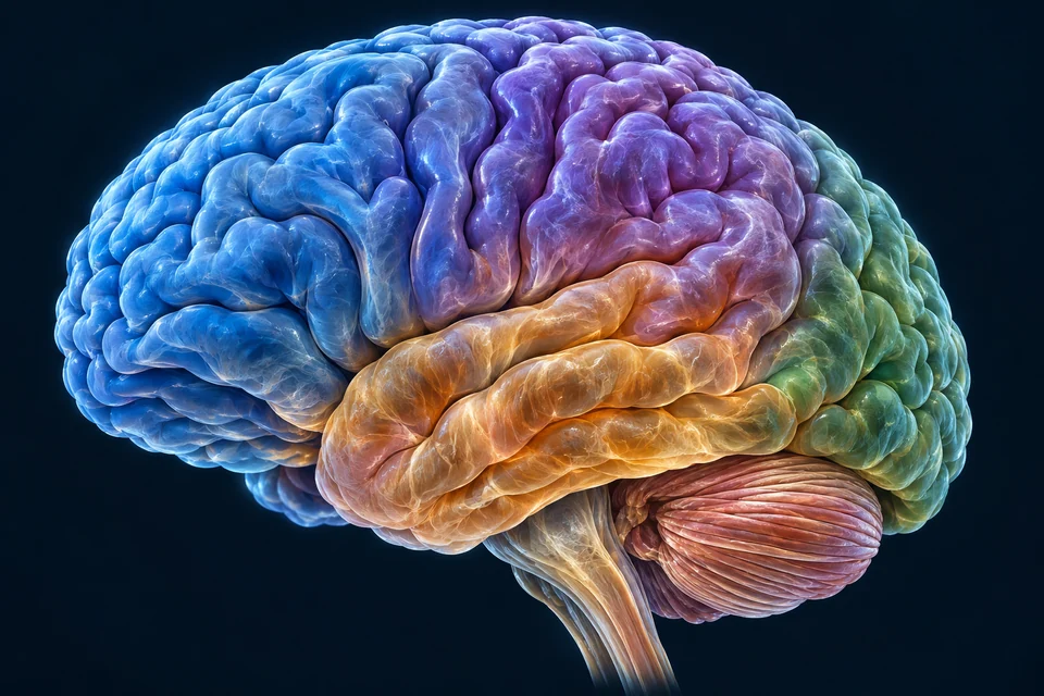
Brain
Structure, fluid spaces, blood delivery, signals, memory and sleep.
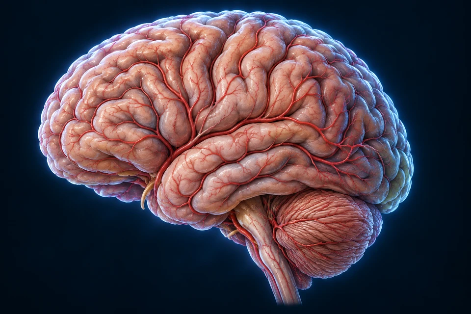
Stroke
Sudden warning signs, interrupted delivery, bleeding, emergency checks and treatment principles.
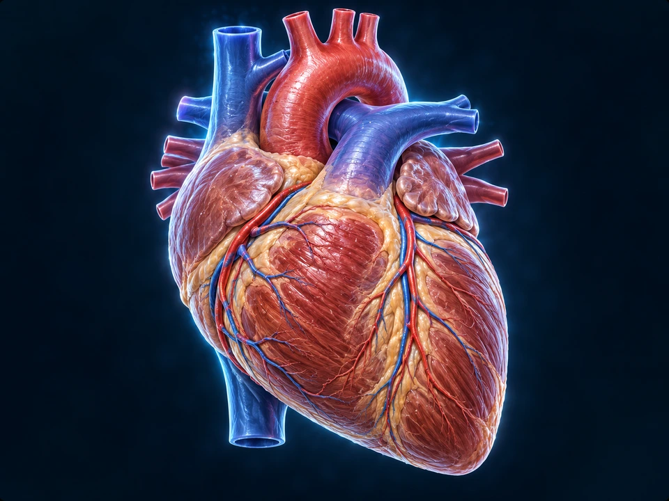
Heart
Chambers, valves, circulation, living muscle, electrical timing and pump regulation.
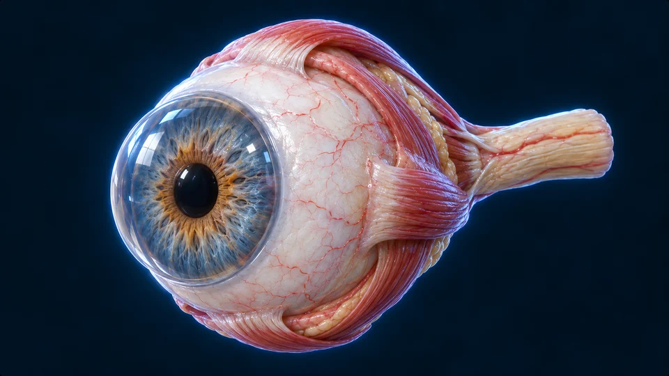
Eyes & Vision
Focus, retinal circuits, real eye imaging, tears, movement and visual fields.
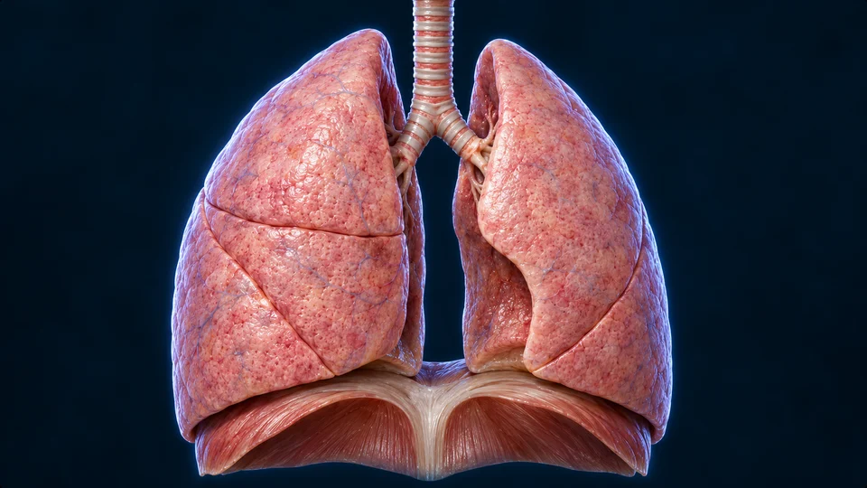
Lungs & Breathing
Airway, alveolar cells, gas exchange, pleura, muscle mechanics, feedback and real laryngoscopy.
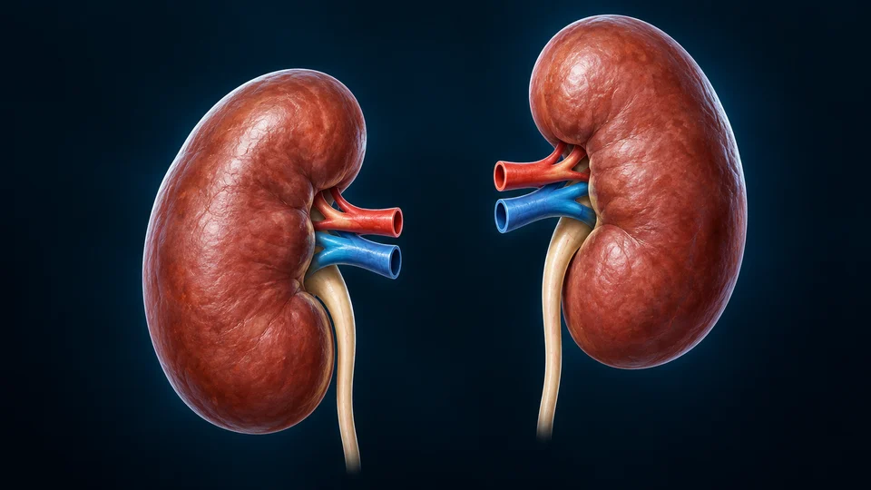
Kidneys
Regions, nephrons, filtration, tubular recovery, urine concentration, endocrine balance and real tissue.
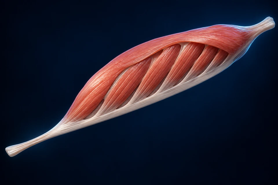
Muscles & Movement
Cellular architecture, activation, force control and sensory feedback.
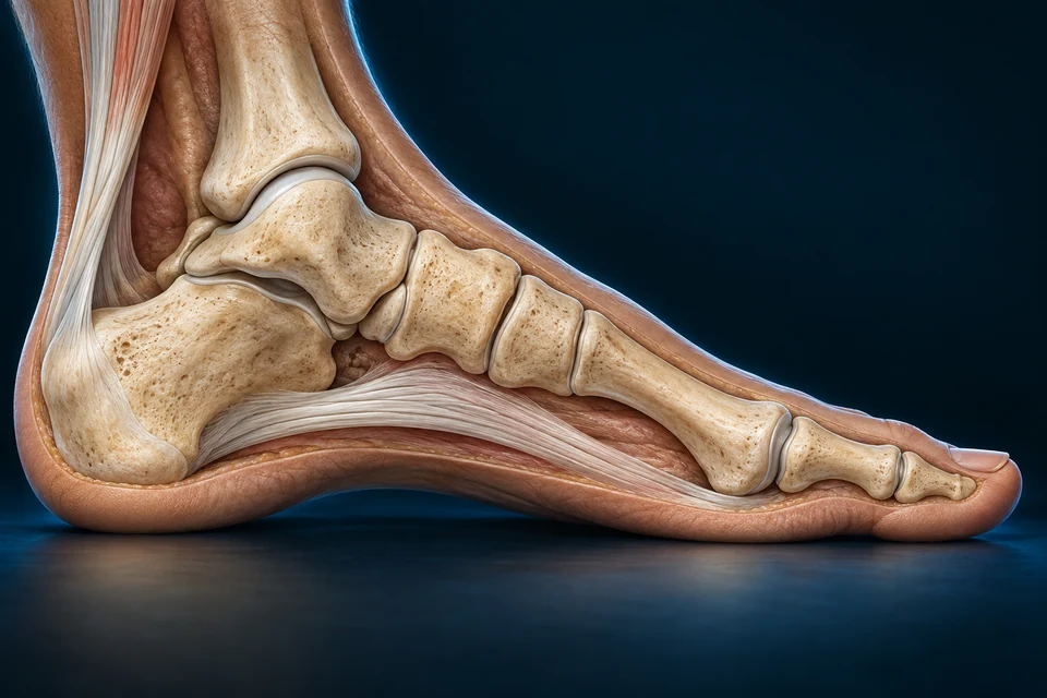
Hand, Foot & Arches
Hand and foot frameworks, tendon routes, arches, walking and living contact tissues.
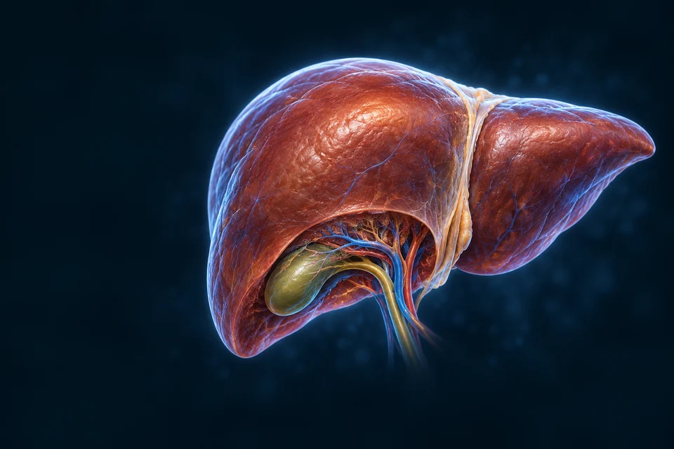
Liver
Blood and bile routes, lobules, working cells, fuel regulation and metabolic connections.
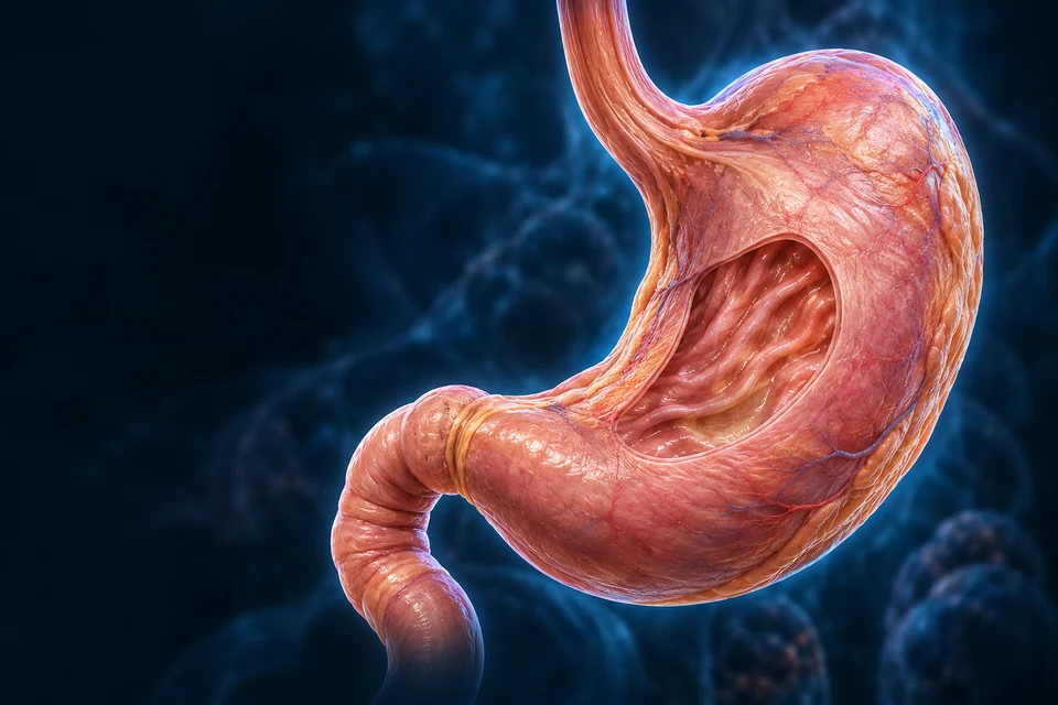
Stomach
Regions, wall layers, glands, secretion, protection and controlled onward delivery.
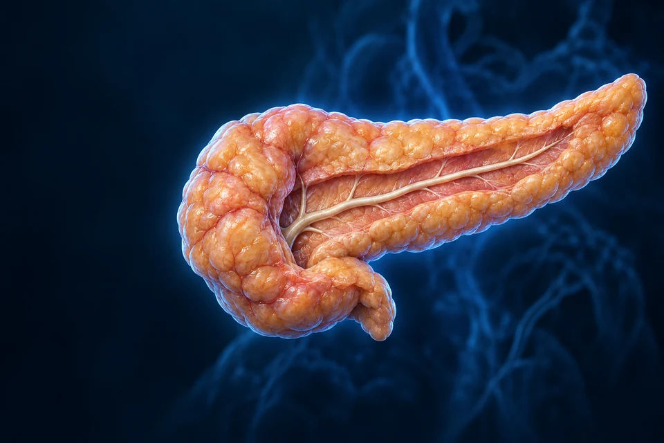
Pancreas
Digestive ducts, acinar cells, islet blood routes, enzyme activation and hormone feedback.
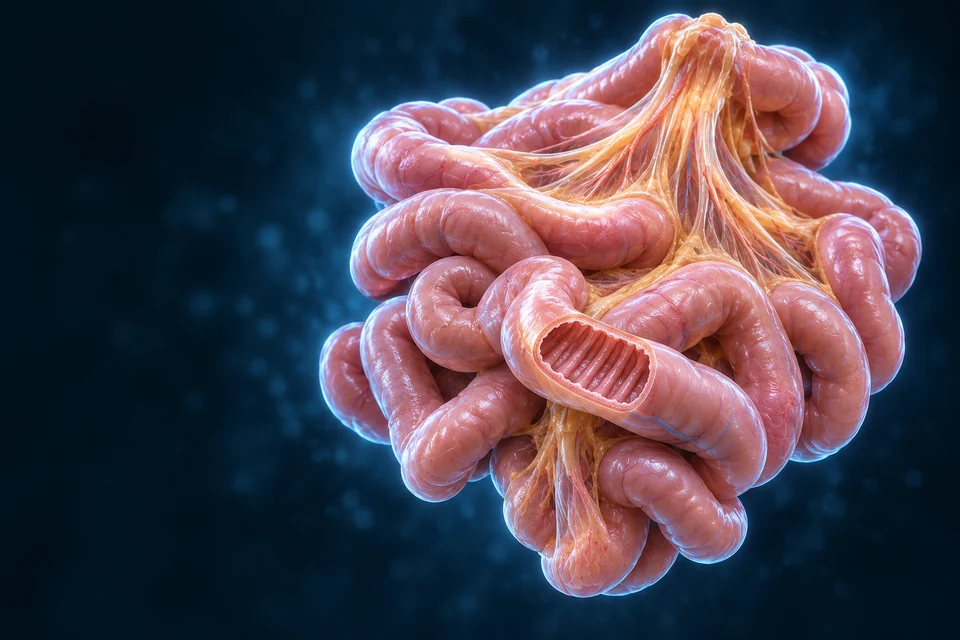
Small Intestine
Folds, villi, renewing cells, selective membranes and separate blood and lymph routes.
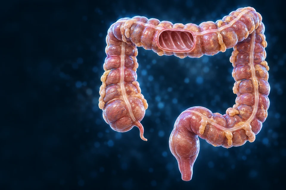
Large Intestine
Regions, crypts, mucus, fluid recovery, microbial products and coordinated movement.
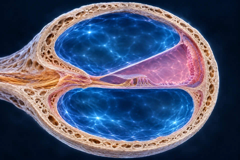
Hearing & Balance
Ear compartments, sensory epithelia, sound processing and coordinated head-motion inputs.
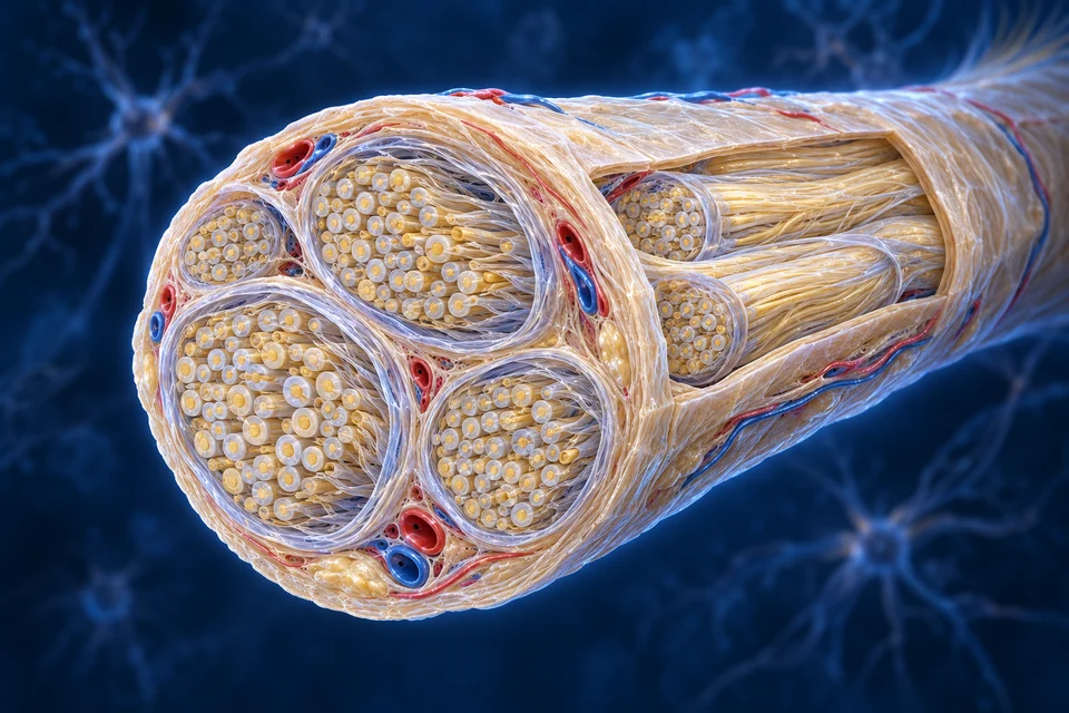
Nervous System
Nerve bundles, supporting cells, myelin, signals, roots, regional routes and visceral regulation.
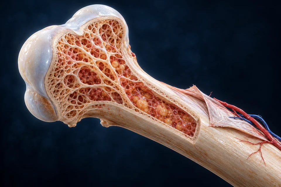
Bone & Joints
Explore normal structure, tissue, mechanisms and connected learning.
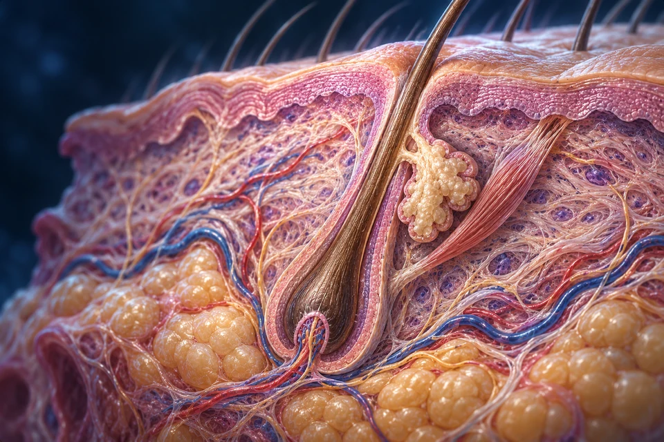
Skin, Hair & Nails
Explore normal structure, tissue, mechanisms and connected learning.
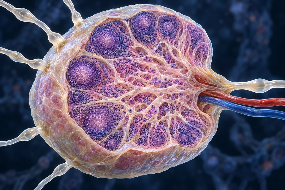
Lymph & Immunity
Explore normal structure, tissue, mechanisms and connected learning.
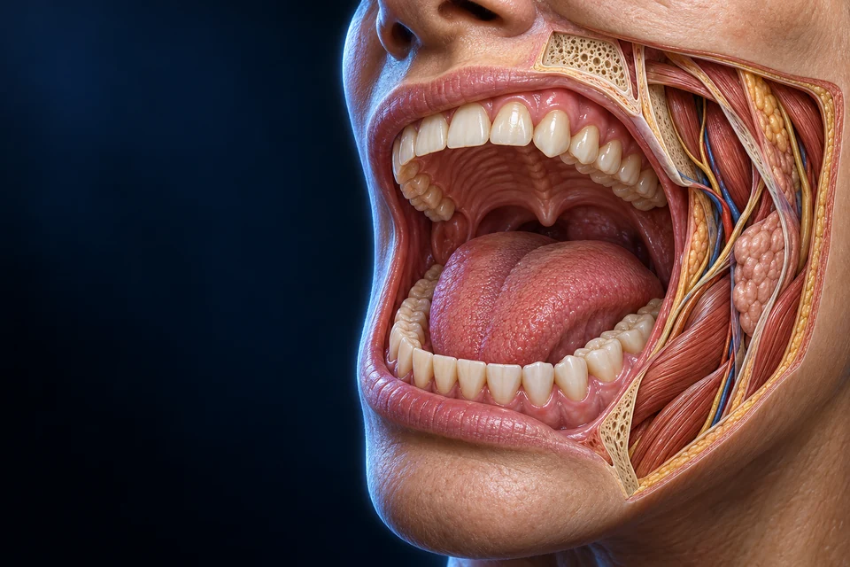
Mouth & Swallowing
Explore normal structure, tissue, mechanisms and connected learning.
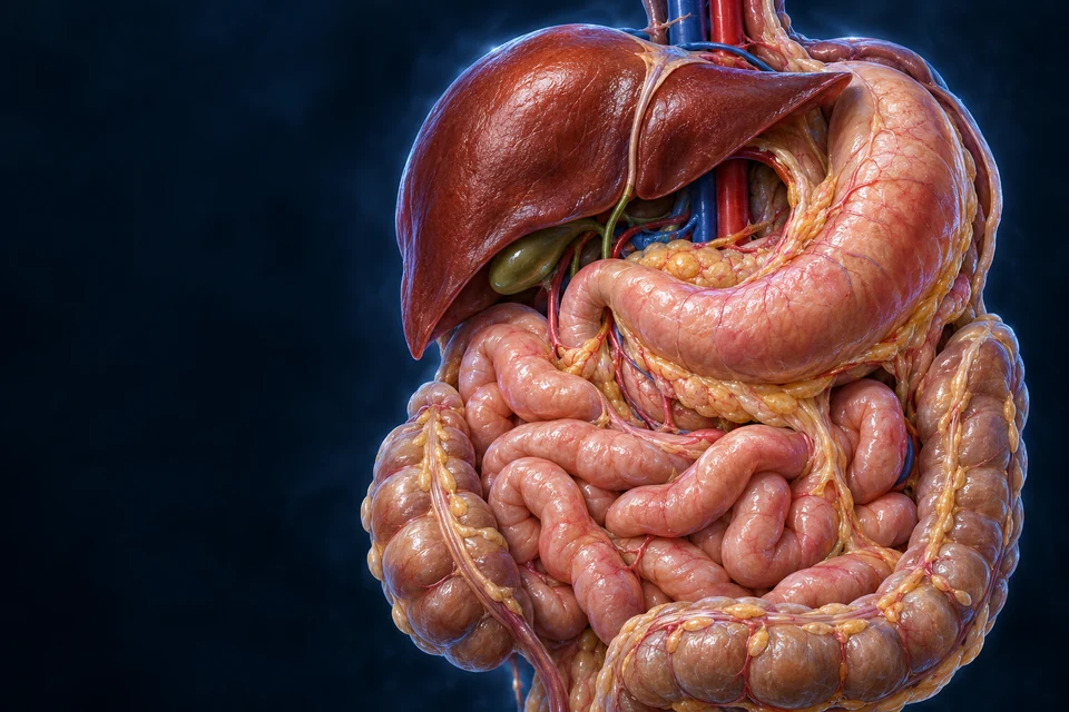
Digestive System
Follow food, secretions, supporting surfaces and different tissue exits.
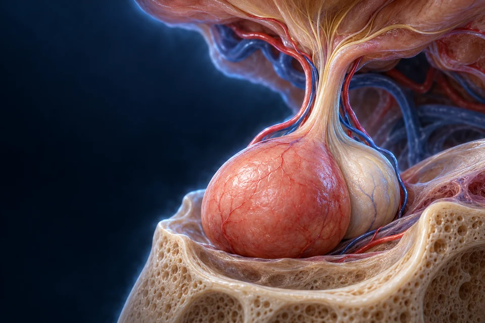
Hypothalamus & Pituitary
Compare neural and portal-blood delivery, tissue secretion and target response.
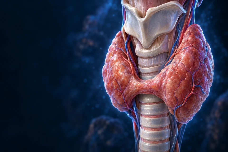
Thyroid & Calcium
Connect gland structure, thyroid hormone formation and calcium regulation.
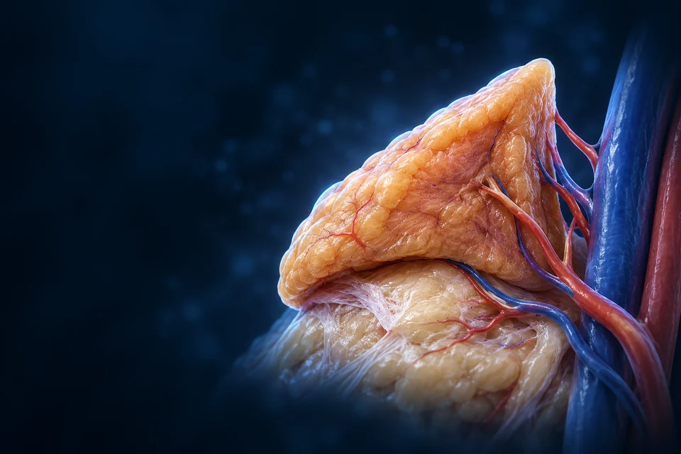
Adrenal Glands
Connect cortical tissue, neural secretion and distinct hormonal control.
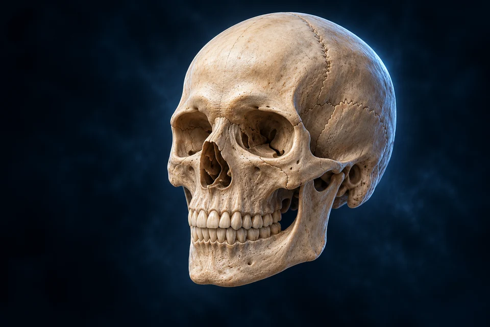
Skull & Cranial Spaces
Connect the cranial enclosure, facial spaces and living bone.
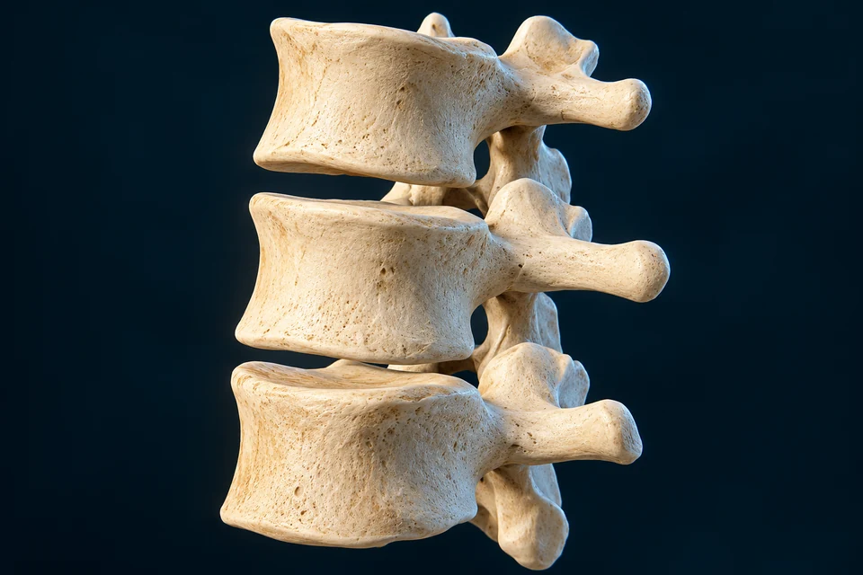
Vertebral Column
Connect the regional column, disc tissues and protected neural spaces.
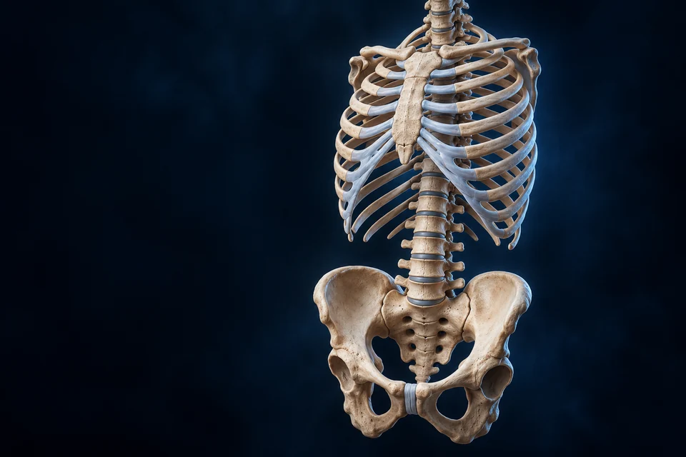
Thoracic Cage & Pelvis
Connect the chest wall, breathing partition and load-sharing pelvis.
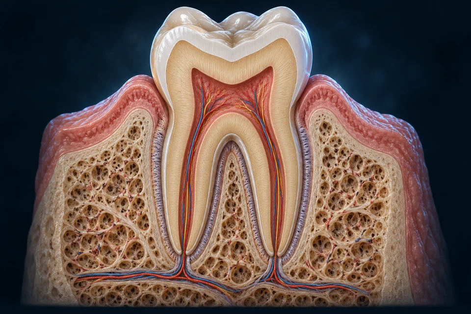
Teeth & Periodontium
Connect crown shape with forming mineral, living pulp and root attachment.
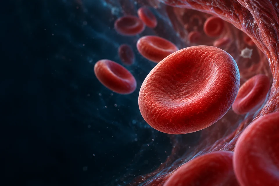
Blood & Vessels
Connect moving cells with living vessel walls, tissue exchange, local repair and return.
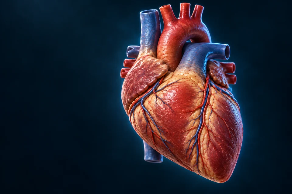
Vascular Routes
Follow branching supply, converging return, regional names and the portal connection.
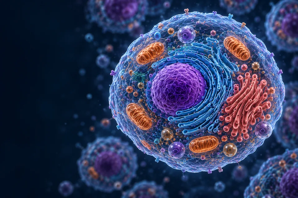
The Integrated Body
Connect organ journeys, tissue boundaries, body fluids, feedback and ways of seeing.
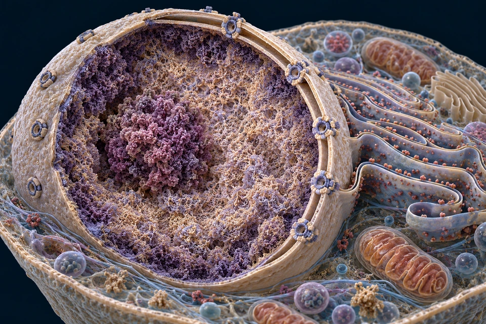
DNA, Genes & Genome
Locate nuclear DNA, follow selected messages, compare molecular evidence and chromosome copying.
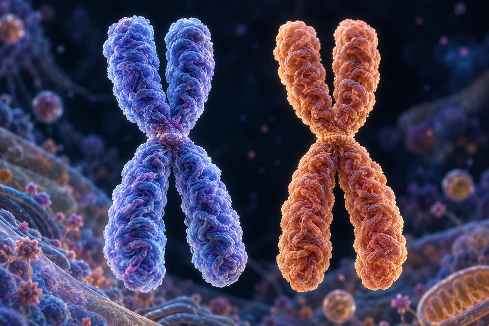
Inheritance & Cell Division
Distinguish parental partners from copied sisters, compare two divisions, and understand bounded inheritance models.
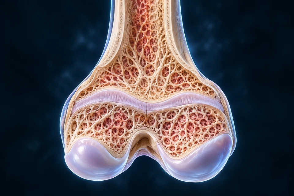
Growth, Puberty & Aging
Follow growth cartilage, tooth succession, tissue maintenance, focusing and changing physiological reserve.
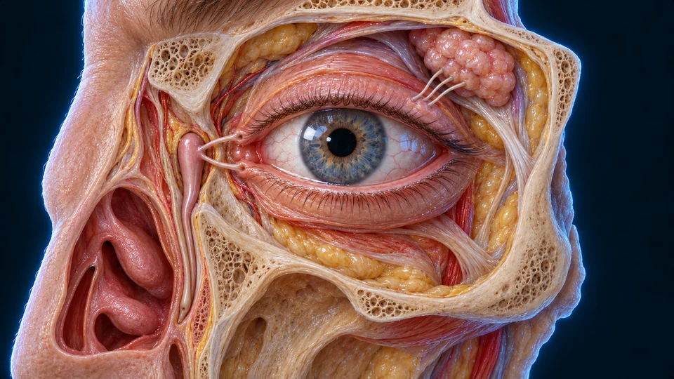
Eye Support & Movement
Connect coordinated gaze, pupil control, blinking, tear supply and nasal drainage.
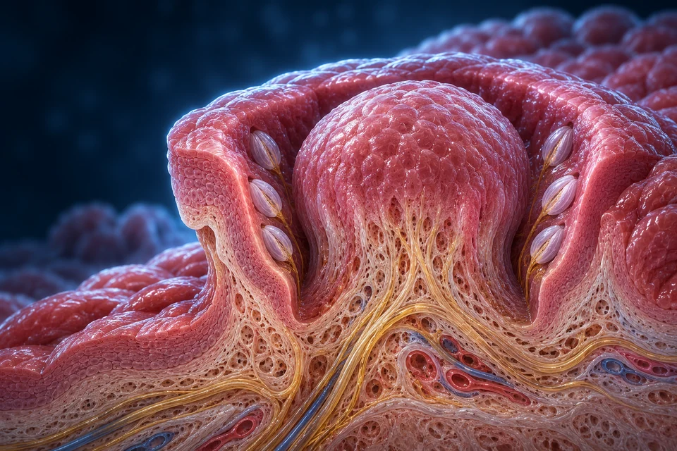
Smell & Taste
Connect nasal access, sensory cells, tongue papillae, taste buds and the information behind flavour.
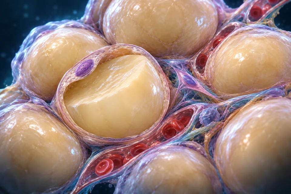
Nutrient Use & Metabolism
Connect nutrient delivery, living reserves, cellular work and heat.
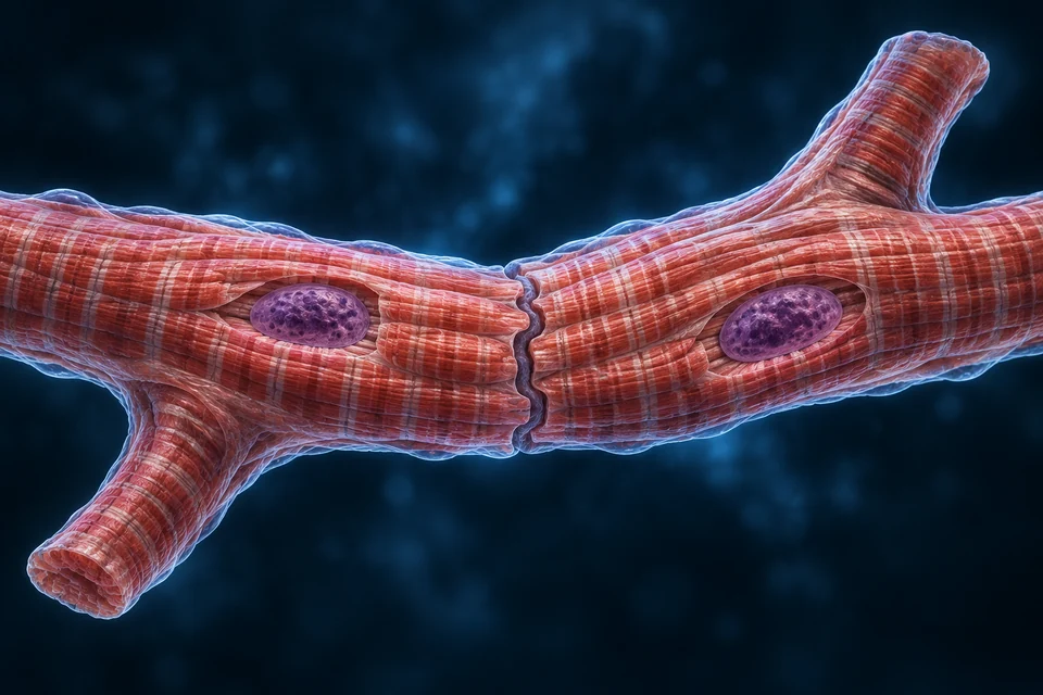
Cardiac Tissue & Coordination
Connect the working wall, its supply, valve support and cellular coordination.
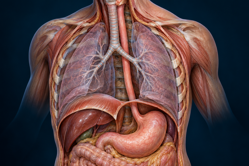
Esophagus
Protective layers, coordinated peristalsis and a controlled entrance to the stomach.
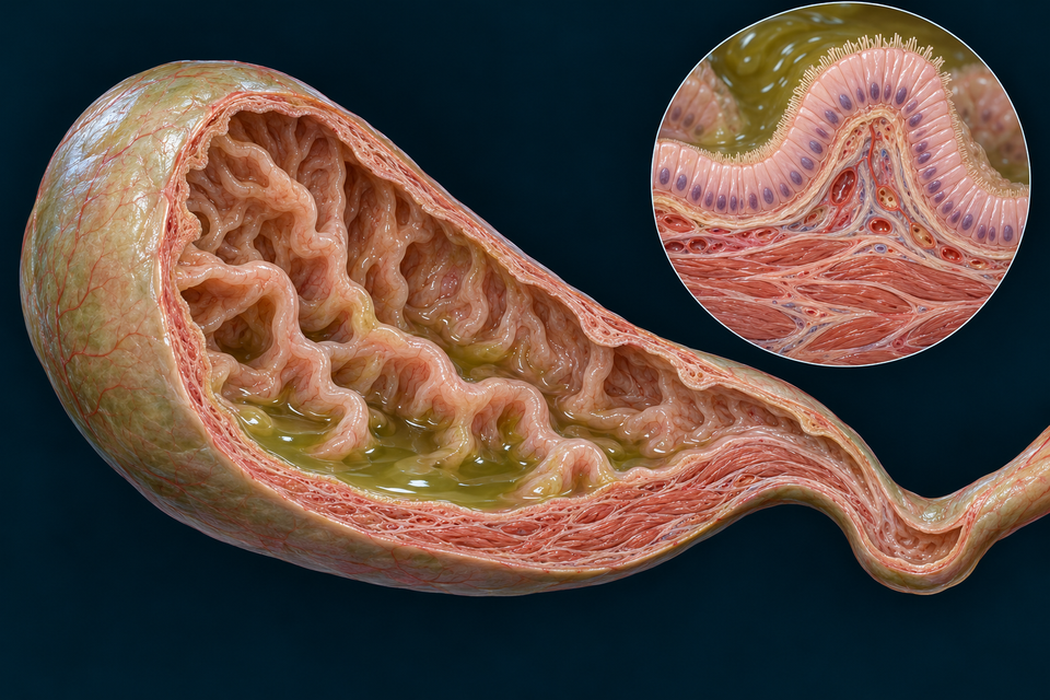
Gallbladder & Bile Routes
A living reservoir, fluid recovery and coordinated delivery to the intestine.
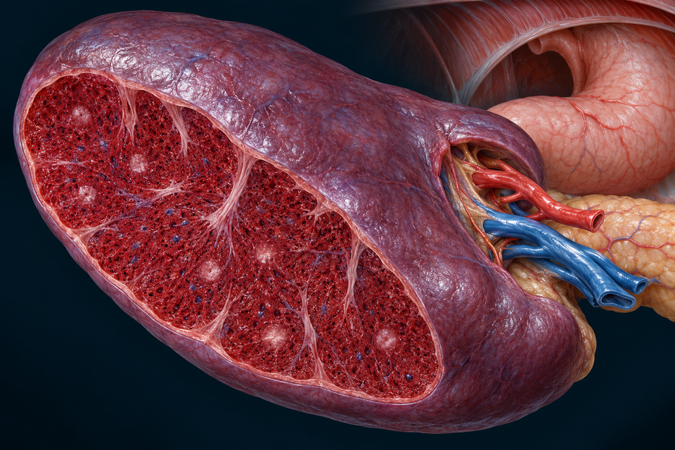
Spleen
Blood-cell handling, immune regions and useful material returned to circulation.
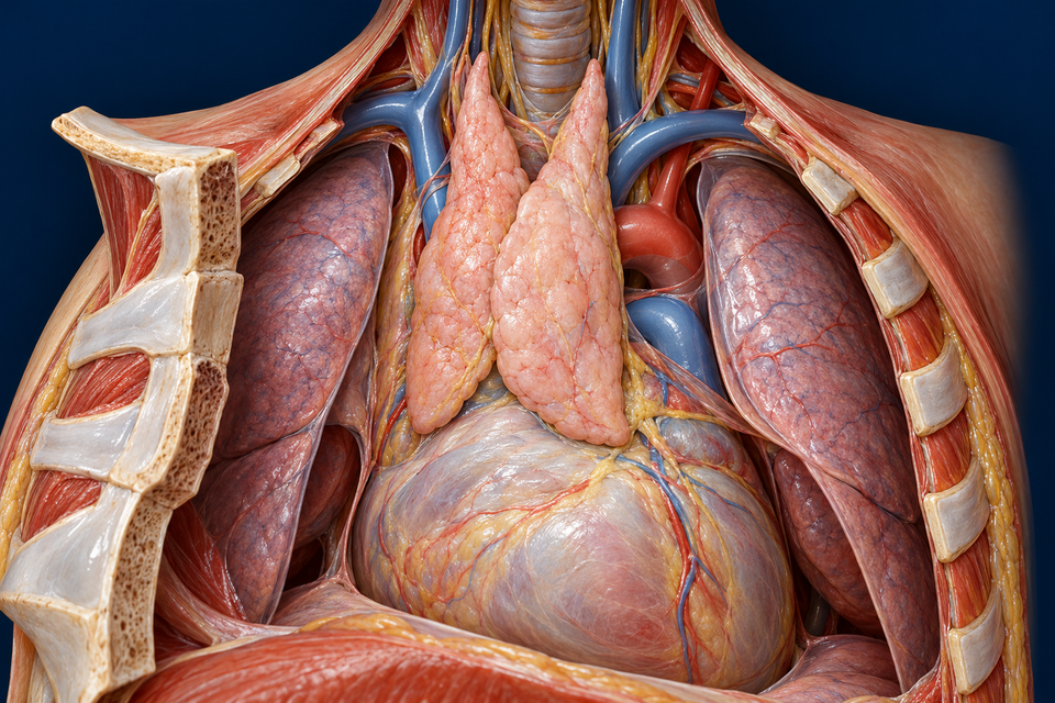
Thymus
A protected tissue environment for T-cell development, selection and immune restraint.
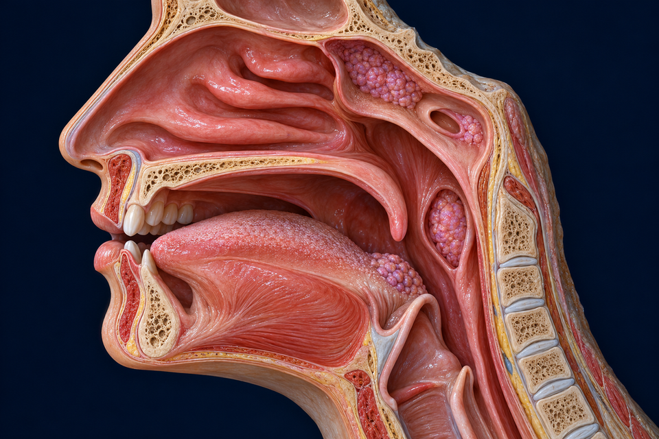
Nose & Nasal Airways
Follow air through a folded passage and meet the living surface that conditions and clears it.
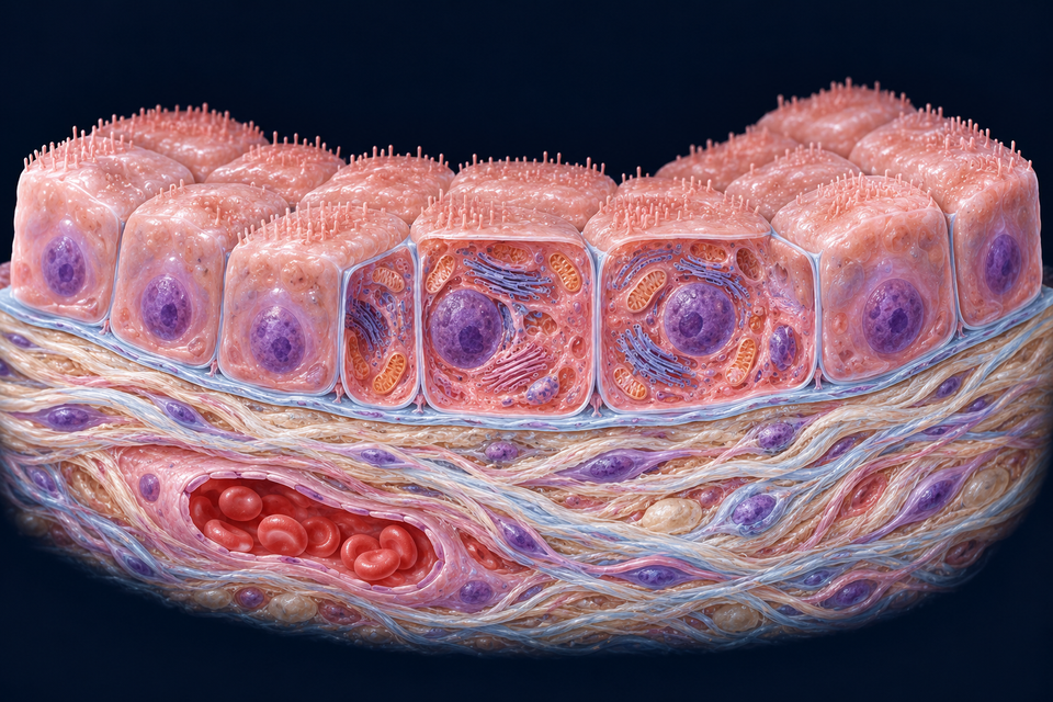
Epithelial Tissues & Barrier Design
Compare living boundaries, cell layers, directional transfer and specialised surfaces across organs.
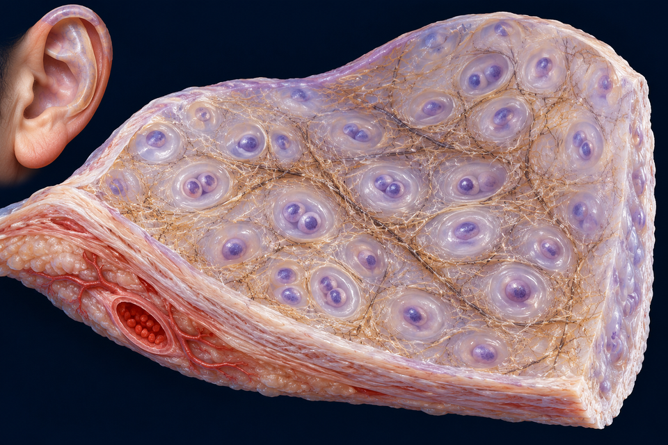
Cartilage: Cells, Matrix & Movement
Compare living cells, hydrated matrix, flexible support and fibre-rich arrangements across cartilage types.
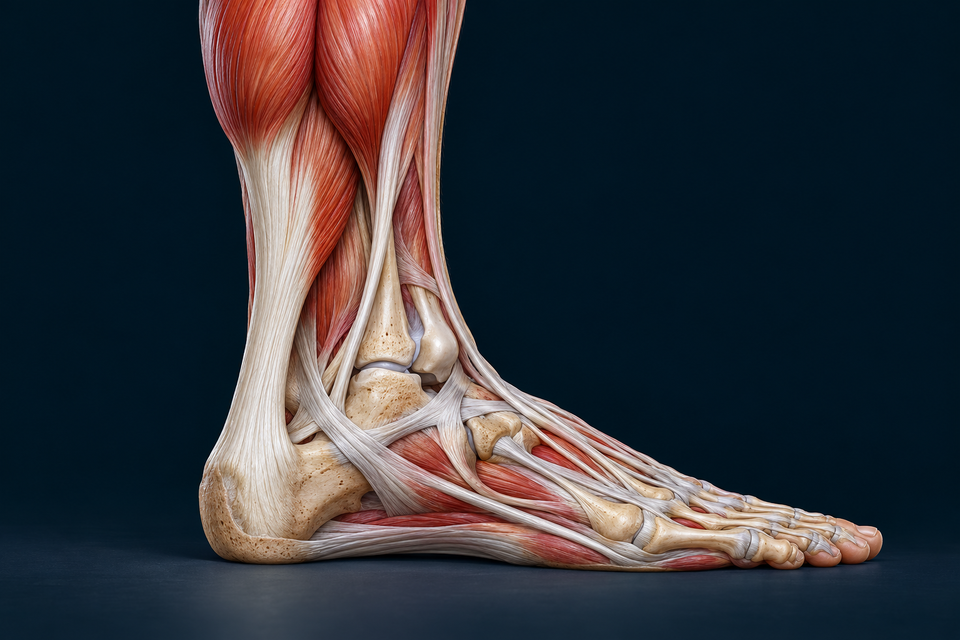
Tendons & Levers
Follow the force from muscle to bone, then compare graded attachments, three body levers and the role of a moment arm.
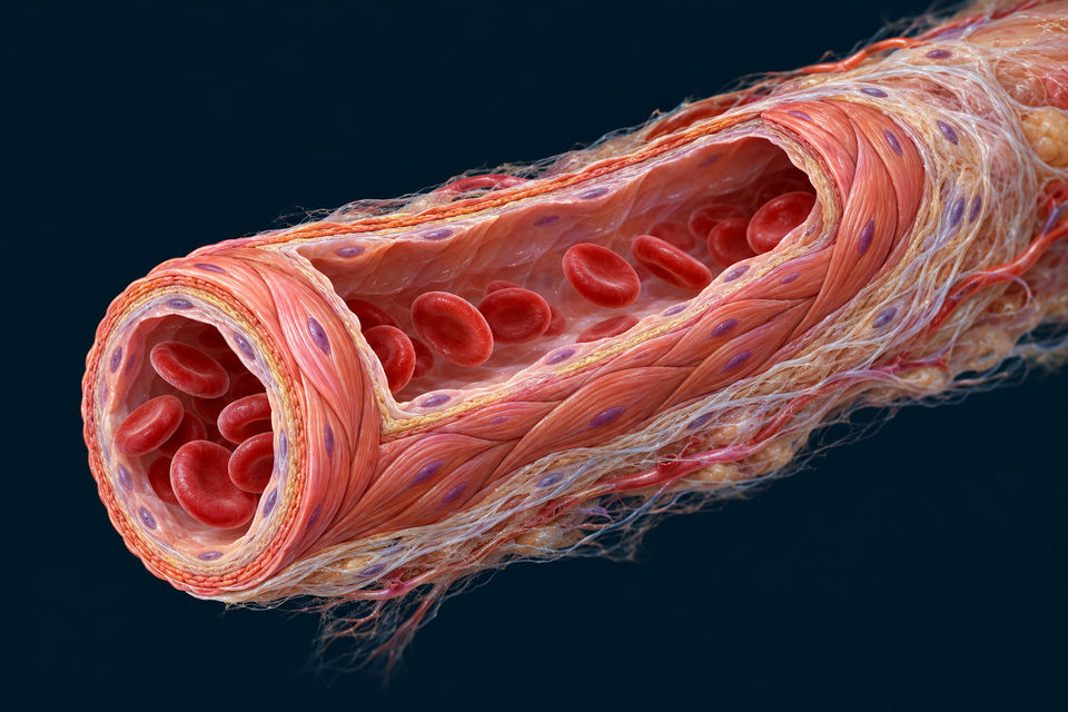
Blood Pressure & Flow
Separate pressure from flow, explore vessel-wall resistance and follow the sensing and feedback that coordinate normal circulation.
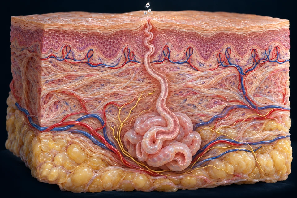
Temperature & Heat Balance
Connect metabolic heat, skin blood flow and evaporation with brain coordination, shivering and environmental exchange.
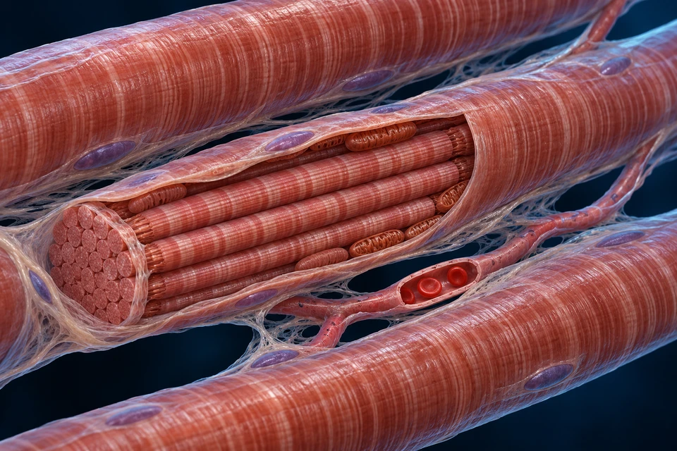
Oxygen Delivery & Active Muscle
Connect lung exchange, blood flow and muscle oxygen use, then compare activity onset, lactate turnover and recovery.
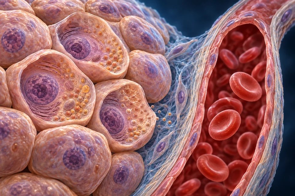
Fluid Balance & Osmoregulation
Distinguish water amount from concentration, then connect cell boundaries, thirst and renal water recovery.
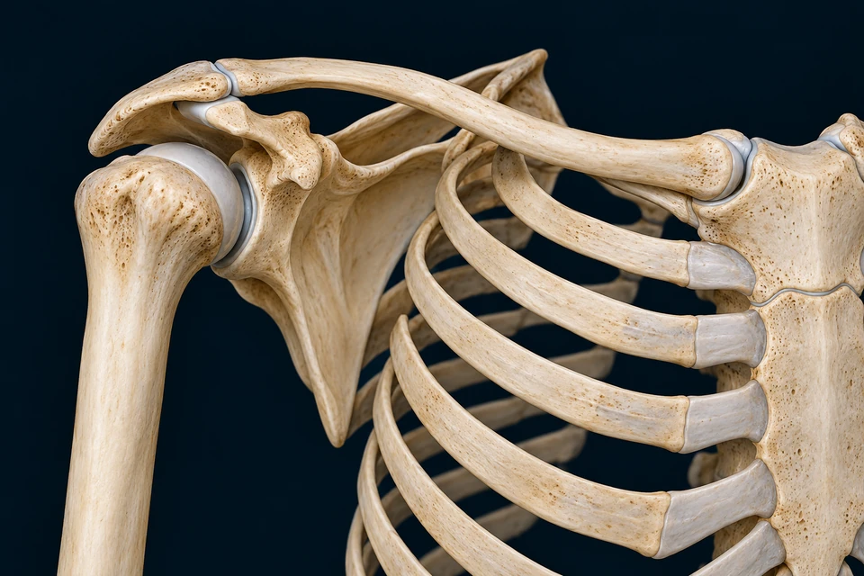
Shoulder Complex & Arm Elevation
Read the moving base, open the joint and compare both faces of the rotator cuff.
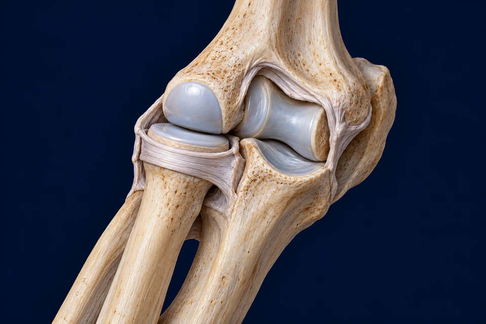
Elbow & Forearm Rotation
Match the contacts, follow the rotating radius and compare radial and ulnar muscle attachments.
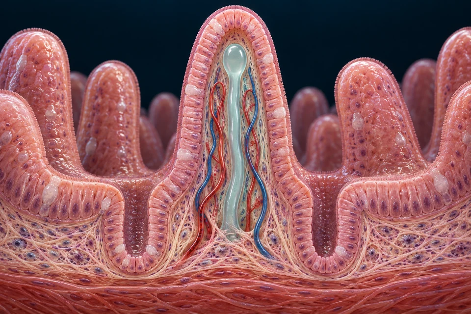
Lipid Digestion & Transport
Follow digestion, cell re-assembly and lymphatic delivery, then distinguish three very different lipid-carrying structures.
This Beta includes the published chapters shown above and source-labelled 3D references. Use the main navigation to find related topics. Generated illustrations teach selected broad relationships and are not specimen images or diagnostic tools. Sources, authentic-image credits and specific limits are included with each chapter.