Bone marrow
Inside selected bones
Blood-cell production and major B-cell developmental context.
Primary lymphoid organ; this chapter does not replace a marrow atlas.
CHAPTER / RETURN, RECOGNITION AND RESTRAINT
Fluid returns. Cells meet. Responses adapt.
A distributed network connects tissue fluid with circulation and brings immune information to cells that can respond. Explore its organs, cellular encounters and normal control.
02 / NODE
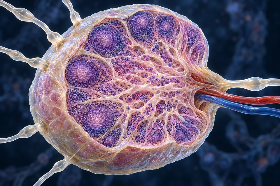
Several afferent channels enter the convex surface. Lymph crosses communicating sinus compartments and leaves at the hilum; nearby blood vessels carry a different fluid.

Afferent lymph carries fluid and tissue-derived material into the node. This route differs from the hilar blood vessels.
Gold outlines locate a teaching context, not measured flow, a cell count or a diagnosed immune state. The complete body-wide drainage atlas remains separate from this node view.
Outer follicles, deep cortex and inner medulla provide different neighbourhoods. Their populations communicate; the coloured tissue is not a map of individually verified B and T cells.
The realistic node is shown once. Enlarging it reveals the same teaching asset rather than repeating its image across several sections.
Teaching references: NCBI · Lymph-node anatomy · NLM · Node organisation and cellular traffic
01 / ORGANS
Lymphatic vessels return tissue fluid while lymphoid organs organise cellular development and encounters. Blood, lymph and exposed epithelial surfaces connect these tasks.
Inside selected bones
Blood-cell production and major B-cell developmental context.
Primary lymphoid organ; this chapter does not replace a marrow atlas.
Anterior mediastinal context, behind the sternum
Supports developing T cells and selection.
Primary organ; tissue composition changes with age.
Along regional lymphatic routes
Connect incoming tissue drainage with immune-cell encounters.
Secondary organs; neck, axillary, thoracic, abdominal and inguinal groups differ.
Left upper abdomen, next to stomach
Processes blood and supports blood-borne immune responses.
Secondary organ; it is not a node filtering afferent lymph.
Tonsillar and selected intestinal sites
Coordinates surveillance near exposed epithelial surfaces.
Not all mucosal tissues contain identical permanent nodules.
A selected location directory complements the realistic regional plates below. It is not a complete body-wide anatomical map; that primary visual remains staged.
Teaching references: OpenStax21.1 · Lymphatic organs and selected organisation · NIAID · Selected immune organs and cell roles
03 / ENTRY
Tissue material arrives through lymphatic channels. Many circulating lymphocytes enter through high endothelial venules within the node, then migrate through tissue and later leave through lymphatic routes.
Tissue → afferent vessel → subcapsular / deeper sinuses → efferent vessel
Carries fluid and tissue-derived material into a regional meeting site.
A broad route, not every microscopic channel traced.
Blood → specialised venule → lymphoid tissue
Supports encounters between circulating cells and locally arriving information.
Not a red artery opening directly into a lymph sinus.
Teaching references: NLM · Specialised venules and lymphocyte entry · NLM · Node organisation and cellular traffic
04 / INITIAL
Blood microvessels exchange fluid with their surroundings. A delicate initial lymphatic collects interstitial fluid and material through specialised endothelial arrangements.
Blood remains inside a separate microvascular channel.
Fluid exchange with surrounding tissue does not mean red blood cells normally pour into the lymphatic tube.
A closed local end begins an initial lymphatic route.
Networks vary in shape. This tube has no open cut end that directly swallows a blood vessel.
Fluid and extracellular material surround the vessels.
Lymphatic return supports volume and protein balance. It is not an optional overflow after universally fixed venous reabsorption.

Collected interstitial fluid is called lymph once inside.
Its composition depends on tissue and circumstances. A clear rendering is not a laboratory analysis or a guarantee that lymph contains no cells.
Overlapping endothelial arrangements help permit entry.
Initial vessels differ from collecting vessels with muscular walls and internal valves. Exact cell junctions in the artwork are not independently verified.
In many tissues at steady state, lymphatic return is important for sustained fluid and protein balance. The old picture of a fixed daily amount that is mostly reabsorbed at every venous capillary end is too universal.
Pressure, endothelial barriers and tissue conditions affect exchange. No universal daily litre quota, measured flow or completely sealed compartment is assigned here.
Teaching references: NLM · Lymphatic vascular structure and transport · NLM · Contemporary exchange and lymph return
05 / TRANSPORT
An initial lymphatic has a delicate entry wall. Downstream collecting vessels have muscular segments and internal valves; contractions and movements of surrounding tissues help move lymph.
Net transport reference: left → right · the static leaflet position is not an instantaneous flow measurement.

Lymphatic-muscle contractions can help drive lymph. They cooperate with surrounding tissue forces; one frozen image cannot supply a measured contraction cycle.
Lymphatic-muscle contractions can help drive lymph. They cooperate with surrounding tissue forces; one frozen image cannot supply a measured contraction cycle.
Forward and reverse pressure conditions act on the leaflets differently. Closure limits backflow; a valve does not actively push fluid by itself.
Skeletal movement, breathing-related changes and other external forces can support transport. These influences vary with location and circumstances.
The controls change the learning focus on the same anatomy, not the valve position or a measured time sequence.
The leaflet is a pressure-responsive gate. Force can come from the muscular vessel wall and from outside the vessel. Sequential valve boundaries help organise collecting segments; this single-pair view does not show an entire serial network.
Compare it with the delicate initial vessel, which collects tissue fluid through different endothelial arrangements. Do not copy the thickened collecting wall or internal valve into that initial-vessel image.
Mechanism reference: Lymphatic pumping physiology · Facts checked 18 October 2026. AI-assisted, not physician review.
Interstitial surroundings → delicate initial vessel
Entry arrangements differ from the internal valves of collecting vessels.
Muscular segment + valve pairs → forward transport
Intrinsic contractions and surrounding movement can both contribute.
Collectors / nodes / trunks → ducts → venous blood
There is no second central lymph heart pumping every segment.
Supporting mechanism map, not exact anatomical geometry or a measured time course. All steps are visible before highlighting.
Supporting transport comparison beside the realistic initial-vessel plate. Valve pairs and contraction are explained without depicting that thin initial tube as a muscular collector.
Teaching references: NLM · Lymphatic vascular structure and transport · NLM · Contemporary exchange and lymph return
06 / DUCTS
The thoracic duct commonly carries drainage from the lower body and left upper regions. Right upper territories often return through right-sided trunks or a right lymphatic duct.
Lower limbs / abdomen + left upper territories → thoracic duct → left venous junction context
This broad teaching pattern differs from the right upper territory.
Course, terminal branches and venous openings vary.
Right head/neck, upper limb and upper thorax → right-sided venous return
A small right-sided duct or separate trunks can participate.
It does not drain the entire right half of the body.
Lumbar / intestinal trunks → lower collecting route
A cisterna chyli may be recognisable in the lower thoracic/upper abdominal context.
It is not a large identical sac present in everyone.
Vessels in the meninges contribute to brain-border drainage; this is not the same as ordinary lymphatic capillaries throughout brain parenchyma. Detailed CNS drainage contributions remain an active research area.
Teaching references: NCBI · Thoracic duct and variation · NLM · Thoracic duct / cisterna-chyli variability · NLM · Meningeal lymphatic context
07 / SPLEEN
The spleen lies in the left upper abdomen beside the stomach. Blood-rich red pulp and lymphoid white-pulp regions have different roles in circulation and immune coordination.
Lymphoid tissue supports responses to blood-borne material.
T-cell and B-cell neighbourhoods relate to arterial context. A pale generated region does not identify every immune population.
An organ in the left upper abdomen lies beside the stomach.
The isolated view explains tissue organisation; it is not a complete body locator or a measured individual organ.
A connective envelope and inward supports organise the organ.
Subtle trabeculae carry support and vascular context. Their exact extent in this generated cutaway is illustrative.
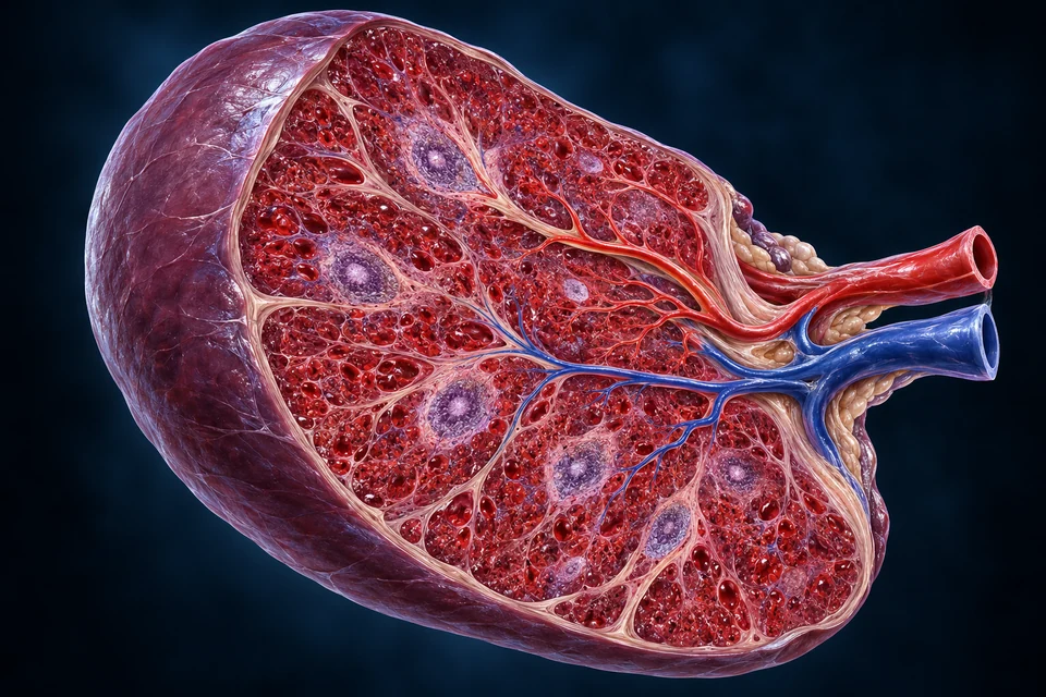
Arterial blood supplies the organ at the hilum.
Blood enters this circulation separately from lymph draining tissues elsewhere. Fine internal branches are not a complete angiogram.
Venous blood leaves and joins portal circulation.
The splenic vein connects with the portal system. Its blue colour is an orientation convention, not a statement that it transports lymph.
Blood-rich tissue participates in red-cell assessment and removal.
Macrophages and specialised circulation help process aged or altered blood cells. This is not a simple mechanical sieve with identical holes.
Red pulp contributes to handling aged or altered red cells; white pulp organises immune responses to blood-borne material. The splenic vein joins portal circulation.
A human spleen should not be taught by relabelling a node cortex and medulla. Fine circulation and marginal/perifollicular compartments also differ between species.
Teaching references: NCBI · Normal spleen anatomy · NLM · Human versus rodent splenic compartments
08 / THYMUS
Precursors reach the thymus from blood-forming tissues. Thymocytes interact with epithelial and other cells as they develop in a cortical–medullary environment.
A densely cellular outer region supports developing thymocytes.
Thymocytes interact with specialised epithelial and other cells. No coloured nucleus verifies a particular developmental or receptor state.
Connective extensions carry support between lobular portions.
Real medullary tissue is continuous across adjoining regions. A two-dimensional cut cannot establish every connection.
Concentric epithelial profiles occur in thymic medullary tissue.
The visible form is illustrative, not a verified Hassall corpuscle specimen. Its presence does not prove that every selection event occurs inside it.

An outer envelope surrounds thymic tissue.
Connective extensions divide lobular portions. The thymus is not a lymph node with afferent lymph filtering through follicles.
A less densely packed internal region supports further selection.
Negative selection is not restricted exclusively to one region. Some self-reactive cells are removed while other outcomes include regulatory-cell development.
Blood supports the living tissue and cellular traffic.
Vessels are separate from developing lymphocytes. Precise microvascular routes and developmental changes remain outside this illustration’s verification scope.
Selection helps establish useful recognition of peptide–MHC context and limit harmful self-reactivity. Outcomes are more complex than “all cells pass” or “all self-reactive cells die”; regulatory-cell development is part of the wider story.
Medullary tissue connects across adjoining lobular regions in three dimensions. With age, much thymic tissue is replaced by fat, although tissue can remain; a young tissue model is not every adult appearance.
Teaching references: NCBI · Thymus organisation · NCBI · Thymic development and selection
09 / CELLS
Blood counts, tissue microscopy and molecular markers reveal different aspects of immune cells. This selected role comparison does not identify individual cells from generated colour or texture.
Blood and recruited tissue sites
Rapid phagocytic and other responses help manage local challenges.
Not every inflammatory cell or multilobed generated nucleus is independently identified.
Circulation and varied tissue populations
Engulf material, help clear debris and coordinate responses.
Not all tissue macrophages simply arose from a recent circulating monocyte.
Barrier tissues and lymphoid meeting sites
Capture material and present processed antigen to suitable T cells.
Antigen presentation is a function; the visible shape alone does not establish identity.
Lymphoid tissues and selected long-lived niches
B cells recognise antigen; differentiated plasma cells secrete antibodies.
B cells and plasma cells are related but not interchangeable states.
Develop in thymus; act across tissues
Helper, cytotoxic and regulatory populations perform different jobs.
A helper T cell does not make antibodies or become a plasma cell.
Blood and tissue contexts
Innate lymphocytes can respond to altered cellular signals.
They are not simply untrained cytotoxic T cells.
Distinct circulating or tissue-resident populations
Selected granule-containing cells contribute signalling and specialised responses.
These are different cell types, not one universal inflammatory cell.
Teaching references: NIAID · Selected immune organs and cell roles · NLM · Normal immune-cell and complement coordination
10 / BARRIERS
Skin, mucosal epithelium, mucus, local secretions and normal clearance mechanisms reduce unwanted entry. These barriers also communicate with tissue-resident immune populations.
Renewing epithelial and lipid barrier
Limits water loss and unwanted penetration while retaining normal routes for appendages.
Not impermeable, and not one uniform thickness.
Mucus + cilia + local cellular context
Traps and clears material from conducting passages.
Alveolar exchange has a different architecture.
Epithelial interface + organised local sampling
Balances nutrient uptake and surveillance near luminal material.
Normal contents are not all treated as foreign invaders.
Teaching references: OpenStax21.2 · Barriers and innate coordination · NLM · Normal immune-cell and complement coordination
11 / MIGRATION
Local signals change endothelial and leukocyte behaviour. Rolling, firm adhesion, passage across the vessel wall and tissue-directed movement are related stages, not blood simply leaking every cell out.
Circulating cell → repeated endothelial contacts
Local context supports slowing near an appropriate site.
Activated interactions → stable attachment
Attachment differs from immediate passage through an open hole.
Across endothelial boundary → tissue side
Cells cross through regulated routes; not every vessel is equally permissive.
Local signals → directed migration
The simplified stages overlap and do not specify a universal duration.
Supporting mechanism map, not exact anatomical geometry or a measured time course. All steps are visible before highlighting.
Supporting normal recruitment sequence. Cell type, vessel context and timing vary; no exact adhesion-molecule network, clinical inflammation grading or experimental manipulation is claimed.
Teaching references: OpenStax21.2 · Barriers and innate coordination · NLM · Normal immune-cell and complement coordination
12 / HANDLING
Phagocytes can enclose material within a membrane compartment and process it internally. Antigen presentation and chemical signalling can connect this handling with other cellular responses.
Material at cell surface → membrane enclosure
Selected receptors and cellular responses help organise uptake.
Not every material or immune cell uses the same route.
Enclosed compartment → interaction with degradative machinery
Handling occurs within specialised intracellular compartments.
Not an organism floating freely in the cytoplasm by definition.
Processed information / local signals → appropriate partners
Professional antigen-presenting cells can support T-cell recognition in the correct molecular context.
Phagocytosis alone does not guarantee effective antigen presentation.
Teaching references: NCBI Immunobiology · Selected immune principles · NLM · Normal immune-cell and complement coordination
13 / RECOGNITION
An antigen is something recognised by an immune receptor, not necessarily an entire organism. B-cell receptors can bind suitable antigen directly; conventional T cells typically recognise peptides presented with MHC.

The presenting cell displays suitable peptide with MHC II. This is a conventional CD4 example, not a whole pathogen being passed into a T cell.
The presenting cell displays suitable peptide with MHC II. This is a conventional CD4 example, not a whole pathogen being passed into a T cell.
The receptor responds to peptide–MHC context. A nearby T cell with a different specificity need not respond to the same display.
Co-stimulation and local signals help determine the outcome. The preparation shows an encounter; neither the highlight nor the cell colour proves that an immune response has begun.
The controls change the learning focus on the same anatomy, not the cell’s identity or a measured time sequence.
Specific recognition is only part of the activation decision. A naive conventional CD4 T cell recognises a peptide–MHC II display, while co-stimulatory and local signals influence its response. Contact alone is not proof that it will divide or differentiate.
The branches belong to a representative immune cell, not a neuron. No process here is an axon and the neighbouring cells do not fuse. Continue with B cells and antibody effects and memory and tolerance rather than applying this one CD4 image to every immune interaction.
Mechanism reference: Conventional helper T-cell activation · Facts checked 18 October 2026. AI-assisted, not physician review.
Suitable antigen → B-cell receptor
Binding and accompanying signals contribute to an appropriate response.
Peptide–MHC II → suitable CD4 T cell
Professional antigen-presenting cells coordinate recognition with other signals.
Peptide–MHC I → suitable CD8 T cell
Activated effectors can act on presenting target cells; initial activation has additional requirements.
Supporting mechanism map, not exact anatomical geometry or a measured time course. All steps are visible before highlighting.
Supporting recognition comparison. Co-stimulation and local signals influence activation; binding alone is not a universal guarantee of an effective response. Non-conventional lymphocyte recognition is outside this introductory scope.
Teaching references: OpenStax21.3 · T-cell recognition · OpenStax21.4 · B cells and antibodies · NCBI Immunobiology · Selected immune principles
14 / ANTIBODIES
Activated B cells can generate antibody-secreting plasma cells and memory populations. Antibodies bind suitable targets; what happens next depends on their class, location and cooperating systems.
Binding can limit a target’s interaction with a cell or tissue
This can act without an antibody directly consuming the target.
Not every bound molecule is neutralised.
Recognition tags support uptake by appropriate phagocytes
Binding to antibody portions helps coordinate clearance.
A receptor-mediated effect, not a magic dissolving arrow.
Soluble proteins participate in marking and downstream responses
Some pathways link with antibodies; others can activate without them.
Complement is not made solely by B cells or active only after antibody binding.
Antibody-producing cells and memory B cells are different outcomes. Not all B-cell responses require the same T-cell help; this overview does not turn one common pathway into an exception-free rule.
Teaching references: OpenStax21.4 · B cells and antibodies · NLM · Normal immune-cell and complement coordination
15 / MEMORY
After a response, some adaptive populations persist and can change a later response. Regulation and tolerance limit inappropriate activity toward self and harmless contexts.
Selected B- and T-cell populations persist
Later recognition can be faster or different in quality.
It does not guarantee lifelong protection against every changing target.
Some plasma cells occupy supportive long-lived niches
Antibody secretion can continue without the original response staying fully active.
A plasma cell is not identical to a memory B cell.
Selection and peripheral control act at different sites
Limiting damaging responses is as important as mounting useful ones.
Not all self-reactivity is solved by a single thymic checkpoint.
Innate and adaptive activity overlap and communicate. They are not two mutually exclusive switches, and “innate” should not be simplified into an absolute claim that no altered response history can ever occur.
Teaching references: OpenStax21.3 · T-cell recognition · OpenStax21.4 · B cells and antibodies · NLM · Normal immune-cell and complement coordination
16 / GUT
Intestinal lacteals receive much of the dietary long-chain lipid packaged into chylomicrons. That route differs from nutrients entering intestinal blood and then portal circulation.
Enterocyte processing → chylomicrons → lacteal / lymph → venous blood
Lipid-rich intestinal lymph is called chyle.
Not all absorbed nutrients or all fatty acids follow one pathway.
Selected absorbed nutrients → intestinal blood → portal vein → liver
This gives the liver early access to many water-soluble absorbed substances.
The portal vein does not collect the lacteal directly.
Teaching references: NLM · Lymphatic vascular structure and transport · OpenStax21.1 · Lymphatic organs and selected organisation
17 / EVIDENCE
A real section depends on species, age, preparation and cutting plane. A rounded follicle is a tissue profile, not a complete three-dimensional node or a labelled molecular cell map.
Duke 0077_E biopsy / Masson; UMich027 human H&E
Descriptions support comparing capsule, cortex and inner compartments.
A biopsy record is not automatically labelled an entirely healthy specimen.
0076_E adult human thymus H&E; 0003_E human spleen H&E
Adult involution and blood-versus-lymph tissue organisation can be compared.
Adjacent monkey/rabbit examples are not substituted for human evidence.
Record descriptions checked; slide pixels not acquired or presented here. The rendered plates are original educational illustrations, not microscopy, clinical imaging or diagnostic evidence.
Teaching references: Duke · Identified lymphoid tissue records · NLM · Human versus rodent splenic compartments
18 / SCOPE
Fluid return, immune-cell traffic, organ organisation, barriers, recognition and regulation form a connected normal-function foundation. Clinical branches remain separate roadmap work.
Organ → compartment → selected cell role → normal mechanism
Four distinct anatomy/tissue views and visible comparisons teach different levels without repeating the same primary image.
Source-supported teaching scope; human medical review not performed.
Body-wide anatomy map, complete cell atlas and clinical branches
Exact duct/collector anatomy, molecular pathways and licensed microscopy require separate work.
Disease, diagnosis and treatment are not implied complete.
Teaching references: NIAID · Selected immune organs and cell roles · NLM · Lymphatic vascular structure and transport
EDITORIAL RECORD
Broad lymph-node, initial-vessel, splenic and young thymic compartments; fluid return, separate cellular entry, selected immune roles, normal barrier, migration, recognition, antibody effects, memory and regulation. Authoritative references are used critically rather than repeating every simplified or historical claim.
AI-assisted visual/content production. Fine generated cellular, vascular, junctional and epithelial details remain illustrative / not independently verified. Human medical review: not performed. Educational only; not diagnostic, clinical or surgical material.
Revision: lymph-immunity-english-2026-10-18-eyes-foundation-v1. Stable local key: ahb_ch_lymph_immunity_english_v1. No invented canonical CMS/atlas entity or 3D dataset. Five retained Golden-family text-free masters and their twenty-five original labels are preserved. Two new text-free collecting-vessel and immune-encounter preparations use separate semantic anatomy anchors and editable English explanations. Generated fine cellular and sinus topology is not acquired microscopy or a traced patient vascular map. Migration physical-phone acceptance: Pending, non-blocking.