CHAPTER / DIGESTIVE & ENDOCRINE

Pancreas

One gland. Two destinations.

Trace digestive fluid toward the intestine and hormones into blood. Move from the organ to its tissues, then see how a cell directs its secretion.

Compare the two tissue exits →

Chapter overview film planned. No playable video is currently available.

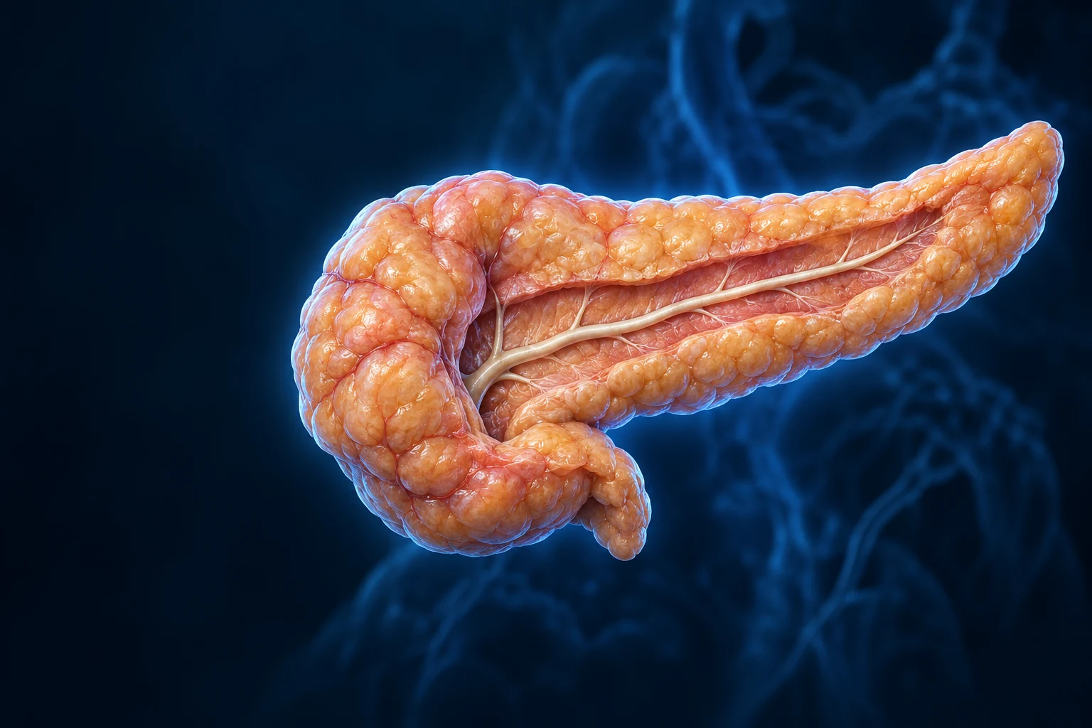
Original elongated adult pancreas exhibit: a broad head image left, tapering body and tail image right, with a selected opened duct strip. Fine internal detail illustrative.
Original pancreatic exhibit · fine detail illustrative

02 / ORGAN IN CONTEXT

A gland behind the stomach.

The pancreatic head lies within the duodenal curve. Its neck and body cross the upper abdomen, and its tail reaches toward the spleen. Most of the gland lies against the posterior abdominal wall.

Anterior expanded pancreas context: head within the duodenal curve on image left, body crossing toward the tail and spleen on image right; lifted translucent stomach reveals the gland behind it.
Original Golden-family anterior preparation: person right is image left. Stomach lifted/translucent for context. Broad neighboring structures and duct direction source-supported; terminal outlet and fine vessels/branches illustrative / not independently verified, not a surgical map. Bile duct omitted from this selected view, not absent anatomically.
Structure notes · further context
Head
Its inferior uncinate process extends behind major mesenteric vessels; that hidden relationship is not resolved in this view.
Body
A short neck connects the head to the body. Most of the pancreas lies against the posterior abdominal wall; this expanded preparation does not show all neighboring vessels.
Tail
The tail extends into the splenorenal ligament. This differs from the mostly retroperitoneal remainder of the gland.
Main pancreatic duct
A common pattern joins the distal bile duct before the major papilla. The bile duct is omitted here; connections and accessory ducts vary.
Duodenum
Its opened wall is a teaching cutaway. Contents of the stomach enter this intestine; they do not pass through pancreatic tissue.
Stomach
Partial transparency reveals the relationship rather than implying that the organs normally overlap as exposed surfaces.
Spleen
The spleen is an anatomical neighbor, not the destination of pancreatic digestive fluid.

Behind the stomach, beside the duodenum · atlas detail

Anterior expanded pancreas context: head within the duodenal curve on image left, body crossing toward the tail and spleen on image right; lifted translucent stomach reveals the gland behind it.

Original Golden-family anterior preparation: person right is image left. Stomach lifted/translucent for context. Broad neighboring structures and duct direction source-supported; terminal outlet and fine vessels/branches illustrative / not independently verified, not a surgical map. Bile duct omitted from this selected view, not absent anatomically.

Open full-resolution image ↗ · Image only; editable labels remain in this atlas detail.

The duct and its neighbors

The main pancreatic duct commonly joins the distal bile duct near the major duodenal papilla. Accessory ducts and terminal connections vary. The anatomy view omits the bile duct; omission does not mean absence.

Explore the separate bile route →

The blood and its route

Branches of the splenic and pancreaticoduodenal arteries supply the gland. Venous blood drains toward the portal system. Digestive contents take a duct route instead; this plate is not a map of surgical vascular variants.

Neighboring organs and the usual digestive duct route: OpenStax · Accessory digestive organs · NCBI · Pancreas anatomyDirected claim/source correspondence check: 17 October 2026. Retained source records and accessible independent references support the teaching; not physician review or validation of every generated detail.

03 / DIFFERENT DESTINATIONS

Digestive fluid is not a hormone signal.

Exocrine tissue sends a secretion into the intestine. Endocrine tissue releases hormones into blood. Both belong to one gland, but they are not interchangeable outputs.

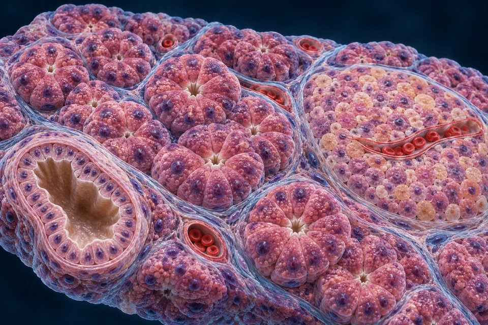
Two secretions. Separate exits.Compare the acinar units and epithelial duct with the cell cluster and red-cell-containing vessel. The digestive lumen and blood are separate compartments.

Prepared pancreatic tissue with secretory acini, a separate epithelial duct, a paler non-acinar islet and red-cell-containing blood capillaries.
Original expanded tissue exhibit, not microscopy. Acini/ducts versus islet/capillary relationships source-supported. Fine cell shape, counts, exact duct connections and capillary geometry illustrative / not independently verified. No generated cell is identified as alpha or beta from color; no fixed human core-and-mantle arrangement.
Structure notes · further context
Acini
Acinar cells make digestive enzymes. The sizes and numbers shown are expanded for teaching and do not measure an actual tissue section.
Duct system
Small ducts connect acinar lumens to larger ducts; duct cells add bicarbonate-rich fluid. These lumens are separate from blood vessels.
Pancreatic islet
Human endocrine cell types are intermixed in variable arrangements. Their identities require appropriate evidence, such as specific staining; this rendering does not establish them.
Capillaries
Islet cells are closely associated with capillaries. Blood does not empty into the digestive duct; the exact red network is illustrative.
Supporting tissue
Thin septa help organize the gland. Fine strands here suggest support rather than a verified collagen-fiber reconstruction.

Two kinds of tissue, two kinds of exit · atlas detail

Prepared pancreatic tissue with secretory acini, a separate epithelial duct, a paler non-acinar islet and red-cell-containing blood capillaries.

Original expanded tissue exhibit, not microscopy. Acini/ducts versus islet/capillary relationships source-supported. Fine cell shape, counts, exact duct connections and capillary geometry illustrative / not independently verified. No generated cell is identified as alpha or beta from color; no fixed human core-and-mantle arrangement.

Open full-resolution image ↗ · Image only; editable labels remain in this atlas detail.

Highlights locate tissue relationships, not measured secretion or a visible moving fluid. The two route summaries describe functional destinations; small acinar–duct connections and the direction of flow in this static capillary are not traced here.

Islands within a secretory landscape.

Most pancreatic tissue consists of enzyme-producing acini and their duct system. Smaller endocrine islets are scattered among them. Connective tissue supports the lobules, ducts, nerves and vessels.

Exocrine: through ducts

Acini + duct cells → duct system → duodenum

Enzymes break down food; bicarbonate-rich fluid buffers incoming acid. This secretion reaches the intestinal lumen.

See the digestive-facing cell →

Endocrine: into blood

Islet cells → nearby capillaries → target tissues

Hormones carry instructions to other tissues. They do not travel down the pancreatic duct with digestive juice.

See the islet and its targets →

University of Leeds · Pancreatic tissues · OpenStax · Endocrine and exocrine roles. Directed tissue and output check: 17 October 2026. Source checking is not physician review.

Acini communicate with lumens

Secretory cells face a tiny shared space that drains into ducts. Pancreatic duct epithelium adds fluid and bicarbonate along the route.

Islets communicate with blood

Endocrine cells lie close to capillaries. Human cell types do not have one fixed concentric arrangement; a pale island in a routine section is not enough to identify every hormone-producing cell.

Acinar/duct and islet/capillary destinations remain separate: University of Leeds · Pancreatic tissues · OpenStax · Endocrine pancreasDirected claim/source correspondence check: 17 October 2026. Retained source records and accessible independent references support the teaching; not physician review or validation of every generated detail.

05 / CELL POLARITY

Make proteins. Package them. Release toward the lumen.

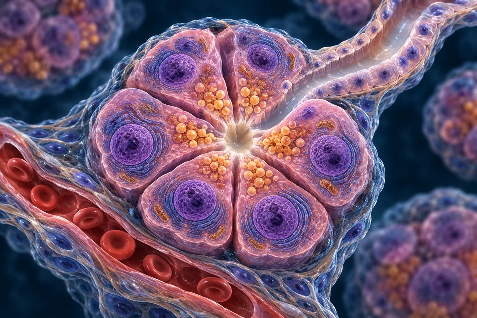
Acinar cells contain abundant protein-making machinery and apical secretory granules. Their organization directs enzyme secretion toward the digestive route rather than into the surrounding blood.

Cut-open acinar unit with outer basal nuclei, granules toward the small central lumen, an upper duct outlet and a separate external blood capillary.
Original enlarged acinar preparation, not electron microscopy. Broad basal-to-apical organization and separate digestive versus blood destinations source-supported. Fine organelles, granules, duct transition and exact proportions illustrative / not independently verified. The supporting pathway map supplies the flow direction.
Structure notes · further context
Acinar cell
Its broad basal region faces supporting tissue; its apical membrane faces the gland lumen. Not every visible membrane fold is independently verified.
Basal region
Rough endoplasmic reticulum supports production of secreted proteins. The blue membrane texture is illustrative rather than a literal organelle map.
Secretory granules
After appropriate stimulation, granules fuse with the apical membrane. Many proteases are packaged as inactive precursors; not every digestive enzyme needs the same activation step.
Acinar lumen
It drains onward into the duct system, not into the red capillary. The front wall is removed to expose this teaching relationship.
Duct outlet
Duct cells add bicarbonate-rich fluid along the exocrine route. This enlarged view does not resolve the complete branching tree.
Blood-side capillary
This capillary does not receive enzyme release from the apical surface. Blood supplies the tissue; islet hormones use a distinct endocrine route.

An acinar cell has a digestive-facing side · atlas detail

Cut-open acinar unit with outer basal nuclei, granules toward the small central lumen, an upper duct outlet and a separate external blood capillary.

Original enlarged acinar preparation, not electron microscopy. Broad basal-to-apical organization and separate digestive versus blood destinations source-supported. Fine organelles, granules, duct transition and exact proportions illustrative / not independently verified. The supporting pathway map supplies the flow direction.

Open full-resolution image ↗ · Image only; editable labels remain in this atlas detail.

  1. Build and package

    Secreted proteins are synthesized through the endoplasmic-reticulum/Golgi pathway and packed into granules.

  2. Release at the apical surface

    Appropriate hormonal and neural stimulation triggers exocytosis into the acinar lumen.

  3. Carry and modify

    Ducts convey the proteins with bicarbonate-rich fluid toward the duodenum.

Supporting sequence. Fine organelle appearance is illustrative; the generated cells are not an electron micrograph. Digestive and endocrine granules serve different products and destinations.

Follow the general ER/Golgi secretory pathway →

Acinar secretion and ductal modification: University of Leeds · Pancreatic tissues · NCBI · The exocrine pancreasDirected claim/source correspondence check: 17 October 2026. Retained source records and accessible independent references support the teaching; not physician review or validation of every generated detail.

Prepare the proteins. Trigger release. Modify the fluid.

Locate the enzyme-producing acini and the duct lining, then connect the structures with the signals and membrane functions that make a digestive secretion possible.

Proteins and alkaline fluid have complementary jobs.

Acini supply digestive proteins; duct epithelium adds bicarbonate-rich fluid. Their luminal outputs join on the way to the intestine. The separate capillary supports tissue and carries signals, not the collected digestive juice.

Prepared pancreatic tissue with secretory acini, a separate epithelial duct, a paler non-acinar islet and red-cell-containing blood capillaries.
Original expanded tissue exhibit, not microscopy. Acini/ducts versus islet/capillary relationships source-supported. Fine cell shape, counts, exact duct connections and capillary geometry illustrative / not independently verified. No generated cell is identified as alpha or beta from color; no fixed human core-and-mantle arrangement.
Structure notes · further context
Acini
Acinar cells make digestive enzymes. The sizes and numbers shown are expanded for teaching and do not measure an actual tissue section.
Duct system
Small ducts connect acinar lumens to larger ducts; duct cells add bicarbonate-rich fluid. These lumens are separate from blood vessels.
Pancreatic islet
Human endocrine cell types are intermixed in variable arrangements. Their identities require appropriate evidence, such as specific staining; this rendering does not establish them.
Capillaries
Islet cells are closely associated with capillaries. Blood does not empty into the digestive duct; the exact red network is illustrative.
Supporting tissue
Thin septa help organize the gland. Fine strands here suggest support rather than a verified collagen-fiber reconstruction.

Two kinds of tissue, two kinds of exit · atlas detail

Prepared pancreatic tissue with secretory acini, a separate epithelial duct, a paler non-acinar islet and red-cell-containing blood capillaries.

Original expanded tissue exhibit, not microscopy. Acini/ducts versus islet/capillary relationships source-supported. Fine cell shape, counts, exact duct connections and capillary geometry illustrative / not independently verified. No generated cell is identified as alpha or beta from color; no fixed human core-and-mantle arrangement.

Open full-resolution image ↗ · Image only; editable labels remain in this atlas detail.

Gold rings locate the selected anatomical domains. They do not trace a continuous duct connection, identify a channel protein or measure secretion. The acinar view establishes polarity; the tissue view shows the complementary epithelial compartments. Molecular mechanisms are explained alongside the retained anatomical reference.

  1. 1

    Acinar product

    Digestive proteins → shared lumen

    Manufacture and storage prepare a protein-rich secretion.

  2. 2

    Duct contribution

    Bicarbonate-rich fluid → duct lumen

    Fluid carries the products; bicarbonate helps buffer incoming acid.

  3. 3

    Intestinal destination

    Duct system → duodenum

    The combined secretion cooperates with bile and intestinal digestion.

Mechanism evidence: NCBI · Digestive enzymes and acinar secretion · NCBI · Ductal water and ion secretion · Original experimental study · Duct bicarbonate and acinar contents

Understand the partnership across scales.

Manufacture, storage and release are different jobs

Secreted proteins pass through endoplasmic reticulum and Golgi before packaging into granules. Polarity concentrates the release apparatus toward the shared lumen while the basal region faces supporting tissue.

Stored proteins can be released when needed without rebuilding the whole cell. Some proteases are packaged as inactive precursors; that does not make every pancreatic enzyme inactive. Follow the existing activation lesson for that distinction.

Mechanism evidence: NCBI · Digestive enzymes and acinar secretion

Calcium couples stimulation to apical exocytosis

Receptor activation can generate intracellular messengers that release calcium from endoplasmic-reticulum stores. Physiological calcium signals vary in space and time and help trigger fusion of secretory granules with the apical membrane.

Isolated human acinar cells have shown calcium signals and secretion after CCK or acetylcholine stimulation. That demonstrates cellular capability; it does not measure the relative neural and hormonal contributions during an ordinary human meal.

Mechanism evidence: NCBI · Digestive enzymes and acinar secretion · Original human acinar-cell experiment · CCK and calcium

Duct cells supply and secrete bicarbonate

Bicarbonate can enter across the blood-facing membrane through sodium-coupled transport. Carbon-dioxide hydration inside the cell also contributes, with acid extrusion supporting intracellular acid-base balance.

At the luminal side, chloride/bicarbonate exchange and CFTR-dependent conductance cooperate. CFTR can itself carry bicarbonate; it should not be presented only as a chloride-recycling route. Transporter contributions vary along the duct, with stimulation and between experimental species.

Mechanism evidence: NCBI · Ductal water and ion secretion · Original isolated-duct experiment · CFTR bicarbonate conductance · Original mouse-duct experiment · Slc26a6 and CFTR

Secretin changes ductal activity

Acid entering the duodenum stimulates secretin release into blood. In duct epithelium, receptor-linked cAMP signalling promotes anion and fluid secretion; calcium-linked neural input can modify the response.

This is a regulated epithelial contribution, not secretin flowing through the duct or acting as a digestive enzyme. The static atlas locates the responding tissue without claiming to show receptor activation.

Mechanism evidence: NCBI · Ductal water and ion secretion · NCBI · Whole-organ pancreatic secretion

A meal recruits overlapping signals

Vagal and local neural pathways help prepare and sustain secretion. Intestinal fat and protein digestion products stimulate CCK, while acid favours secretin release. These inputs interact rather than operating as isolated on/off switches.

Acinar enzyme output and duct fluid output can reinforce digestive delivery. The balance changes through a meal; a selected state here highlights one question, not a universal time sequence or a measured secretion rate.

Mechanism evidence: NCBI · Whole-organ pancreatic secretion · Original human acinar-cell experiment · CCK and calcium

Buffering supports delivery and intestinal digestion

Bicarbonate-rich fluid supports transport of protein-rich acinar secretion. Experimental work also links bicarbonate to the handling and solubilization of acidic secretory contents in the acinar lumen.

In the duodenum, buffering gastric acid helps create conditions for digestive enzymes. Bicarbonate does not perform the enzymes’ bond-cleaving reactions, and pancreatic secretion is not the only influence on intestinal pH.

Mechanism evidence: Original experimental study · Duct bicarbonate and acinar contents · NCBI · Ductal water and ion secretion

A human organ is not an isolated experimental duct

The CFTR conductance and Slc26a6 cooperation studies linked here used animal duct preparations. They support mechanisms, but their exact fluxes, transporter ratios and experimental concentrations are not universal human constants.

The human acinar study addresses a different scale and preparation. The illustrations show broad tissue organization, not molecular localization evidence or a measured map of every duct branch.

Mechanism evidence: Original isolated-duct experiment · CFTR bicarbonate conductance · Original mouse-duct experiment · Slc26a6 and CFTR · Original human acinar-cell experiment · CCK and calcium

Check the destination and the mechanism.

Does the duct merely carry finished enzyme juice?

No. Its epithelium adds and modifies bicarbonate-rich fluid, complementing acinar protein secretion.

Are acinar release and beta-cell release the same destination?

Both use regulated secretion, but acinar contents enter a digestive lumen. Beta-cell insulin is released toward extracellular fluid and blood.

Does a calcium signal mean calcium is the digestive product?

No. Intracellular calcium helps regulate exocytosis; the secreted granules contain digestive proteins.

Does CFTR only recycle chloride?

No. It can also conduct bicarbonate. Its contribution and interaction with exchangers depend on the physiological and experimental setting.

Directed normal-physiology source check: 21 October 2026. Human cell experiments, animal duct preparations and generated atlas images have different evidential limits. No individual diagnosis, fluid target or treatment is provided. AI-assisted source checking is not physician review, fine-anatomy certification or a physical-phone test.

06 / CHEMICAL DIGESTION

Different substrates need different tools.

Pancreatic enzymes work alongside stomach and intestinal enzymes. They reduce food molecules to smaller components; digestion continues at the intestinal surface before absorption.

Amylase

Acts on: Starch

Cuts internal carbohydrate bonds, producing smaller sugars for further intestinal digestion.

It does not digest cellulose or complete every step to absorbable glucose.

Lipase + colipase

Acts on: Dietary triglycerides

Produces fatty acids and monoglycerides at the fat–water interface.

Bile salts help disperse fats and transport products; bile is not a pancreatic enzyme.

Proteases

Acts on: Proteins and peptides

Trypsin, chymotrypsin and related enzymes cut peptide chains; intestinal enzymes continue the work.

Many pancreatic proteases leave the gland as inactive precursors, rather than digesting its own tissue.

Nucleases

Acts on: DNA and RNA in food

Break nucleic-acid chains into smaller nucleotide components.

These digestive reactions take place in the intestine, not inside endocrine islets.

This selected family map is not a complete enzyme catalog. Pancreatic juice, liver bile and intestinal brush-border enzymes cooperate without becoming the same secretion.

Continue with intestinal digestion →

Digestive substrates, enzyme families and downstream work: OpenStax · Accessory digestive organs · University of Leeds · Pancreatic tissuesDirected claim/source correspondence check: 17 October 2026. Retained source records and accessible independent references support the teaching; not physician review or validation of every generated detail.

07 / ACTIVE AT THE RIGHT PLACE

A precursor becomes an enzyme.

Many protein-digesting enzymes are secreted as inactive precursors called zymogens. Activation in the intestine separates their manufacture from their digestive action.

  1. Pancreas releases trypsinogen

    The inactive precursor travels down the exocrine duct route.

  2. Enteropeptidase starts the switch

    This intestinal brush-border enzyme cleaves trypsinogen to form active trypsin.

  3. Trypsin extends activation

    It activates additional trypsinogen and other pancreatic protease precursors.

Not every enzyme uses this switch

Amylase and pancreatic lipase do not both wait for the trypsinogen activation step. “All pancreatic enzymes are inactive” would be misleading.

Several safeguards work together

Packaging, compartmentalization and pancreatic trypsin inhibitor help limit inappropriate activity. Detailed disease mechanisms, diagnosis and treatment are outside this normal-function overview.

Deeper learning · when the safeguards fail

Digestive enzymes act on food in the intestine. When enzymes damage pancreatic tissue, they can contribute to pancreatitis, an inflammatory condition. This helps explain why separate compartments and controlled activation matter; it is not a complete account of every cause or pathway.

Gallstones can obstruct pancreatic drainage, but they are not the only cause of pancreatitis. The normal preparations here do not show a diagnosed obstruction or determine whether a reader has a disease.

Compare the normal bile and pancreatic delivery routes →

Protease activation and the boundary with tissue injury: OpenStax · Accessory digestive organs · NLM MeSH · Enteropeptidase · NIDDK · Pancreatitis mechanism · NIDDK · Pancreatitis causesDirected claim/source correspondence check: 17 October 2026. Retained source records and accessible independent references support the teaching; not physician review or validation of every generated detail.

08 / COORDINATED DELIVERY

The intestine asks for what it needs.

Acid and nutrient products in the small intestine trigger different but cooperating signals. Hormones travel in blood; neural pathways coordinate the response as well.

Highlighted: Secretin + neural coordination → Bicarbonate-rich fluid.

Secretin + neural coordination

S cells sense acidic contents. Bicarbonate buffers acid and creates a more suitable environment for intestinal enzymes.

CCK + cholinergic pathways

I cells release the hormone CCK. Hormonal and vagal/cholinergic pathways cooperate; this is not an exclusive one-signal switch.

Hormones and nerves coordinate rather than act alone. CCK and secretin are signals, not digestive enzymes. This supporting diagram does not model secretion rates.

Bicarbonate buffers acidic gastric contents. It is not an enzyme and does not itself break the food’s protein or fat bonds. The amount and composition of secretion change together rather than following a fixed illustrated rate.

Different intestinal signals coordinate bicarbonate and enzyme output: OpenStax · Accessory digestive organs · NCBI · Whole-organ pancreatic secretionDirected claim/source correspondence check: 17 October 2026. Retained source records and accessible independent references support the teaching; not physician review or validation of every generated detail.

09 / ENDOCRINE CELL ROLES

Small cell groups send far-reaching signals.

The major islet hormones coordinate fuel handling and local signaling. Cell identity is established by evidence such as specific staining, not by the color of an unlabelled generated cell.

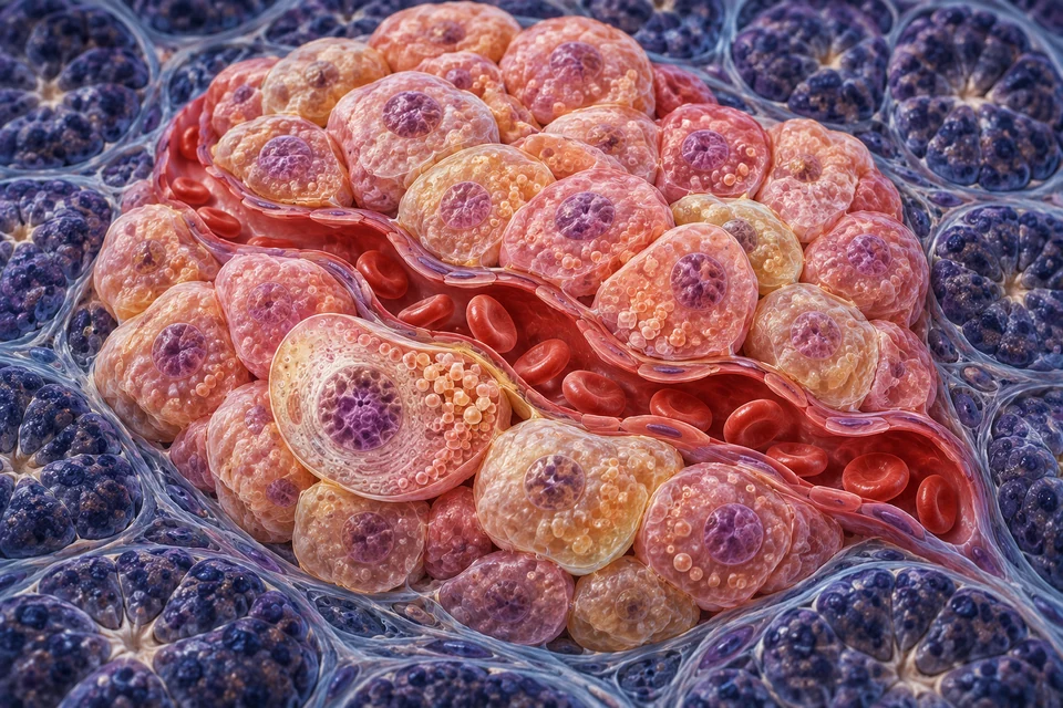
Prepared endocrine cell cluster around a red-cell-containing capillary, surrounded by darker acinar tissue; no central digestive lumen or fixed colored cell-type core.
Original islet–capillary relationship exhibit, not microscopy. Nearby endocrine cells and blood route source-supported. Cell identities, organelles, pores, numbers and fine vascular geometry illustrative / not independently verified. No universal human arrangement or color-based hormone identification.
Structure notes · further context
Endocrine cell context
Generated color does not identify an alpha, beta, delta or PP cell. Hormone-specific staining or other evidence is needed for that identity.
Capillary boundary
Real islet capillaries are fenestrated; individual pores and lining-cell geometry are not independently verified in this image.
Blood in the capillary
This red-cell-containing compartment is not an acinar lumen or pancreatic duct. The editable supporting map confirms the destination.
Surrounding exocrine tissue
Islets sit among exocrine tissue without joining its duct lumens. Fine adjacent cellular organization is illustrative.

An islet communicates with blood · atlas detail

Prepared endocrine cell cluster around a red-cell-containing capillary, surrounded by darker acinar tissue; no central digestive lumen or fixed colored cell-type core.

Original islet–capillary relationship exhibit, not microscopy. Nearby endocrine cells and blood route source-supported. Cell identities, organelles, pores, numbers and fine vascular geometry illustrative / not independently verified. No universal human arrangement or color-based hormone identification.

Open full-resolution image ↗ · Image only; editable labels remain in this atlas detail.

Beta cells

Insulin

Promotes fuel use and storage after nutrient intake; helps restrain liver glucose production.

Alpha cells

Glucagon

Supports blood glucose chiefly by signaling the liver to mobilize glycogen and make glucose.

Delta cells

Somatostatin

Provides local inhibitory signals that help coordinate other islet secretions.

PP cells

Pancreatic polypeptide

Participates in regulation of digestive activity; its functions extend beyond this glucose-focused overview.

Selected roles, not a fixed percentage or complete endocrine-cell census. Human islet arrangements vary; the tissue plate does not claim a universal beta-cell core surrounded by alpha cells.

Selected endocrine cell roles and variable human organization: OpenStax · Endocrine pancreas · Cabrera et al. · Human islet architectureDirected claim/source correspondence check: 17 October 2026. Retained source records and accessible independent references support the teaching; not physician review or validation of every generated detail.

10 / FEEDBACK ACROSS ORGANS

A message changes how fuel is handled.

Insulin and glucagon help balance nutrient storage with glucose supply. They change target-cell activity; the pancreas does not store and dispense the body’s entire glucose supply.

Highlighted: Insulin → Liver, muscle and adipose tissue.

After a meal: Insulin

Insulin favors glycogen storage and lowers liver glucose production. In muscle and fat, it promotes GLUT4 recruitment for glucose uptake.

As the glucose stimulus falls, glucose-driven insulin secretion falls too. Nutrients, gut hormones and nerves also modify the response.

Between meals: Glucagon

Glucagon supports hepatic glycogen breakdown and new glucose production. It does not simply make every body cell release glucose.

Restored glucose and local islet signals help limit the response. This is a conceptual feedback loop, not a glucose prediction or a fasting instruction.

Insulin is not required for glucose entry into every tissue. Hepatic glucose transport is different from insulin-sensitive GLUT4 recruitment in muscle and fat.

Liver: store or supply

Hepatic glycogen metabolism and new glucose production respond to the hormonal balance. Muscle glycogen mainly fuels that muscle rather than releasing free glucose to blood.

Follow hepatic fuel handling →

Gut, nerves and other tissues

Gut hormones such as GIP and GLP-1 amplify nutrient-linked insulin secretion. Neural input and local islet communication also matter. Glucose feedback is not a single isolated on/off switch.

Explore renal homeostatic links →

Hormonal fuel handling and tissue-specific glucose transport: OpenStax · Endocrine pancreas · Chadt & Al-Hasani · Liver, muscle and adipose transportersDirected claim/source correspondence check: 17 October 2026. Retained source records and accessible independent references support the teaching; not physician review or validation of every generated detail.

Send information. Move membranes. Coordinate fuel.

Keep the existing meal-feedback comparison above. Now move from the islet to a muscle-cell surface and a hepatocyte to understand how the message changes tissue work.

  1. Islet → blood → receptorsA hormonal message is different from its nutrient supply.
  2. Muscle: change transporter locationGLUT4 travels in membranes before facilitating glucose entry.
  3. Liver: change storage and supplyGlucose transport, phosphorylation and production remain distinct.

A hormone carries information. It is not the glucose supply.

Islet secretion → circulation → receptor-dependent response

Prepared endocrine cell cluster around a red-cell-containing capillary, surrounded by darker acinar tissue; no central digestive lumen or fixed colored cell-type core. Selected teaching context: Send a signal. Islet secretion → circulation → receptor-dependent response.  Gold rings locate structures, not measured activity. No directional membrane-delivery arrow is shown in this context.
Original islet–capillary relationship exhibit, not microscopy. Nearby endocrine cells and blood route source-supported. Cell identities, organelles, pores, numbers and fine vascular geometry illustrative / not independently verified. No universal human arrangement or color-based hormone identification. Selected teaching context: Send a signal. Islet secretion → circulation → receptor-dependent response. Read the preparation: Locate the endocrine-cell cluster beside a separate red-cell-containing vessel. Cell colours do not identify alpha versus beta cells; this preparation establishes the route, not a fixed human islet arrangement. Gold rings locate structures, not measured activity. No directional membrane-delivery arrow is shown in this context.
Structure notes · further context
Endocrine cell context
Generated color does not identify an alpha, beta, delta or PP cell. Hormone-specific staining or other evidence is needed for that identity.
Capillary boundary
Real islet capillaries are fenestrated; individual pores and lining-cell geometry are not independently verified in this image.
Blood in the capillary
This red-cell-containing compartment is not an acinar lumen or pancreatic duct. The editable supporting map confirms the destination.
Surrounding exocrine tissue
Islets sit among exocrine tissue without joining its duct lumens. Fine adjacent cellular organization is illustrative.

An islet communicates with blood · atlas detail

Prepared endocrine cell cluster around a red-cell-containing capillary, surrounded by darker acinar tissue; no central digestive lumen or fixed colored cell-type core. Selected teaching context: Send a signal. Islet secretion → circulation → receptor-dependent response.  Gold rings locate structures, not measured activity. No directional membrane-delivery arrow is shown in this context.

Original islet–capillary relationship exhibit, not microscopy. Nearby endocrine cells and blood route source-supported. Cell identities, organelles, pores, numbers and fine vascular geometry illustrative / not independently verified. No universal human arrangement or color-based hormone identification. Selected teaching context: Send a signal. Islet secretion → circulation → receptor-dependent response. Read the preparation: Locate the endocrine-cell cluster beside a separate red-cell-containing vessel. Cell colours do not identify alpha versus beta cells; this preparation establishes the route, not a fixed human islet arrangement. Gold rings locate structures, not measured activity. No directional membrane-delivery arrow is shown in this context.

Open full-resolution image ↗ · Image only; editable labels remain in this atlas detail.

Beta and alpha cells release different peptide signals. Blood distributes those messages, while target cells respond according to their receptors and metabolic machinery. Insulin is not a package of glucose, and neither peptide needs to become an intact vesicle travelling into a muscle cell.

Read the image: Locate the endocrine-cell cluster beside a separate red-cell-containing vessel. Cell colours do not identify alpha versus beta cells; this preparation establishes the route, not a fixed human islet arrangement.

Mechanism evidence: OpenStax · Endocrine pancreas · Rorsman & Ashcroft · Human and mouse beta-cell secretion

Different inputs. Different signalling machinery.

Supporting biochemical comparison beside the anatomical preparations, not a molecular shape reconstruction, complete pathway or measured time series.

Glucagon: hepatic signalling

G-protein-coupled receptor → cAMP–PKA

This selected branch helps explain liver glucose supply. Other glucagon receptor pathways also exist; this is not a complete signalling network or a muscle-glycogen instruction.

Mechanism evidence: Endotext · Glucagon receptor and hepatic actions

Follow the message through the tissue response.

A receptor is not a glucose doorway

Insulin recognition starts at an insulin receptor. Its kinase activity changes phosphorylation of the receptor and intracellular substrates. IRS proteins help connect recognition to downstream effectors, including PI3K-dependent pathways; AKT is an important part of metabolic signalling. These names identify supported biochemical steps, not shapes diagnosed from generated texture.

GLUT4 has a different task: it is a facilitative glucose transporter. Insulin does not pass through GLUT4 to turn it on, and a GLUT4-containing vesicle is not an insulin secretory granule. Receptor recognition, signal transduction and membrane transport can be explained together while remaining distinct processes.

Mechanism evidence: Reactome · Human insulin-receptor signalling · Reactome · GLUT4 membrane recruitment

Membrane traffic changes the available transport capacity

A substantial GLUT4 pool cycles within intracellular membranes. Insulin changes the balance of trafficking towards the cell surface. Vesicle movement, docking and fusion require coordinated machinery; a short drawing of an arrow cannot specify every intermediate or the speed of these events.

When a vesicle fuses, its membrane becomes continuous with the plasma membrane. The transporter remains embedded in a membrane, while the vesicle lumen becomes continuous with extracellular space. The opened vesicle and fusion pocket are expanded cutaway examples, not a claim that the fibre has permanent open holes.

Endocytosis and recycling retrieve surface transporters as well. Resting tissue is not a cell with zero surface GLUT4; insulin-stimulated tissue is not a cell in which retrieval stops completely.

Mechanism evidence: Reactome · GLUT4 membrane recruitment · Foley et al. · GLUT4 endocytosis and recycling

Entry, phosphorylation and use are separate steps

A facilitative carrier permits glucose movement down an appropriate concentration gradient; it does not directly pump each glucose molecule using ATP. Adding surface carriers changes transport capacity, not the chemical identity of glucose or an unlimited guarantee of entry.

Once inside muscle, hexokinase converts glucose to glucose-6-phosphate. That product is used in pathways such as glycolysis and glycogen synthesis and is not simply carried back out as free glucose by GLUT4. Metabolism, supply and transport therefore work together. Energy is required for processes such as membrane traffic and phosphorylation even though facilitated glucose transport itself is not an ATP-driven pump.

Mechanism evidence: Chadt & Al-Hasani · Tissue-specific glucose transport · Reactome · GLUT4 membrane recruitment

Hepatic transport is coupled to a different metabolic setting

In liver, GLUT2 supports glucose exchange across the hepatocyte membrane rather than copying insulin-responsive GLUT4 recruitment. Entry or exit depends on the physiological and concentration context. Insulin still matters greatly through regulation of metabolic work; insulin-independent membrane transport does not mean an insulin-independent liver.

Glucokinase phosphorylates glucose, and the resulting glucose-6-phosphate can feed storage or metabolism. Conversely, hepatic glucose production requires pathways that ultimately provide free glucose for release. Transport, phosphorylation, synthesis and breakdown are connected but cannot be collapsed into a single opening-and-closing gate.

Mechanism evidence: Chadt & Al-Hasani · Tissue-specific glucose transport · Reactome · Human insulin-receptor signalling

Why glucagon can mobilise hepatic fuel

Glucagon binding activates a G-protein-coupled receptor. A major branch couples Gs to adenylyl cyclase, which generates cAMP; cAMP activates protein kinase A. Selected enzyme and gene-regulatory effects help explain increased hepatic glucose supply. This is one branch, not every glucagon response or a complete molecular map.

Glycogenolysis releases material from an existing glycogen store; gluconeogenesis makes glucose from suitable non-carbohydrate precursors. They are different sources. An acute rise in glucagon can strongly promote glycogen breakdown when a store is available. Substrate supply and the changing physiological setting limit the response, so the same hormone does not predict one fixed output.

Mechanism evidence: Endotext · Glucagon receptor and hepatic actions · Chadt & Al-Hasani · Tissue-specific glucose transport

The same storage polymer serves different tissue roles

Liver glycogen can support glucose availability to other tissues. Skeletal-muscle glycogen mainly supports the muscle fibre itself. Muscle does not simply follow a glucagon command to empty its glycogen as free glucose into blood.

Insulin can favour glycogen storage in both tissues, but their glucose handling is not identical. The liver–muscle distinction is about their metabolic machinery and roles, not a different chemical definition of glycogen. The existing storage chapter follows the broader carbon and fuel relationships.

Mechanism evidence: Chadt & Al-Hasani · Tissue-specific glucose transport · OpenStax · Endocrine pancreas

Glucose feedback includes local, gut and neural information

Glucose is an important input to beta-cell secretion, and the existing secretion lesson explains the metabolic/electrical/Ca2+ sequence. Nutrient-linked GIP and GLP-1 amplify insulin responses; local somatostatin, insulin-related signals and neural activity also affect islet coordination. One displayed vessel or colour cannot prove those cellular identities or interactions.

Glucagon regulation is not simply the beta-cell chain played backwards. Its response depends on glucose, amino acids and other inputs. A meal containing different nutrients need not suppress every alpha-cell signal, and basal secretion continues between meals. This normal-physiology account does not specify a diet or treatment.

Mechanism evidence: OpenStax · Endocrine pancreas · Rorsman & Ashcroft · Human and mouse beta-cell secretion · Endotext · Glucagon receptor and hepatic actions

Signal strength, tissue response and blood concentration are not the same measurement

A circulating hormone concentration, receptor activation, transporter surface availability and net glucose balance describe different levels of the system. The presence of one receptor is not proof of a complete response, and an image highlight is not a measure of uptake or hormone release.

Some tissues take up glucose through transporters that do not require this GLUT4 recruitment mechanism. Contraction is another input in muscle. Liver and kidneys contribute to endogenous glucose production in different physiological contexts. These qualifications prevent a two-hormone comparison from being mistaken for a complete metabolic model or a personal glucose prediction.

Mechanism evidence: Chadt & Al-Hasani · Tissue-specific glucose transport · Reactome · GLUT4 membrane recruitment · Endotext · Glucagon receptor and hepatic actions

Check the distinctions.

Does insulin enter a muscle cell through GLUT4?

No. Insulin binds an insulin receptor and initiates signalling. GLUT4 transports glucose; it is not the insulin receptor.

Does a GLUT4 vesicle deliver free glucose as its cargo?

No. Its membrane carries transporters. Fusion supplies those proteins to the cell surface, after which they can facilitate glucose transport under suitable gradients.

Does the liver need to recruit GLUT4 in the same way as muscle?

No. Hepatic transport involves GLUT2 and a different metabolic context. Insulin still regulates important hepatic processes.

Do insulin and glucagon form a completely exclusive on/off pair?

No. Basal secretion, local and gut inputs, substrate availability and different tissue responses continue together. The examples isolate important relationships rather than predict a person’s glucose.

Normal physiology · source checks 21 October 2026. Original wordless muscle-membrane illustration with retained qualified islet/liver preparations. Receptor families and protein functions are independently explained; generated shapes are not molecular measurements. Not physician review, physical-phone acceptance, glucose prediction or treatment/diet guidance.

11 / FROM FUEL TO ELECTRICAL SIGNAL

How a beta cell links glucose to secretion.

In the core glucose-stimulated pathway, metabolism changes the cell’s electrical state. Calcium then links that change to release of stored insulin.

Prepared endocrine cell cutaway with intracellular granules, a single open membrane fusion profile, extracellular space and a separate red-cell-containing capillary.
New original premium 3D cell-to-blood context, not microscopy or diagnostic imaging. Membrane fusion and separate extracellular/blood compartments are source-supported broad relationships. Cell identity is assigned for teaching, not established by generic texture. Fine organelles, granule contents, membrane thickness, endothelial pores and dimensions remain illustrative; molecular channel gating is explained separately.
Structure notes · further context
Nucleus
This is an original teaching illustration, not a cell identified in authentic histology.
Mitochondrial context
The mechanism below explains ATP/ADP and electrical coupling. This image does not measure mitochondrial function.
Secretory granules
Granule contents, counts and core appearance are illustrative, not measured insulin crystals.
Fusion profile
The selected open profile explains exocytosis, not a literal recording of molecular fusion machinery or its timing.
Extracellular space
Tiny points represent released contents. They are not intact granules or a measured molecular transport map.
Capillary blood
Red cells stay within the vessel. The illustration does not establish endothelial pores or an exact across-wall route.

Release begins at a cell boundary, not a digestive duct · atlas detail

Prepared endocrine cell cutaway with intracellular granules, a single open membrane fusion profile, extracellular space and a separate red-cell-containing capillary.

New original premium 3D cell-to-blood context, not microscopy or diagnostic imaging. Membrane fusion and separate extracellular/blood compartments are source-supported broad relationships. Cell identity is assigned for teaching, not established by generic texture. Fine organelles, granule contents, membrane thickness, endothelial pores and dimensions remain illustrative; molecular channel gating is explained separately.

Open full-resolution image ↗ · Image only; editable labels remain in this atlas detail.

The prepared cell gives a spatial setting for secretion; it is not a specimen identified by staining. Small released points illustrate secreted contents, not intact storage granules entering blood. The membrane channels in the core pathway below are explained independently; the illustration does not identify particular channel proteins.

SUPPORTING CELL MECHANISM / NOT A TIMING SIMULATION

  1. Glucose metabolism raises the ATP-to-ADP ratio inside the beta cell.

  2. Reduced potassium efflux changes the electrical state of the membrane.

  3. The changed voltage opens voltage-dependent calcium channels.

  4. The intracellular calcium signal helps trigger granule fusion.

  5. Stored insulin is secreted by exocytosis toward extracellular fluid and blood.

Selected step 1: Metabolize glucose

This core stimulus–secretion chain is simplified. Glucose transport, amplifying pathways and gut/neural inputs are not fully modeled; no rodent-specific transporter is asserted as a universal human rule.

PRECURSOR / COMPARTMENT / RELEASABLE STORE

How does your body make insulin?

A beta cell first makes a precursor, then folds, transports and processes it. Preparing the stored product is different from making a granule ready to release it. Follow both the molecule and the membranes around it.

  1. Build and foldAn entry signal guides a precursor into the ER; folding establishes disulfide links.
  2. Process and storeGranule enzymes trim the connecting region. The lumen supports processing and storage.
  3. Prepare and releaseDocking and priming support regulated fusion. Released molecules enter extracellular fluid and then blood.
Terms used in this lesson
Preproinsulin
The precursor including its ER-entry signal peptide.
Proinsulin
The precursor after signal-peptide removal; the future B and A chains are still connected through the C region.
C-peptide
The connecting peptide liberated by productive proinsulin processing, not the early signal peptide.
Lumen
The space enclosed by an organelle or granule membrane, distinct from surrounding cytosol.
Disulfide bond
A covalent link between cysteine residues; insulin retains two between A and B and one within A.
PC1/3
A prohormone convertase central to human proinsulin processing; the name does not mean a mixture of two enzymes.
CPE
Carboxypeptidase E, which trims terminal basic residues after endoproteolytic cleavage.
ZnT8
A granule membrane zinc transporter, not an insulin transporter.
Priming
Preparation for regulated membrane fusion, not another name for precursor cleavage.

How does a beta cell start making insulin?

A ribosome builds a precursor with an entry signal. The chain enters the ER, and removing that signal leaves proinsulin. The signal peptide is not C-peptide.

Where: ribosome on the cytosolic side of the ER membrane → ER lumen.

Prepared endocrine cell cutaway with intracellular granules, a single open membrane fusion profile, extracellular space and a separate red-cell-containing capillary.
New original premium 3D cell-to-blood context, not microscopy or diagnostic imaging. Membrane fusion and separate extracellular/blood compartments are source-supported broad relationships. Cell identity is assigned for teaching, not established by generic texture. Fine organelles, granule contents, membrane thickness, endothelial pores and dimensions remain illustrative; molecular channel gating is explained separately.
Structure notes · further context
Nucleus
This is an original teaching illustration, not a cell identified in authentic histology.
Mitochondrial context
The mechanism below explains ATP/ADP and electrical coupling. This image does not measure mitochondrial function.
Secretory granules
Granule contents, counts and core appearance are illustrative, not measured insulin crystals.
Fusion profile
The selected open profile explains exocytosis, not a literal recording of molecular fusion machinery or its timing.
Extracellular space
Tiny points represent released contents. They are not intact granules or a measured molecular transport map.
Capillary blood
Red cells stay within the vessel. The illustration does not establish endothelial pores or an exact across-wall route.

Release begins at a cell boundary, not a digestive duct · atlas detail

Prepared endocrine cell cutaway with intracellular granules, a single open membrane fusion profile, extracellular space and a separate red-cell-containing capillary.

New original premium 3D cell-to-blood context, not microscopy or diagnostic imaging. Membrane fusion and separate extracellular/blood compartments are source-supported broad relationships. Cell identity is assigned for teaching, not established by generic texture. Fine organelles, granule contents, membrane thickness, endothelial pores and dimensions remain illustrative; molecular channel gating is explained separately.

Open full-resolution illustration ↗ · Image only; editable labels and explanations remain in this lesson.

Read the compartments, not the texture as a molecular assay. The detailed preparation locates the cell, granules, open fusion profile, extracellular gap and separate blood vessel. The orange granule contents are not measured insulin crystals, and no protein identity is inferred from their shapes.

Cargo stays inside a membrane-bound route
CYTOSOLRibosome outside ERER LUMENProinsulin folds insideQuality controlor disposalTrafficGOLGIYounggranuleStoredcargoCargo / membrane remodeling

Compartment-level schematic. Small outlined carriers represent membrane-bound traffic and remodeling, not insulin poured into cytosol. The dashed quality-control branch names further handling or disposal, not one exact transmembrane route. Young and stored granules are conceptual maturation states, not fixed sizes or an open pipe.

Evidence: Insulin Biosynthesis, Secretion, Structure, and Structure-Activity Relationships · The Role of TRAPγ/SSR3 in Preproinsulin Translocation Into the Endoplasmic Reticulum · Direct identification of prohormone conversion site in insulin-secreting cells

Read the full explanation and its evidence limits ↓

Follow the complete route.

How does a beta cell start making insulin?

A ribosome builds a precursor with an entry signal. The chain enters the ER, and removing that signal leaves proinsulin. The signal peptide is not C-peptide.

Where: ribosome on the cytosolic side of the ER membrane → ER lumen.

The insulin gene provides the information for a single precursor chain called preproinsulin. Translation takes place on ribosomes, not inside the nucleus. The new chain includes an amino-terminal signal peptide that helps route it into the ER. Signal-recognition and membrane-translocation machinery coordinate this entry; the Sec61 channel and associated factors participate in the process. Signal peptidase acts on the luminal side to remove the signal peptide, leaving proinsulin.

The word “pre” therefore marks an entry segment that is removed early. It is not C-peptide. The connecting region remains within proinsulin through folding and onward trafficking, and is removed later during maturation. Signal-peptide removal and C-peptide liberation are different cleavages in different parts of the secretory pathway.

Look closely: the ribosome is outside the ER lumen, while the newly translocated proinsulin is inside. The compartment map distinguishes cytosolic translation from luminal cargo; it does not depict a measured protein shape.

Go deeper: preproinsulin is a short secretory protein. Experiments support both co-translational entry, coupled to synthesis, and post-translational entry. TRAP/SSR proteins assist translocation, but this course does not impose a universal percentage for either route. Work on TRAPγ includes cultured cell models and isolated human islets; it is not a film of every entry event in an intact human pancreas.

Evidence: Insulin Biosynthesis, Secretion, Structure, and Structure-Activity Relationships · The Role of TRAPγ/SSR3 in Preproinsulin Translocation Into the Endoplasmic Reticulum · Direct identification of prohormone conversion site in insulin-secreting cells

Check your understanding

Is the signal peptide released later as C-peptide? No. They are different regions. The signal peptide is removed at ER entry; C-peptide comes from the connecting region of proinsulin.

Why does proinsulin need to fold first?

Folding establishes disulfide links before the connecting region is removed. Quality control is different from the productive processing that makes insulin.

Where: ER lumen.

Proinsulin is one continuous polypeptide, with the B region connected through the C region to the A region. Folding brings selected cysteines together. The native structure contains three disulfide bonds: B7–A7 and B19–A20 connect the future B and A chains, while A6–A11 lies within the A region. These are covalent links, not the peptide-backbone connections that later processing enzymes cut.

The ER provides a folding environment and protein-handling machinery. Molecular chaperones and oxidoreductases help manage newly synthesized proteins. Correct folding supports onward transport. Molecules that fold incorrectly can be retained and directed toward additional handling or disposal rather than being treated as finished insulin. Experimental work shows that ER-associated degradation can clear particular misfolded proinsulin forms. This is quality control, not the productive conversion that releases C-peptide.

Look closely: before and after C-peptide removal, the two interchain disulfide links remain. The A and B chains do not drift apart and later find each other in blood.

Go deeper: experiments with engineered proinsulins help identify how disulfide formation contributes to transport competence. Such experiments do not establish a rigid folding timetable for every native human molecule. A numbered teaching sequence indicates relationships, not the only allowed order of all molecular folding events.

Evidence: Disulfide Mispairing During Proinsulin Folding in the Endoplasmic Reticulum · Insulin Biosynthesis, Secretion, Structure, and Structure-Activity Relationships · Chaperone-Driven Degradation of a Misfolded Proinsulin Mutant in Parallel With Restoration of Wild-Type Insulin Secretion

Check your understanding

Does cutting out C-peptide also cut the A–B disulfide bonds? No. Processing cleaves selected peptide bonds and trims residues. Mature insulin retains the two disulfide bridges between A and B, plus the bridge within A.

Is a new granule already a finished store?

No. Luminal cargo moves through membrane-bound traffic into an immature granule. Cargo and membrane sorting continue as the compartment matures.

Where: ER export → Golgi → trans-Golgi network → immature secretory granule.

Transport-competent proinsulin continues through the secretory pathway. At the Golgi's outgoing region, cargo and membrane components are directed toward the regulated secretory route. A newly formed granule is immature: it has entered the storage pathway, but its contents and membrane composition are still changing.

Sorting is more than a perfect one-time gate at the Golgi. Experiments in isolated rodent islets show that granule contents can be refined after entry. Insulin condensation helps retain stored cargo, while some other proteins are removed from the maturing compartment. The regulated store is therefore built through coordinated entry, retention and removal rather than by a single universal sorting receptor.

Proinsulin is luminal cargo. It is not first poured into the cytosol and then pumped back through ZnT8. The membrane route preserves the distinction between protein-containing lumens and cytosol even as carriers bud and fuse. That distinction will matter again when a granule finally fuses with the plasma membrane.

Look closely: the ER, Golgi and granule are separate membrane-bound compartments. A trafficking arrow connects them; it is not an open pipe extending continuously across the cell.

Go deeper: immunocytochemical work in human and rat islets localizes proinsulin strongly to Golgi and young granules and supports young granules as a major processing site. This does not make every Golgi compartment an insulin-conversion chamber. Some teaching accounts allow initial processing near the trans-Golgi boundary; the important shared conclusion is that maturation continues prominently in young secretory granules.

Evidence: Direct identification of prohormone conversion site in insulin-secreting cells · Distinct molecular mechanisms for protein sorting within immature secretory granules of pancreatic beta-cells · Cryo-EM structures of human ZnT8 in both outward- and inward-facing conformations · Insulin Biosynthesis, Secretion, Structure, and Structure-Activity Relationships

Check your understanding

Is an immature granule just a mature granule drawn smaller? No. “Immature” describes ongoing cargo processing and compartment remodeling, not a universally fixed diameter or shape.

How does proinsulin become insulin?

A proton pump supports the granule environment. Proteases cut and trim the precursor, producing insulin and C-peptide without breaking insulin’s A–B disulfide bridges.

Where: maturing secretory-granule lumen and its surrounding membrane.

The granule membrane contains a vacuolar-type proton pump, V-ATPase. ATP-dependent proton transport helps make the lumen acidic relative to the cytosol. A suitable luminal environment supports proinsulin-processing enzymes. Acidity does not itself cut the protein into insulin, and the pump is not a protease.

Processing uses complementary enzyme actions. Prohormone convertases are endoproteases: they cleave within the precursor at basic-residue junctions. Carboxypeptidase E, or CPE, removes basic residues left at newly exposed carboxyl ends. Productive processing liberates C-peptide and leaves mature insulin's A and B chains joined by their previously formed disulfides. The signal peptide was removed earlier and is not part of this granule reaction.

Human evidence matters. PC1/3 is central to human proinsulin processing. A functional study of isolated human islets found mature insulin production despite manipulations that inhibited PC2-dependent processing, whereas suppressing PC1/3 disrupted proinsulin processing. Human tissue studies have reported differing PC2 staining patterns. A mandatory “PC1/3 first, PC2 second” chain is therefore not a universal human rule. PC2 remains important in comparative accounts, particularly mouse work, without becoming an obligatory second gate in this human-focused map.

Look closely: the H+ arrow crosses the granule membrane. The PC1/3-supported cleavage and CPE-trimming label is inside the lumen. They represent two different operations.

Go deeper: complete processing of one proinsulin molecule produces one insulin molecule and one C-peptide molecule. That molecular relationship is not a claim that every granule contains only fully processed cargo, every secretion event is identical, or their concentrations in blood must match. No personal laboratory interpretation follows from this diagram.

Evidence: Conversion of proinsulin to insulin occurs coordinately with acidification of maturing secretory vesicles · Stimulation by ATP of proinsulin to insulin conversion in isolated rat pancreatic islet secretory granules. Association with the ATP-dependent proton pump · Measuring pH in insulin secretory granules by phasor-based fluorescence lifetime imaging of a genetically encoded sensor · Revisiting Proinsulin Processing: Evidence That Human β-Cells Process Proinsulin With Prohormone Convertase (PC) 1/3 but Not PC2 · Deletion of Carboxypeptidase E in β-Cells Disrupts Proinsulin Processing but Does Not Lead to Spontaneous Development of Diabetes in Mice · Insulin Biosynthesis, Secretion, Structure, and Structure-Activity Relationships · Heterogeneous Expression of Proinsulin Processing Enzymes in Beta Cells of Non-diabetic and Type 2 Diabetic Humans · Distinct molecular mechanisms for protein sorting within immature secretory granules of pancreatic beta-cells

Check your understanding

Is PC1/3 a mixture of “PC1” and “PC3” that replaces CPE? No. PC1/3 is one convertase name with historical synonyms. CPE performs the separate end-trimming step.

What does zinc do inside an insulin granule?

ZnT8 transports zinc into the granule. Zinc supports insulin association and compact storage; it is not an insulin-making enzyme or an insulin transporter.

Where: granule membrane, granule lumen and the surrounding cytosol.

ZnT8, encoded by SLC30A8, transports zinc from the cytosol into secretory granules. It is a membrane transporter, not an insulin receptor or a channel for insulin to enter the granule. Human ZnT8 structural and functional studies support zinc/proton exchange. The proton pump and ZnT8 therefore have different roles: one helps establish an acidic lumen, while the other supports zinc accumulation. This diagram does not assign a universal ion-exchange ratio.

Zinc supports the association of insulin molecules, including hexameric assemblies, and compact storage. Granule maturation also includes processing and remodeling. These overlapping changes should not be reduced to a single moment when a shiny crystal suddenly appears. The primary illustration's orange spheres are teaching texture, not individual insulin hexamers or measured crystals.

Look closely: ZnT8 is in the membrane; insulin and C-peptide are cargo in the lumen. A dense-storage symbol identifies a storage state, not the position of a particular transporter.

Go deeper: in a ZnT8-knockout mouse study, the usual dense cores were strongly altered even though proinsulin conversion and secretion were preserved under the studied conditions. Thus dense-core appearance, chemical processing and secretory performance are not interchangeable measurements. This animal result does not predict a person's physiology, and it does not turn zinc transport into advice about supplements.

Mature granules are a maintained storage pool rather than a row of synchronized packets. Granule contents and release readiness need not be identical across the population. No fixed human granule lifespan, granule number or universal core appearance is assigned here.

Evidence: Cryo-EM structures of human ZnT8 in both outward- and inward-facing conformations · Insulin crystallization depends on zinc transporter ZnT8 expression, but is not required for normal glucose homeostasis in mice · Insulin Biosynthesis, Secretion, Structure, and Structure-Activity Relationships · Distinct molecular mechanisms for protein sorting within immature secretory granules of pancreatic beta-cells · Glucose-Dependent Granule Docking Limits Insulin Secretion and Is Decreased in Human Type 2 Diabetes

Check your understanding

Can an orange dot in the generated illustration prove that a granule contains a particular zinc–insulin crystal? No. Molecular identity and ultrastructure require appropriate experimental evidence.

Is mature insulin automatically ready to be released?

No. Cargo maturation, granule availability, docking, priming and fusion are distinct. Fusion opens the granule to extracellular fluid, not directly into blood.

Where: intracellular store → plasma membrane → extracellular fluid → capillary blood.

Having processed insulin is not the same as being ready to release it immediately. Granules move into the surface-associated population, interact with docking machinery and acquire fusion competence through priming. Human beta-cell imaging distinguishes newly docked granules from those that have become release competent. Some granules fuse after a short surface residence, so the course does not assign every granule a long, identical waiting period.

SNARE proteins and accessory factors support membrane fusion. Calcium-sensitive machinery connects a rise in cytosolic calcium to release, but no single protein is presented as the only human sensor or a visible feature of the primary artwork. The retained glucose–KATP–voltage–calcium lesson explains the upstream trigger. This lesson explains the prepared cargo and membrane event to which that trigger connects.

At fusion, the granule membrane becomes continuous with the plasma membrane and its lumen communicates with extracellular fluid. Soluble released molecules, not a membrane-wrapped granule, reach the space outside the beta cell. The adjacent capillary remains a separate blood compartment: secretion must cross the tissue-to-vessel interface before joining the circulation. Insulin's onward receptor actions belong to the existing target-action course.

Look closely: follow three different places in the retained image: the open fusion profile, the extracellular gap and the red-cell-containing vessel. The gap is not already blood. Red cells remain in the vessel; they are not drawn taking insulin packets inside themselves.

Go deeper: fixed human pancreatic sections show vascular-facing organization of beta cells, and isolated human cells on extracellular-matrix-coated surfaces show preferential fusion toward that interface. These findings support a functional relationship with nearby capillaries, not a universal geometric arrangement or a direct live measurement of transit through every human vessel wall.

Evidence: Glucose-Dependent Granule Docking Limits Insulin Secretion and Is Decreased in Human Type 2 Diabetes · Synaptotagmin-7 Functions to Replenish Insulin Granules for Exocytosis in Human Islet β-Cells · Structural and functional polarisation of human pancreatic beta cells in islets from organ donors with and without type 2 diabetes · Insulin Biosynthesis, Secretion, Structure, and Structure-Activity Relationships

Check your understanding

If a granule has mature insulin, must it fuse as soon as calcium rises? No. Cargo maturation, granule availability, priming and fusion regulation all matter. A storage label alone does not establish release probability.

Sources, species and actual access

Source checks dated 23 October 2026. Human islets, cultured cells, engineered proteins and animal experiments answer different questions. The older textbook is not used to override newer human processing evidence. No human medical review or comprehensive publication-integrity certification is claimed.

  • Insulin Biosynthesis, Secretion, Structure, and Structure-Activity Relationships

    Evidence context: Endotext chapter by Michael Weiss, Donald F. Steiner and Louis H. Philipson; last update 1 February 2014. Integrates human insulin structure with comparative experimental literature.

    Access: NCBI direct access returned a verification/reCAPTCHA page, which was not bypassed. Indexed chapter text and the official Endotext PDF were accessible. Relevant synthesis, ER-entry, folding and processing passages were inspected; not every page of the 50-page PDF was read.

    Supports: Preproinsulin is the initial translation product; signal-peptide cleavage in the ER produces proinsulin. C-peptide is distinct from the signal peptide. Native insulin has two A–B interchain disulfides and one A-chain intrachain disulfide. Complete processing of one proinsulin produces an insulin molecule and a C-peptide molecule. Secretory-route and storage context.

    Limits: Older generalized PC1/3–PC2 processing accounts are not a universal human sequence; use P08 and P09 for the human distinction. Do not export model-derived timing, detailed maturation order, concentration relationships or therapeutic material into this course. The update date is 2014, not the date a search engine crawled the PDF.

    Integrity: Authoritative named textbook chapter, with its age disclosed. No claim of exhaustive publication-integrity screening. Later human processing evidence is explicitly prioritized for the PC2 question.

  • The Role of TRAPγ/SSR3 in Preproinsulin Translocation Into the Endoplasmic Reticulum

    Evidence context: Rat INS832/13 beta-cell line, human HEK293T cells and isolated human islets; experimental TRAP/SSR perturbation.

    Access: Substantial indexed introduction, results, figure-3 context and human-islet passages were retrieved. Direct PMC access returned verification rather than article body; no bypass. Online publication was 2 December 2021; print citation is 2022.

    Supports: Preproinsulin entry involves ER translocation machinery including Sec61-associated TRAP. Co- and post-translational entry are supported experimental routes. Signal-peptide removal and ER translocation are distinct from later connecting-peptide processing.

    Limits: No universal human percentage for co- versus post-translational entry. Cultured-cell and isolated-islet perturbations are not direct observation of every entry event in an intact human pancreas.

    Integrity: A targeted exact-title or DOI search for correction, retraction and erratum found no relevant notice. This is not an exhaustive integrity certification.
  • Disulfide Mispairing During Proinsulin Folding in the Endoplasmic Reticulum

    Evidence context: Engineered human proinsulins studied in rat INS1 beta cells, human 293T cells and biochemical folding assays.

    Access: Indexed abstract and relevant results/methods were retrieved. Direct PMC access returned verification, not readable full text.

    Supports: Native proinsulin disulfides are B7–A7, B19–A20 and A6–A11. Mispairing and interchain disulfide formation influence ER transport competence. Peptide-backbone cleavage is different from breaking disulfides.

    Limits: Do not equate engineered constructs with every native human folding event. Do not impose a fixed order of native disulfide formation or a three-bond all-or-none export checkpoint.

    Integrity: Targeted DOI notice searches found no relevant notice. Retraction results for unrelated articles with DOIs db06-1595 and db05-1622 were not misattributed to this paper.

  • Chaperone-Driven Degradation of a Misfolded Proinsulin Mutant in Parallel With Restoration of Wild-Type Insulin Secretion

    Evidence context: Misfolded Akita-proinsulin experiments in heterologous and beta-cell model systems. Human HEK293/293T context was verified; no unverified beta-cell species is assigned here.

    Access: Indexed abstract, introduction, bibliographic metadata and PubMed MeSH were retrieved. The complete methods were not independently read.

    Supports: Chaperone-associated handling can promote disposal of a misfolded proinsulin form through ER-associated degradation. Quality-control disposal is different from productive insulin/C-peptide processing.

    Limits: Mutant-protein model, not a measured normal-human rejection fraction. No therapeutic conclusion or patient guidance. Do not treat this as direct evidence of the complete normal-human quality-control network.

    Integrity: Exact-title notice search found no relevant notice. A linked Science Translational Medicine commentary is commentary, not a correction or retraction.

  • Direct identification of prohormone conversion site in insulin-secreting cells

    Evidence context: Immunocytochemical studies of human and rat islets. The precise species allocation of each perturbation was not established from the accessible abstract.

    Access: Complete indexed abstract and institutional bibliographic record were read; full methods were not independently accessible.

    Supports: Proinsulin travels through ER/Golgi compartments. Proinsulin enrichment in Golgi and coated young granules helps localize conversion prominently to young granules.

    Limits: Do not attribute all perturbation experiments to humans. Does not justify showing every Golgi cisterna as an active conversion chamber or giving fixed universal human timing.

    Integrity: A targeted exact-title or DOI search for correction, retraction and erratum found no relevant notice. This is not an exhaustive integrity certification.
  • Distinct molecular mechanisms for protein sorting within immature secretory granules of pancreatic beta-cells

    Evidence context: Isolated rodent islets: rat islets for aggregate/condensation analyses and mouse islets for lysosomal-enzyme trafficking experiments.

    Access: Complete indexed abstract and methods excerpts from the primary scanned reprint were retrieved. Direct PMC returned verification; a PubMed request returned 429 and was not retried as a bypass.

    Supports: Sorting continues within immature granules. Insulin condensation contributes to retention; some other proteins can be removed during maturation. Granule biogenesis is not perfectly selective one-time entry at a single Golgi gate.

    Limits: No unique mandatory universal sorting receptor. No direct intact-human timing or exact numerical retention/removal fractions. The different experimental arms must retain their rat/mouse provenance.

    Integrity: A targeted exact-title or DOI search for correction, retraction and erratum found no relevant notice. This is not an exhaustive integrity certification.
  • Conversion of proinsulin to insulin occurs coordinately with acidification of maturing secretory vesicles

    Evidence context: Cultured rat islets, acidic-compartment labeling with DAMP and immunogold localization during granule maturation.

    Access: Indexed abstract and bibliographic metadata were read. Full article-body access was not claimed.

    Supports: Acidification accompanies proinsulin conversion in maturing secretory vesicles. Immature and mature granules differ in luminal environment.

    Limits: Rat experimental evidence is not an exact universal human pH threshold or maturation clock. Correlation/localization does not mean acid alone proteolyzes proinsulin.

    Integrity: A targeted exact-title or DOI search for correction, retraction and erratum found no relevant notice. This is not an exhaustive integrity certification.
  • Stimulation by ATP of proinsulin to insulin conversion in isolated rat pancreatic islet secretory granules. Association with the ATP-dependent proton pump

    Evidence context: Isolated rat-islet secretory-granule fraction, enriched rather than perfectly purified; small mitochondrial and lysosomal contamination reported.

    Access: Complete indexed abstract, including experimental conditions and interpretation, was read. Full body was not independently read.

    Supports: ATP-dependent proton pumping supports a processing-favorable granule lumen. ATP stimulation of conversion is linked to acidification, not direct ATP cleavage of the precursor.

    Limits: Not a complete human granule energy budget or pump-turnover measurement. Do not copy experimental pH, dose or time values as universal human physiology.

    Integrity: A targeted exact-title or DOI search for correction, retraction and erratum found no relevant notice. This is not an exhaustive integrity certification.

  • Revisiting Proinsulin Processing: Evidence That Human β-Cells Process Proinsulin With Prohormone Convertase (PC) 1/3 but Not PC2

    Evidence context: Human pancreatic immunofluorescence/in situ hybridization, isolated human-islet pulse-chase and convertase perturbation, with comparative rodent material.

    Access: Complete indexed abstract was verified. Direct PubMed access returned only footer material. Indexed Crossmark status was found; this is not claimed as independent full-body reading.

    Supports: PC1/3 has a central functional role in human proinsulin processing. Human-islet processing persisted under conditions inhibiting PC2-dependent processing, whereas PC1/3 suppression disrupted processing. A mandatory PC1/3-then-PC2 sequence should not be taught as universal human physiology.

    Limits: The study reported no detectable PC2 in healthy human beta-cell staining, but P09 found heterogeneous PC2 immunoreactivity. Do not turn one assay into an absolute absence claim. Functional perturbation and protein localization answer different questions. Accessible abstract does not justify unverified dosing/method details.

    Integrity: Targeted DOI notice search found an indexed Crossmark record reporting the document current at crawl, and no relevant correction/retraction. Indexed status is not an exhaustive integrity certificate.

  • Heterogeneous Expression of Proinsulin Processing Enzymes in Beta Cells of Non-diabetic and Type 2 Diabetic Humans

    Evidence context: Small human-donor fixed-pancreas immunostaining study of PC1/3, PC2 and CPE, including non-diabetic and type 2 diabetic donors.

    Access: Indexed full abstract and relevant discussion/method-related passages were read. Direct PubMed returned footer material; no full-body access claim.

    Supports: Human beta-cell processing-enzyme staining can be heterogeneous. PC2 immunoreactivity was reported in human beta cells, qualifying a universal absence claim.

    Limits: Expression/localization is not a measurement of catalytic flux or enzyme necessity. Small donor sample, tissue handling, death/fixation and antibody/method differences limit generalization. Only normal-physiology uncertainty is used; disease comparisons do not become clinical instruction.

    Integrity: A targeted exact-title or DOI search for correction, retraction and erratum found no relevant notice. This is not an exhaustive integrity certification.

  • Deletion of Carboxypeptidase E in β-Cells Disrupts Proinsulin Processing but Does Not Lead to Spontaneous Development of Diabetes in Mice

    Evidence context: Beta-cell-specific Cpe deletion in mice, with mouse and human tissue immunostaining for localization/context.

    Access: Indexed abstract, highlights, relevant methods on human pancreas tissue and pertinent results were retrieved. Direct PMC returned verification.

    Supports: CPE removes terminal basic residues in proinsulin maturation. Processing, storage morphology and the capacity for secretion should not be collapsed into one event.

    Limits: Functional deletion results are mouse evidence; human staining is not a human knockout experiment. Do not convert the title phenotype into a prediction about human disease or testing.

    Integrity: A targeted exact-title or DOI search for correction, retraction and erratum found no relevant notice. This is not an exhaustive integrity certification.

  • Cryo-EM structures of human ZnT8 in both outward- and inward-facing conformations

    Evidence context: Engineered human ZnT8 construct (residues 50–369) expressed in HEK293F/BacMam systems, cryo-EM and heterologous vesicular-uptake experiments.

    Access: Full publisher HTML was accessible; relevant abstract, results and methods were inspected. Published version dated 14 August 2020; accepted 29 July 2020.

    Supports: ZnT8 supports zinc transport from cytosolic side toward the granule lumen. Proton-coupled exchange provides a mechanistic context distinct from the V-ATPase proton pump. ZnT8 transports ions, not proinsulin cargo.

    Limits: Not in-situ tomography of a native human beta-cell granule. No exact universal Zn2+:H+ ratio, flux, abundance or crystal occupancy follows. Molecular structures are not new anatomical coordinates on the retained primary illustration.

    Integrity: A targeted exact-title or DOI search for correction, retraction and erratum found no relevant notice. This is not an exhaustive integrity certification.

  • Insulin crystallization depends on zinc transporter ZnT8 expression, but is not required for normal glucose homeostasis in mice

    Evidence context: ZnT8-knockout versus wild-type mice with ultrastructural, secretion and processing analyses under specified experimental conditions.

    Access: PMC full HTML was accessible. Relevant abstract, methods and discussion were inspected.

    Supports: ZnT8 and zinc support insulin crystallization/compact storage in these mouse granules. Loss of the usual crystal/dense-core appearance can be separated from complete loss of processing or secretion.

    Limits: Do not equate all dense cores with a measured insulin crystal. Not a universal human necessity statement or an invariant mouse phenotype across strains and conditions. Do not claim zinc is the protease or that crystallization must precede every successful cleavage.

    Integrity: A targeted exact-title or DOI search for correction, retraction and erratum found no relevant notice. This is not an exhaustive integrity certification.

  • Measuring pH in insulin secretory granules by phasor-based fluorescence lifetime imaging of a genetically encoded sensor

    Evidence context: Rat INS1E cells with an engineered C-peptide–E1GFP sensor and fluorescence-lifetime methods; experimental culture conditions and perturbations.

    Access: Extensive indexed publisher results/methods and publisher-PDF passages were retrieved. Direct Nature HTML failed with a nonretryable access/redirect error and direct PMC returned verification. No claim of independent complete HTML access.

    Supports: Insulin-granule pH is acidic and actively maintained. Granule pH measurements depend on model, state and method and show heterogeneity. V-ATPase activity helps maintain granule acidity.

    Limits: Reported values near pH 5.8 belong to the studied cell line/conditions, not a fixed human setpoint. No universal monotonic glucose-to-acidification rule. No experimental inhibitor doses or methods become learner interventions.

    Integrity: A targeted exact-title or DOI search for correction, retraction and erratum found no relevant notice. This is not an exhaustive integrity certification.

  • Glucose-Dependent Granule Docking Limits Insulin Secretion and Is Decreased in Human Type 2 Diabetes

    Evidence context: Imaging of isolated human beta cells/islets, including non-diabetic and type 2 diabetic donors.

    Access: Complete indexed publisher abstract/highlights and university abstract were read. Direct publisher access returned 403; no bypass or full-body claim.

    Supports: Docking and release readiness are distinct. Newly docked granules are not necessarily instantly releasable. Cargo preparation and membrane-site preparation are separate layers.

    Limits: Do not assign one universal human docking-to-fusion delay or identical release probability. Disease comparisons are not used for clinical guidance.

    Integrity: A targeted exact-title or DOI search for correction, retraction and erratum found no relevant notice. This is not an exhaustive integrity certification.

  • Synaptotagmin-7 Functions to Replenish Insulin Granules for Exocytosis in Human Islet β-Cells

    Evidence context: Human islet/beta-cell synaptotagmin-7 knockdown, imaging, secretion and ultrastructural experiments.

    Access: Indexed abstract and relevant introduction/results/discussion passages were retrieved. Independently accessible full-body reading is not claimed.

    Supports: Granule replenishment and calcium-sensitive exocytotic machinery contribute to human beta-cell release. Predocked and newly arriving granules can participate. SNARE-associated membrane preparation is distinct from luminal precursor processing.

    Limits: Do not make synaptotagmin-7 the only calcium sensor or assign fixed contributions of granule pools. Experimental knockdown does not define every healthy-cell event.

    Integrity: Targeted DOI notice search found no relevant notice. A search result for an unrelated lipoprotein-study erratum was not assigned to this paper.

  • Structural and functional polarisation of human pancreatic beta cells in islets from organ donors with and without type 2 diabetes

    Evidence context: Fixed human pancreatic tissue from surgical/cadaveric sources and isolated human beta cells on laminin-511 surfaces. Structural vascular-polarity and cultured live-fusion experiments are separate evidence arms.

    Access: Full publisher HTML was accessible; relevant methods, results and discussion were inspected. Direct PMC returned verification.

    Supports: Human beta cells have spatial relationships with islet vasculature. Membrane fusion releases luminal cargo into extracellular space rather than sending intact granules into blood. Vascularly organized secretion provides context for onward circulation.

    Limits: Not direct imaging of every molecule crossing human capillary endothelium. Do not imply every beta cell has the same vessel contact or every event occurs at one fixed membrane site. Cultured laminin-surface fusion is not identical to intact-islet transendothelial transport.

    Integrity: A targeted exact-title or DOI search for correction, retraction and erratum found no relevant notice. This is not an exhaustive integrity certification.

AI-assisted normal-physiology teaching. Detailed existing pancreatic anatomy and its coordinates are retained; the new process views are editable supporting schematics, not microscopy, exact molecular folds or a timed simulation. No disease, laboratory interpretation, diet, drug or supplement advice.

Connect secretion with general membrane transport →

Metabolic/electrical coupling, calcium and granule exocytosis: Rorsman & Ashcroft · Human and mouse beta cellsDirected claim/source correspondence check: 17 October 2026. Retained source records and accessible independent references support the teaching; not physician review or validation of every generated detail.

12 / CHECKING REAL TISSUE

An illustration and a specimen answer different questions.

The section plane and stain determine what can be recognized. Real tissue reveals acini, ducts, supporting tissue and islets without the clean separation of an expanded teaching model.

Human pancreas, Azan stain

The University of Minnesota source’s MH 132 specimen provides real exocrine tissue, duct and stromal context. Look for grouped acini and supported duct profiles.

Open the institutional human slide ↗

External reference only. No specimen pixels are copied or embedded; redistribution rights were not established.

Human islet organization

Immunostaining identifies hormone-containing cells, unlike our generated texture. A human-islet study shows why a mouse-style core-and-mantle cartoon should not be treated as universal human anatomy.

Read the human-islet study ↗

A study is not a claim that every islet has an identical arrangement. This normal-function chapter does not diagnose a disorder or recommend treatment.

JOIN THE ORGAN, TISSUE AND CELL

Follow the destination, then the mechanism.

How can one gland have two exits?

A digestive lumen collects acinar secretion. The islet releases signals toward a nearby blood supply. One route is not a shortcut into the other.

Why make some enzymes as precursors?

Production, packaging and activation are distinct processes. Intestinal activation helps separate manufacture from digestive action; not every pancreatic enzyme uses the same switch.

Where does an islet signal do its work?

Insulin and glucagon change the activity of other tissues. A hormone in blood is a message, not a parcel of glucose released by the pancreas.

How does a stored product leave a cell?

Metabolic and electrical coupling helps control insulin release. Membrane fusion is part of secretion; the cell image does not identify individual ion-channel proteins.

WHAT CONNECTS THE LESSON

A lumen for digestion. Blood for signaling.

Follow the organ to its tissues, the acinar cell to its duct and the islet to its targets. Distinct structures make the two outputs possible.

Return to AllHumanBody

EDITORIAL RECORD

Sources & Editorial Status

Source scope. Source-verified educational relationships: gross position and duct route; acinar/duct versus islet/capillary tissue; secretion polarity; selected digestive enzymes and activation; bicarbonate, meal signaling, islet hormones and core glucose feedback. The sources support teaching claims, not every generated pixel.

Review state. Public English normal-function teaching chapter. AI-assisted visual/content production. Fine generated texture, branching, organelles, cell counts and hidden anatomy remain illustrative / not independently verified. Human medical review: not performed. Educational purpose only; not an exhaustive pancreatic or clinical guide.

English revision: pancreas-english-2026-10-17-connected-quality. Stable chapter identity unchanged. No canonical Pancreas knowledge entity or verified chapter-specific 3D mapping is asserted. This chapter is not a clinical-imaging or disease guide. Physical-phone acceptance has not been performed. Labels and supporting mechanisms remain editable English layers separate from text-free anatomy masters.