Turning points
The right colic flexure lies near the liver; the left colic flexure lies near the spleen. The transverse and sigmoid regions are more mobile than a tidy atlas outline suggests. Length and position vary between people.
CHAPTER / DIGESTIVE SYSTEM
Recovery, movement and a living community.
The final bowel recovers fluid, maintains a busy tissue boundary and coordinates contents toward controlled elimination.
Follow regions, tissue and recovery →Chapter overview film planned. No playable video is currently available.
01 / ORGAN AND ROUTE
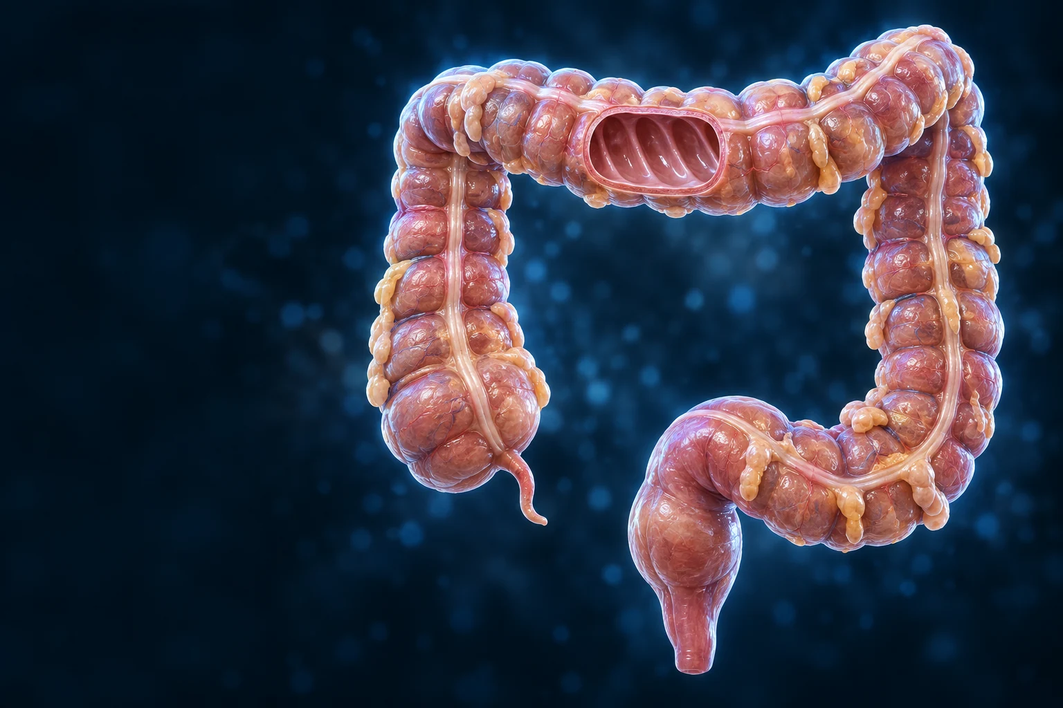
Terminal ileal contents enter the cecum, continue through ascending, transverse, descending and sigmoid colon, then reach the rectum and anal canal. Most nutrient absorption has already taken place upstream.

The right colic flexure lies near the liver; the left colic flexure lies near the spleen. The transverse and sigmoid regions are more mobile than a tidy atlas outline suggests. Length and position vary between people.
Mesenteric vessels supply and drain the wall; lymphatics and nerves also support it. Venous drainage links much of this region with portal circulation. Fine arterial branching and individual surgical variants are not mapped here.
Follow the preceding bowel →02 / FROM SHAPE TO WALL
Haustra give much of the colon its segmented outer appearance. Taeniae coli concentrate longitudinal muscle into three bands. The mucosal lining and its supporting layers surround one continuous digestive space.
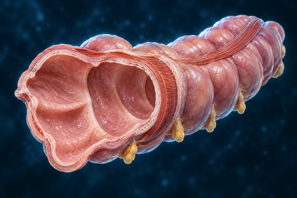
Mucosa faces the contents, followed by submucosa, muscularis externa and an outer covering. Circular and longitudinal muscle arrangements support mixing and propulsion. Enteric nerve networks coordinate local activity.
The taeniae spread into a more continuous longitudinal coat toward the rectum. The rectum does not simply repeat the haustral colon pattern. Not every external surface has the same peritoneal covering.
03 / WALL TO MUCOSA
The colonic mucosa has straight tubular crypts beneath a broadly flat luminal surface. Absorptive cells have microvilli, but the tissue does not carry the fingerlike villi seen in the small intestine.
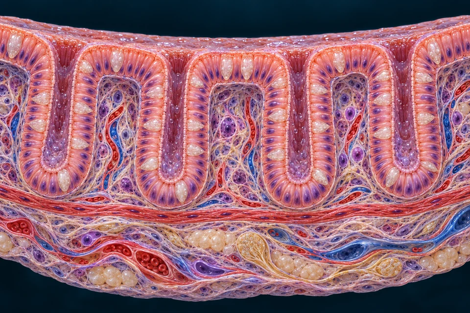
Crypt stem cells generate descendants that replace lost epithelial cells. Supporting lamina propria surrounds glands and contains vascular and immune components. Exact cellular distributions require real tissue evidence.
A crypt is an inward glandular space. A microvillus is a membrane projection of one cell. A haustrum is an organ-scale sacculation. These features cannot be exchanged just because they all modify a surface.
Compare small-intestinal surfaces →04 / CELL ROLES
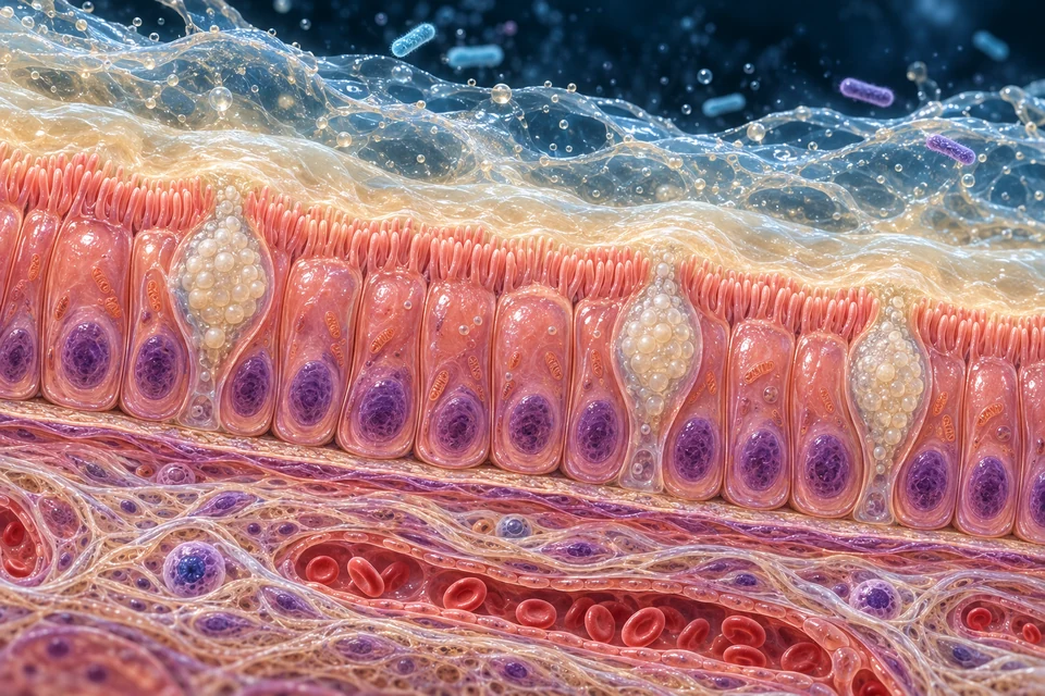
Different cell populations support a surface that exchanges material while separating luminal microbes from internal tissues. Pale generated cells are not independently identified histological cell types.
Water/solute handling and barrier support
Surface and crypt epithelium
Specialized membranes and junctions contribute to selective transport. Microvilli are a cell-scale feature, not whole mucosal villi.
Mucus
Colonic epithelium
Mucus helps lubricate contents and organize protection over the epithelial boundary.
Epithelial renewal
Crypt base region
New descendants differentiate and replace cells lost from the surface. No universal exact turnover clock is asserted.
Digestive/metabolic signals
Scattered epithelial cells
Distal gut signals such as PYY and GLP-1 participate in coordination beyond the colon.
Surveillance and tissue support
Within and beneath the epithelium
The mucosa encounters a large microbial community while maintaining a regulated barrier. This is not a full immune-cell census.
Selected roles, not a complete census. Regional composition varies; no absolute “one cell type never occurs anywhere in the normal colon” claim is made.
05 / FUNCTIONAL RECOVERY
The colon retrieves water and electrolytes from material delivered by the small bowel. Water moves with osmotic conditions established by solute transport; it is not pulled by a dedicated water pump.
Mixing keeps contents near the lining. Sodium and other ion pathways contribute to net absorption; both absorptive and secretory processes matter.
Membrane transport and epithelial permeability shape osmotic conditions. Water can move through and between cells in response to these conditions.
Net recovery helps consolidate stool. Stool contains water, unabsorbed material, microorganisms, shed cells and digestive products; it is not simply unused food.
Supporting mechanism sequence, not an exact transit clock, fixed stool-composition ratio, diagnostic chart or fluid-treatment advice.
06 / LUMINAL COMMUNITY
Many colonic microbes use material not digested or absorbed upstream. Fermentation generates short-chain fatty acids and other products. Community composition and metabolism vary with context; there is no single fixed “ideal” mixture.
Some carbohydrates resist digestion or escape absorption upstream.
Different microbes exchange intermediate products; this is a community process.
Acetate, propionate and butyrate are important products. Amounts and proportions vary.
Butyrate can supply energy for colonocytes. The basic idea is supported by human tissue and wider research; it is not a promise that one food or supplement treats disease.
Absorbed products can enter blood and participate in wider metabolism. Microbial production is not the same pathway as intestinal chylomicron packaging into lymph.
Focused relationship: local epithelial fuel
Supporting mechanism diagram, not a microbial census or microscope image. No fixed ideal microbiome, species ratio, vitamin sufficiency, probiotic claim or therapeutic recommendation is implied.
Mucus, epithelial junctions and immune activity help organize separation between luminal microbes and internal tissues. This is a regulated relationship, not a completely sterile lumen or an absolute wall through which nothing passes.
Microbial processing can generate gases as well as usable metabolites. Different organisms exchange intermediate products, so one named microbe does not explain an entire pathway. The chapter makes no supplement or disease-prevention recommendation.
07 / MOVEMENT OVER DIFFERENT DISTANCES
Colonic motor patterns redistribute contents and sometimes move them longer distances. Signals after eating can alter activity, but eating does not instantly send that same meal from mouth to outlet.
Local redistribution ↔ repeated surface contact
Shorter-range motor patterns mix contents and support contact for absorption. Not every contraction advances material toward the outlet.
Coordinated propulsion → distal bowel
Stronger propulsive events can move contents toward the rectum. Mass movement is not a continuously running conveyor or a fixed meal timer.
Rectal filling → sensory and motor coordination
Rectal distension triggers sensation and reflex responses while sphincters and pelvic-floor activity contribute to continence.
Focused process: Mix and retain
Supporting functional comparison. Actual colonic motor patterns are varied; these are not three sealed compartments or a fixed-time animation.
08 / RECTUM AND COORDINATED OUTLET
Rectal filling supplies sensory input. Internal smooth-muscle tone, external striated-sphincter activity and pelvic-floor support work with local reflexes and central control. An urge is not identical to immediate elimination.
Source-supported functional map — not a precise anatomical cutaway or a clinical procedure.
Sensory input and local reflex coordination.
Smooth muscle. Resting tone and reflex relaxation contribute to control.
Striated muscle and puborectalis support continence and controlled release.
Distension supplies sensory input
Internal sphincter can transiently relax through the rectoanal inhibitory reflex. External sphincter activity and other mechanisms help maintain continence.
Storage with coordinated muscle activity
Resting internal smooth-muscle tone, external striated-sphincter activity and puborectalis/pelvic-floor support cooperate. Holding is not merely closing one valve.
Relaxation + propulsive effort
When appropriate, external sphincter and puborectalis relaxation work with rectal/abdominal pressure and reflex activity to permit passage. These are coordinated events, not a forced clinical instruction.
Focused state: Rectal filling
The main gross-anatomy plate identifies the rectum. A detailed sphincter/pelvic-floor cutaway remains a visual gap; this functional map does not claim to show exact muscle boundaries, angles or attachments.
The internal anal sphincter is a continuation of thickened circular smooth muscle. The external sphincter is striated skeletal muscle with somatic control, including pudendal innervation. These are distinct from the gross rectal reservoir.
Puborectalis contributes to anorectal support and continence. Its coordinated relaxation can help permit elimination. This chapter does not show a validated muscle-attachment map or prescribe posture, straining or pelvic-floor treatment.
09 / REGULATION
Enteric neural circuits organize wall activity. Autonomic pathways influence it, while somatic control participates in the striated outlet muscles. Sensory signals connect local events with conscious awareness and behavior.
Gastric and intestinal signals can increase colonic motor activity after eating. This is coordinated feedback, not proof that new food has already reached the colon.
Enteroendocrine products including PYY and GLP-1 participate in digestive and metabolic coordination. Their actions overlap with other hormones and neural circuits; they are not simple on/off switches or treatment advice.
Connect with pancreatic metabolic control →10 / RELATED SYSTEMS
The bowel recovers fluid from its lumen and eliminates remaining material. Kidneys filter blood and regulate urinary losses. Both influence water and salt balance, but their routes and controls are different.
Absorbed colonic products can enter portal blood and reach the liver. This connects local microbial metabolism with broader processing without making every luminal compound a guaranteed nutrient.
Follow portal circulation →Barrier and immune functions connect the bowel with wider host defense. The appendix has lymphoid tissue, while the whole mucosal surface encounters microbial signals. A fuller immune-system chapter remains a separate dependency.
Compare the urinary route →11 / REAL TISSUE EVIDENCE
Real tissue sections show crypts, epithelial populations and layered muscle in ways a generated preparation cannot independently prove. A tissue slice and a spread-out organ are different kinds of evidence.
Histology Guide’s MHS 274 shows colonic wall layers, absence of villi, crypts, goblet cells and the longitudinal muscle bundles. Use it for tissue context, not an individual diagnosis.
Open MHS 274 Colon ↗External reference only. No specimen pixels copied; redistribution rights were not established for those images.
Detailed anorectal artwork, clinical imaging and disease comparison remain gaps. Diagnosis, treatment and therapeutic microbiome claims belong to a later evidence/clinical phase. These gaps do not erase the verified normal-function relationships taught here.
Read the institutional tissue context ↗EDITORIAL RECORD
Broad regional route, haustra/taeniae and mucosal distinctions, selected cell roles, normal fluid handling, fermentation, movement and functional elimination/control. These claims have independent sources; every generated pixel has not been verified.
AI-assisted visual/content production. Fine generated cells, fibers, nuclei, gland openings, hidden connections, vessel branches and proportions remain illustrative / not independently verified. Human medical review: not performed. Educational purpose only; not an exhaustive colorectal or clinical guide.
English revision: large-intestine-english-2026-10-01-deep-v1. Chapter identity: ahb_ch_large_intestine_english_v1. No canonical colonic CMS entity or verified chapter-specific 3D mapping is asserted. Physical-phone acceptance has not been performed. Labels and supporting diagrams remain editable English code, separate from anatomical masters.
Return to the preceding absorption lesson →