NOSE / THE 30-SECOND PICTURE

Nose & Nasal Airways

A folded passage. A living conditioner.

The nose directs air over a moist vascular surface before it continues into the throat. Tissue warms and humidifies, secretions retain material, and motile cilia help carry surface material toward clearance.

Air enters a folded, living passage.

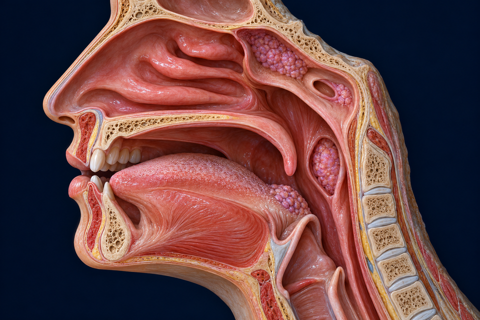
Retained original opened lateral upper-throat anatomy with a nasal entrance and mucosa-covered conchal shelves above the hard palate, continuing posteriorly toward the nasopharyngeal setting beside the soft palate.
Retained original generated text-free anatomical preparation. The selected lateral wall is opened; this is not a strict median CT slice. Route cues identify broad breathing direction, not computed airflow or proof of every narrow meatus. The surrounding lymphoid tissue is retained, not newly diagnosed.
Structure notes
Nasal entrance / vestibular context
A nostril leads into an entrance region before the respiratory cavity proper. The small field does not identify every vestibular hair or cartilage boundary.
Mucosa-covered conchal region
Curved side-wall shelves increase the living surface available for air contact. This view supplies a spatial context, not exact identification of every fine bony attachment.
Air passage beside the lining
Air occupies spaces between the mucosal surfaces. It does not travel through the cells, lymphoid masses or a sinus as a compulsory next chamber.
Posterior nasal / nasopharyngeal context
The nasal cavity continues through the choanal region into the nasopharyngeal setting. This broad opened view is not an ostial or surgical reconstruction.
Hard-palate floor
Bony palatal support separates the nasal setting above from the oral setting below. It is not the central right–left nasal septum.
Soft-palate neighbour
The posterior mobile palate participates in coordinated swallowing. Its broad resting context is shown here, not a timed swallowing simulation.

A folded nasal passage above a shared palate · atlas detail

Retained original opened lateral upper-throat anatomy with a nasal entrance and mucosa-covered conchal shelves above the hard palate, continuing posteriorly toward the nasopharyngeal setting beside the soft palate.

Retained original generated text-free anatomical preparation. The selected lateral wall is opened; this is not a strict median CT slice. Route cues identify broad breathing direction, not computed airflow or proof of every narrow meatus. The surrounding lymphoid tissue is retained, not newly diagnosed.

Open original full-resolution illustration ↗ · Text-free medical base; anatomy names, directions and explanations remain independent language layers in the chapter.

Air enters a folded, living passage.

Follow the cyan route from the entrance toward the posterior nasal setting. Mucosal surfaces condition the passing air; the line is a direction guide, not a measured stream through every meatus.

  1. Air meets the surface →Conchal folds bring a large lining into a compact space.
  2. Heat, water and capture →Blood, surface fluid and secretions contribute different functions.
  3. Clearance continuesCilia move surface material separately from the breathing air route.

Normal structure and function: University of Michigan BlueLink · Nasal cavity, sinuses and ear · OpenStax · Normal conducting-airway functions · Stannard & O’Callaghan · Ciliary function and clearance (2006)

01 / SEPTUM · CONCHAE · ORBITS · NEIGHBOURING SINUSES

Two nasal passages sit among several different spaces.

An opened side view explains the passage; the retained anterior Skull preparation adds a central septum and neighbouring bony spaces. The empty orbits and sinus cavities have different identities.

Two nasal passages share a central boundary.

Locate the septal context between the two sides, then compare the lateral conchal regions. The drawing is a selected construction rather than proof of perfect individual symmetry.

Retained original anterior educational craniofacial cutaway with nasal septal and conchal regions between the maxillary spaces, empty orbital sockets above them, frontal sinus cavities and ethmoid air-cell context.
Exact retained qualified Skull atlas base. Broad neighbouring spaces are supported; this is not a patient CT, verified ostial map or universal symmetric sinus arrangement. The deeper sphenoid sinus is outside this anterior view.
Structure notes
Frontal sinus context
Air spaces can occupy the frontal bone above orbital level. Their shape, development and side-to-side form vary.
Orbital socket
This empty bony socket normally houses the eye and supporting tissues. An orbit is not a paranasal sinus.
Maxillary sinus context
An air-containing cavity lies within the upper jaw beneath the orbit. Exact generated tooth-root distances and tiny drainage openings are not verified.
Ethmoid air-cell context
Small spaces lie between upper nasal and medial orbital regions. This is a broad context rather than a certified count or connected labyrinth.
Central septal context
The partition separates right and left nasal passages. Fine cartilage–bone boundaries and individual asymmetry are not measured in this illustration.
Lateral conchal context
Curved lateral shelves border the central nasal airway. The frontal view complements the opened side view without tracing each meatus or ostium.

A central passage with neighbouring bony spaces · atlas detail

Retained original anterior educational craniofacial cutaway with nasal septal and conchal regions between the maxillary spaces, empty orbital sockets above them, frontal sinus cavities and ethmoid air-cell context.

Exact retained qualified Skull atlas base. Broad neighbouring spaces are supported; this is not a patient CT, verified ostial map or universal symmetric sinus arrangement. The deeper sphenoid sinus is outside this anterior view.

Open original full-resolution illustration ↗ · Text-free medical base; anatomy names, directions and explanations remain independent language layers in the chapter.

Connections have a regional pattern

Different sinuses and the tear-drainage route open into different nasal regions. A broad frontal cutaway cannot establish their tiny openings or expose a deep sphenoid sinus.

Read the normal communicating regions →

Preserve the wider anatomy

The existing Skull chapter follows the surrounding bony framework and other cranial spaces. The original atlas base is retained unchanged here.

Continue with skull spaces →

Normal structure and function: University of Michigan BlueLink · Nasal cavity, sinuses and ear · Kenhub · Lateral nasal wall, compared with institutional anatomy

02 / ONE EPITHELIAL LAYER · SURFACE FLUID · LIVING SUPPORT

A layered-looking tissue can still be one epithelial layer.

Compare the staggered nuclei, tall ciliated profiles, pale secretory cells and small basal region. Then look above the cells to the hydrated surface and mucus, and below to separate vascular support.

A surface film can retain exposed particles.

Look for material resting within or on the mucus context. Retention is a physical relationship; it does not establish the object’s identity or complete removal from every inhaled breath.

Original enlarged nasal respiratory mucosa cutaway with tall ciliated columnar cells at staggered nuclear heights, pale non-ciliated goblet cells, small basal cell regions, surface fluid and mucus with trapped particles, and separate supporting vessels beneath the epithelial boundary.
Original generated respiratory-tissue reconstruction, not H&E or electron microscopy. The relative film, cilia, nuclei and vessels teach relationships without certifying molecular identity, cell counts or measured dimensions. An independent arrow denotes qualitative surface transport toward the pharynx.
Structure notes
Mucus-film context
A thin surface medium helps retain exposed material. Its pictured thickness and rheology are not measurements.
Trapped-particle example
One small irregular object provides an exposure example; it is not identified as a particular pathogen or a guarantee of complete filtration.
Motile ciliary field
Fine apical processes extend from the ciliated epithelial cells. The drawing does not certify an axonemal cross-section, beat frequency or every fibre.
Hydrated surface zone
A suitable fluid environment around cilia permits effective movement and coupling with mucus. This is a selected relationship, not an exact molecular boundary or fixed fluid depth.
Secretory goblet-cell example
The pale mucin-rich apical region belongs to a non-ciliated secretory cell incorporated into the lining. Its appearance is not an assay of secretion or a diagnosis.
Basal-cell region
Small lower profiles illustrate cells that do not all reach the free surface. Exact cell phenotype is assigned for teaching, not inferred from colour alone.
Epithelial–support boundary
Tall and basal epithelial profiles meet a continuous supporting boundary. This is not a second airway or a row of vessels.
Separate vascular support
A vessel profile in underlying connective tissue supplies a different route from the surface film. The image is not a branch-by-branch vascular map.

Living cilia, secretions and blood support one surface · atlas detail

Original enlarged nasal respiratory mucosa cutaway with tall ciliated columnar cells at staggered nuclear heights, pale non-ciliated goblet cells, small basal cell regions, surface fluid and mucus with trapped particles, and separate supporting vessels beneath the epithelial boundary.

Original generated respiratory-tissue reconstruction, not H&E or electron microscopy. The relative film, cilia, nuclei and vessels teach relationships without certifying molecular identity, cell counts or measured dimensions. An independent arrow denotes qualitative surface transport toward the pharynx.

Open original full-resolution illustration ↗ · Text-free medical base; anatomy names, directions and explanations remain independent language layers in the chapter.

Gold → surface clearance toward the pharynxAirflow changes direction with breathing; this tissue cue does not.

Movement needs the right environment

Effective clearance depends on mucus properties, hydration and coordinated ciliary function. More visible fluid or longer drawn cilia alone would not prove more effective defence.

Connect secretion, fluid and transport →

Smell is a distinct specialisation

This is respiratory epithelium. Superior olfactory tissue detects chemicals through a different organisation and neural pathway.

Compare the sensory setting →

Normal structure and function: Histology Guide · MH134 human nasal conchae and palate · Stannard & O’Callaghan · Ciliary function and clearance (2006)

GO DEEPER / STRUCTURES · PHYSIOLOGY · SURFACE DEFENCE · RELATED SYSTEMS

Understand what each structure contributes.

A flexible entrance continues into a divided cavity

The visible nose combines a bony upper framework with cartilaginous support around its projecting part. Each nostril opens into a vestibule. That entrance has skin-associated features, including coarse hairs, before the lining transitions toward the cavity’s mucosa.

The septum separates right and left passages. Its major supports include anterior septal cartilage, the vomer posteroinferiorly and the ethmoid’s perpendicular plate posterosuperiorly. A two-dimensional illustration cannot establish every material boundary or the degree of asymmetry in an individual.

Normal structure and function: University of Michigan BlueLink · Nasal cavity, sinuses and ear · OpenStax · Normal conducting-airway functions

Folded side walls change how air meets living tissue

The lateral walls project inward as superior, middle and inferior conchae, also called turbinates. The superior and middle conchae are parts of the ethmoid; the inferior concha is a separate bone. Mucosa adds considerable living tissue over that framework.

These folds provide a large interface in a compact space and influence the distribution and resistance of airflow. The meatuses are spaces associated with the shelves. The conceptual route cue does not calculate turbulence or certify every narrow passage in a generated image.

Normal structure and function: University of Michigan BlueLink · Nasal cavity, sinuses and ear · OpenStax · Normal conducting-airway functions

Heat and water exchange depend on the surface and its blood supply

Incoming air encounters a moist, well-perfused lining. Heat transfers between the vascular tissue and air, and water from the surface supports humidification. Geometry and contact time help explain why conditioning is an organ-level process rather than a single chemical switch.

Exhaled air can return some heat and moisture to the nasal surface. This recovery varies with environmental and physiological conditions; the illustrations do not promise complete filtration, a constant final temperature or protection from every inhaled particle.

Normal structure and function: OpenStax · Normal conducting-airway functions · Review · Nose and paranasal sinus physiology (2001)

A nose contains more than one kind of surface

Most of the cavity’s respiratory region has pseudostratified ciliated columnar epithelium with secretory cells. Cells sit at one epithelial boundary, but their nuclei lie at different heights. Small basal cells do not all reach the free surface, producing an apparent layering without several stacked tiers of whole cells.

The vestibular skin and the specialised superior olfactory mucosa differ from this respiratory example. Motile respiratory cilia help move surface material; olfactory sensory cells use a different organisation to detect chemicals and generate neural information.

Normal structure and function: Histology Guide · MH134 human nasal conchae and palate · University of Michigan BlueLink · Nasal cavity, sinuses and ear

A mucus film and a hydrated surface zone cooperate

Mucin-rich secretions form a protective surface medium that can retain particles and biological material. Goblet cells contribute at the epithelium, while deeper seromucous glands add other secretions. A pale secretory region in the drawing is an explanatory cell example, not a measured secretion volume.

Ciliary movement needs a suitable hydrated environment. Surface-fluid depth, mucus properties, epithelial transport and ciliary function work together; simply drawing longer hairs or a thicker gel would not prove better clearance. The visual focus changes where to look, not the tissue’s real hydration or secretion rate.

Normal structure and function: Stannard & O’Callaghan · Ciliary function and clearance (2006) · Histology Guide · MH134 human nasal conchae and palate

Surface transport has a destination distinct from the air route

Coordinated ciliary activity moves surface material through much of the respiratory-lined nasal region toward the pharynx, where it can be swallowed. This clearance route is different from the rapid direction changes of air during breathing. The vestibular entrance and local boundary regions are not an identical conveyor at every point.

The surface protects against exposure through several complementary processes. Retaining a particle in mucus is not the same as identifying it immunologically, and neither step proves that every particle will be removed before it can enter deeper airways.

Normal structure and function: Stannard & O’Callaghan · Ciliary function and clearance (2006) · Review · Nasal physiology, nasal cycle and ciliary action · OpenStax · Normal conducting-airway functions

Neighbouring air spaces communicate through specific regions

Frontal, maxillary, ethmoidal and sphenoidal spaces occupy different bones and vary greatly in size and form. They communicate with the nasal cavity through openings rather than forming four chambers that every breath must traverse in sequence.

In the usual anatomical pattern, frontal, maxillary and anterior/middle ethmoidal drainage relates to the middle-meatus region; posterior ethmoidal cells relate to the superior meatus, and the sphenoid sinus opens into the sphenoethmoidal recess. The retained anterior bone view supplies broad context, not verified tiny ostia or an exposed deep sphenoid sinus.

Normal structure and function: University of Michigan BlueLink · Nasal cavity, sinuses and ear

Tear drainage and auditory pressure connect different systems

Tears drain through a separate nasolacrimal route into the inferior nasal meatus. This explains a real eye–nose connection without making the nose a container for an eyeball or depicting tears as a secretion of the sinus cavities.

Posteriorly, the nasopharyngeal setting neighbours the auditory-tube openings and the soft palate. Middle-ear pressure communication and swallowing coordination are related functions; neither is a direct open passage from a paranasal sinus into an ear chamber.

Normal structure and function: University of Michigan BlueLink · Nasal cavity, sinuses and ear

A rich vascular lining is more than an anatomical backdrop

Blood supply supports the metabolic and exchange functions of nasal tissue. Contributions come from both ophthalmic and external-carotid branch territories, including ethmoidal, sphenopalatine and facial/palatine routes. Anterior septal branches communicate in a vascular network.

Venous spaces can change the volume of the mucosal lining and influence available airway space. The tissue image shows separate vessel context, not a complete vascular map, a bleeding-risk threshold or a clinical explanation for a particular person’s symptoms.

Normal structure and function: University of Michigan BlueLink · Nasal cavity, sinuses and ear · Review · Nose and paranasal sinus physiology (2001)

Normal control can change the balance between the two sides

Autonomic regulation affects mucosal vascular tone and secretion. A normal nasal cycle can shift relative congestion and resistance between sides over time. People and conditions vary; the chapter does not impose one fixed period or require equal-looking passages in every snapshot.

This physiological control differs from the motile activity of individual cilia and from the neural detection of smell. Keeping those levels separate helps connect tissue volume, surface function and sensation without turning a static image into a diagnosis.

Normal structure and function: Review · Nasal physiology, nasal cycle and ciliary action · Review · Nose and paranasal sinus physiology (2001)

General sensation and smell use different neural roles

Branches of the trigeminal nerve contribute general nasal sensation; selected V1 and V2 territories serve different regions. Olfactory receptor neurons and their connections provide the specialised smell pathway. Irritation, cooling sensation and recognising an odour are not interchangeable descriptions.

The existing Smell & Taste chapter follows chemical access, sensory epithelium and neural processing in greater detail. This chapter adds the surrounding respiratory passage and surface-conditioning context rather than reproducing that sensory lesson.

Normal structure and function: University of Michigan BlueLink · Nasal cavity, sinuses and ear · Review · Nasal physiology, nasal cycle and ciliary action

The surrounding spaces develop and remain variable

The shape of the septum, conchae and adjacent sinus cavities differs among people. Their proportions also change with growth. A symmetric atlas view is a selected teaching construction, not an anatomical standard against which every individual should be graded.

Variation in geometry can affect local relationships and airflow, but a visible asymmetry alone does not establish disease or identify a treatment. The normal chapter keeps precise patient imaging, clinical assessment and procedures outside its scope.

Normal structure and function: University of Michigan BlueLink · Nasal cavity, sinuses and ear · Review · Nose and paranasal sinus physiology (2001)

A specimen records tissue, preparation and a limited field

The linked MH134 record identifies human nasal conchae and palate stained with H&E. It describes respiratory epithelium, seromucous glands, conchal bone and the oral–nasal separation of the palate. The same record notes distortion and partial disruption during preparation.

Use it to compare tissue relationships across scales, not to infer live ciliary motion from a stained section. The new atlas reconstruction is original generated artwork; its visual detail does not make it a microscope image or certify a molecular phenotype.

Normal structure and function: Histology Guide · MH134 human nasal conchae and palate
REAL HUMAN TISSUE / COMPLEMENTARY INSTITUTIONAL OBSERVATION

A stained section shows relationships, not live transport.

Histology Guide identifies MH134 as human nasal conchae and palate in H&E, from the University of Minnesota. Compare the respiratory lining, conchal support and the separate oral surface; the record states that preparation distorted and partly disrupted the tissue.

The external slide remains on its institutional site; its pixels are not redistributed. It is not a universal healthy control or a video of ciliary motion. The three atlas bases on this page are generated teaching illustrations.

Normal structure and function: Histology Guide · MH134 human nasal conchae and palate

Check the relationship.

Does all inhaled air pass through every sinus?

No. The main nasal passage conducts air; sinuses communicate through particular openings rather than acting as obligatory chambers in a serial route.

Do cilia reverse clearance whenever air is breathed out?

No. Surface transport and the changing direction of air are separate processes. Coordinated respiratory cilia generally carry surface material toward the pharyngeal route.

Why do the nuclei appear at different heights?

Respiratory epithelium is pseudostratified: cells share an underlying boundary, while some shorter profiles do not reach the surface. The appearance is not several stacked tiers of identical complete cells.

Is every nasal cilium part of the smell pathway?

No. Motile respiratory cilia and the specialised olfactory sensory setting have different roles; the linked Smell chapter explains that sensory organisation.

Follow the connected systems.

From chemical contact to a nerve signal →

Sources and normal educational scope

Targeted normal anatomy and physiology checks: 20 October 2026. FB019 and FB052 inventories supplied related book coverage; air-conditioning and mucociliary mechanisms were supplemented from independent physiology and institutional tissue sources. Original book text, images and source compositions were not copied.

Retained qualified originals and one new tissue base share independent semantic targets, short names, direction cues and explanations. English is the current chapter language; no translated route is created. This normal lesson provides no sinusitis diagnosis, treatment, drug-delivery or surgical advice. Physician review, precise generated microstructure, exact sinus ostia and physical-phone acceptance are not certified.