Smell
Superior nasal sensory tissue
Olfactory receptor neurons detect patterns of volatile chemicals.
CHAPTER / CHEMICAL SENSES
Surface chemistry. Sensory experience.
Explore how volatile odorants meet nasal sensory tissue and dissolved chemicals meet taste receptors. Follow their different neural routes, then see how smell, taste and texture contribute to flavour.
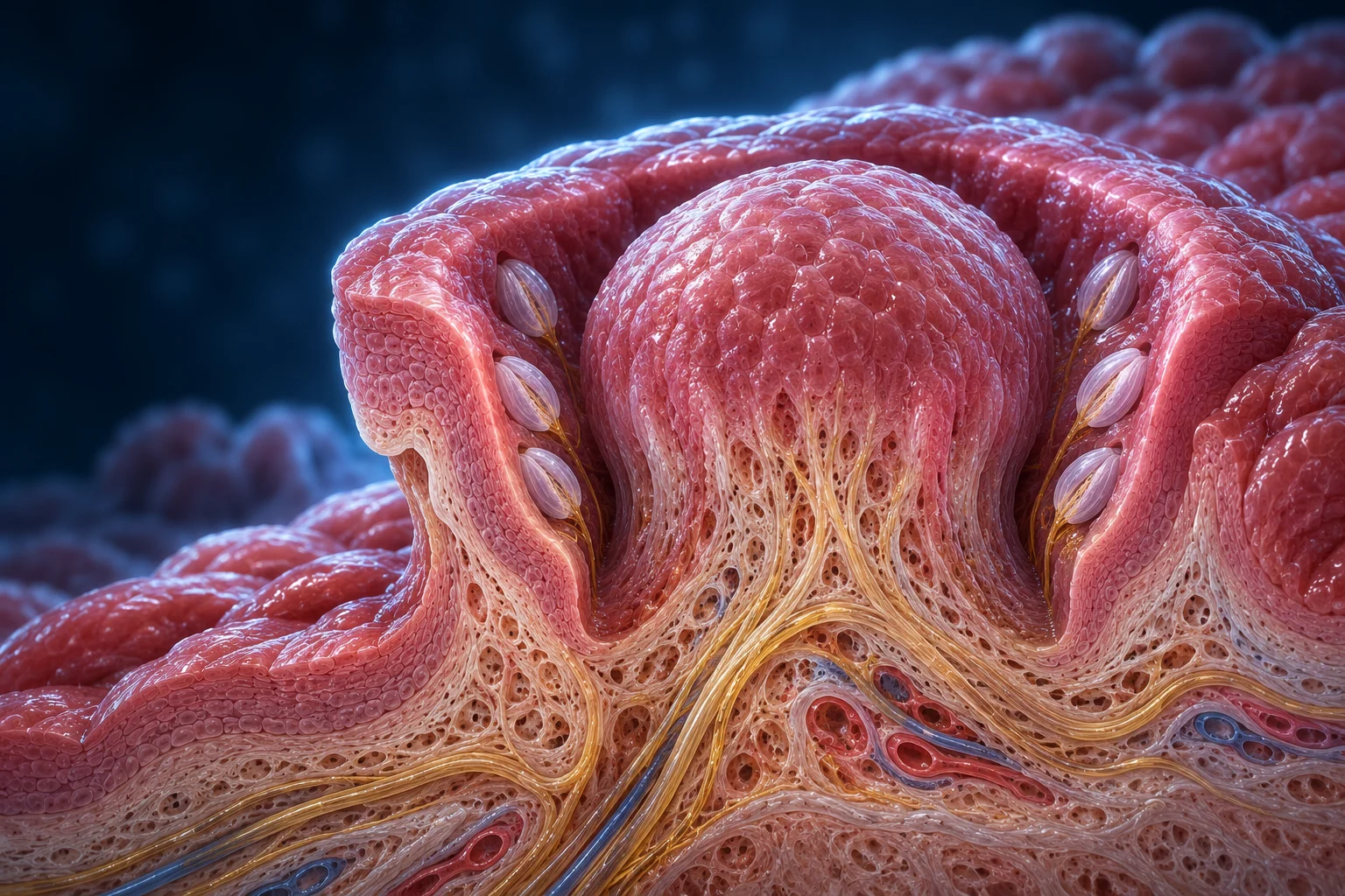
Two different sensory interfaces contribute: a neuron in the nose and epithelial cells in taste buds. Use the four scales to locate them before following their signals.
30-SECOND ORIENTATION / SURFACE → CELL → NEURAL INFORMATION → EXPERIENCE
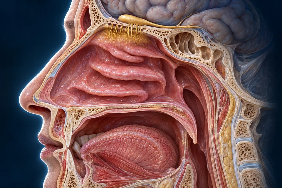
The olfactory bulb lies above a bony separation; sensory tissue lies below. Chemicals are detected at the nasal surface, not carried as food into the brain.
02 / NASAL-CONTEXT
The nasal cavity supplies airspace; olfactory tissue occupies selected superior regions. A bony roof separates the mucosa from the bulb. Most nasal lining is not smell-receptor tissue.
Teaching references: NCBI · Olfactory nerve and bulb relationships · NIDCD / NIH · Normal smell and two delivery routes
CHEMICAL CONTACT / CELL RESPONSE / NEURAL OUTPUT
A molecule arrives at a surface. A living cell responds. Information travels onward. Follow the links between these events, and see why a smell neuron and a taste cell do not send their messages the same way.
No. A chemical changes what a sensory cell does. Ion movement changes its voltage, and electrical or chemical communication carries the information onward.

Read the preparation: The left neuron and enlarged cilium at right show two scales, not two cells joined by a pipe. Only the neuron at left supplies its own axon. Channel silhouettes locate a membrane context, not exact molecular identities.
Supporting compartment diagrams. The detailed tissue preparation above provides the anatomical context; these simplified views isolate direction and communication, not molecular shape, scale, ion count or timing.
A conceptual causal sequence, not a measured timing trace or an exact molecular reconstruction.
Evidence: The Olfactory System, Introduction to Behavioral Neuroscience §8.3 · The Gustatory System, Introduction to Behavioral Neuroscience §8.2
Read the complete explanation and its limits ↓Every explanation stays available here. Opening a question also selects its visual context.
No. A chemical changes what a sensory cell does. Ion movement changes its voltage, and electrical or chemical communication carries the information onward.
Odorants reach mucus around olfactory cilia. Dissolved tastants reach exposed tips of taste cells. Arrival is transport outside the cell, not yet an action potential. The stimulating molecule does not travel along the axon as the message.
A receptor protein is not the same thing as a whole sensory receptor cell. A receptor potential is a graded change in membrane voltage. An action potential is a regenerative electrical event. A transmitter is a chemical carrying information across a cell-to-cell contact. Keeping these levels separate makes the rest of the course easier.
Both olfactory neurons and several types of taste cell can generate electrical spikes. The important architectural difference is that the olfactory neuron has its own axon, whereas the taste epithelial cell communicates with a different sensory neuron.
Keep this boundary: A brighter stimulus symbol is not a measured voltage or a guarantee of stronger conscious perception.
Evidence: The Olfactory System, Introduction to Behavioral Neuroscience §8.3 · The Gustatory System, Introduction to Behavioral Neuroscience §8.2
No. It has arrived at a sensory surface; a cell must transduce that input into a change in electrical activity.
A receptor activates an intracellular messenger pathway. cAMP opens channels, allowing positive ions to enter the cilium and change the neuron’s electrical state.
The receptor is a G-protein-coupled receptor. Its outside-facing stimulus interaction and its cytoplasmic signaling partners are on different sides of one membrane. AC3 produces cAMP inside the cell; cAMP acts on the intracellular face of CNG channels.
Under the relevant physiological conditions, inward sodium and calcium current through CNG channels helps depolarize the cilium. The graded receptor potential is not a spike located at the receptor protein. It changes neuronal excitability, and voltage-gated channels support propagated electrical output in the neuron.
Mouse loss-of-function studies of G_olf and AC3, together with channel experiments, support different links in this chain. They are converging experimental approaches, not one microscopic image proving every arrow or a genetic experiment performed in a person.
Keep this boundary: The illustrated membrane shapes locate a conceptual signaling interface; their colours or folds do not identify experimentally resolved OR, AC3 or CNG proteins.
Evidence: The Olfactory System, Introduction to Behavioral Neuroscience §8.3 · A cyclic nucleotide-gated conductance in olfactory receptor cilia · Mice deficient in G(olf) are anosmic · Disruption of the type III adenylyl cyclase gene leads to peripheral and behavioral anosmia in transgenic mice · The Ca2+-activated Cl− channel TMEM16B shapes the response time course of olfactory sensory neurons
No. In this chain, cAMP is an intracellular messenger made from ATP on the cytoplasmic side.
In olfactory cilia, opening ANO2/TMEM16B can let negatively charged chloride leave. Loss of negative charge can reinforce depolarization, but a larger receptor current does not always mean more spikes.
The effect of a channel depends on the ion gradients and membrane voltage, not just the ion’s name. In this ciliary context, chloride can move out. That is electrically different from the familiar inhibitory chloride entry described in some other neurons.
A 2024 mouse study found that removing TMEM16B could permit better repeated spike firing despite a smaller receptor current. Amplification of receptor current, spike timing and conscious smell strength are therefore not interchangeable quantities.
Response termination is the subsiding of signaling and ion changes. Peripheral adaptation is altered responsiveness during sustained or repeated input. Central habituation concerns later processing. Calcium-dependent feedback and extrusion, including NCKX4 evidence from mice, interact with cAMP metabolism; no single universal reset switch explains every condition.
The NCKX4 source was available at abstract level for this review, so the course does not claim verified exchanger stoichiometry or detailed human kinetics. Human imaging supports a separate central processing level, not one universal time at which every familiar smell fades.
Keep this boundary: A smell fading from notice does not establish that the air is safe. No measured current gain, response duration or conscious intensity is encoded in the illustration.
Evidence: The Ca2+-activated Cl− channel TMEM16B shapes the response time course of olfactory sensory neurons · The Na+/Ca2+ exchanger NCKX4 governs termination and adaptation of the mammalian olfactory response · Olfactory CNG channel desensitization by Ca2+/CaM via the B1b subunit affects response termination but not sensitivity to recurring stimulation · Phosphodiesterase 1C is dispensable for rapid response termination of olfactory sensory neurons · Time course of odorant-induced activation in the human primary olfactory cortex
No. The receptor current and the neuron’s spike-generating machinery interact; their relationship depends on timing and cellular conditions.
For sweet, bitter and umami pathways, intracellular calcium helps excite a taste cell. ATP released through CALHM1/3 channels then signals to a separate sensory nerve ending.
Sweet commonly uses T1R2/T1R3, umami uses T1R1/T1R3, and bitter uses members of the T2R family. Distinct receptor populations can share downstream machinery. A representative Type II cell does not mean all these receptor identities are mixed in every taste cell. Human receptors have been studied in expression systems, while much intact-cell causal evidence comes from mice.
PLCβ2 acts at the cytoplasmic membrane face and generates IP3 from membrane phosphoinositide. IP3 remains within the cytoplasm and opens a calcium-release channel in the ER membrane. Calcium leaves the internal ER store for cytoplasm, not from extracellular fluid directly through TRPM5.
TRPM5 is a monovalent-cation channel, not the calcium-release pore. Mouse evidence also implicates TRPM4, so TRPM5 is not presented as the only depolarizing conductance in every species. Electrical excitation in the taste cell and excitation in the nerve ending are distinct events.
CALHM1/3 forms an ATP-release pathway at the Type II cell’s channel synapse. ATP crosses into the extracellular contact space and activates purinergic receptors in the afferent membrane. The CALHM1 primary experiments help separate a cell’s own stimulus response from successful transmitter output. Type II ATP is not packaged into the conventional synaptic vesicles depicted in the sour comparison.
Keep this boundary: The diagram represents one mechanism-specific epithelial cell and a separate afferent, not a retrospectively identified cell in the old taste-bud artwork or an exact molecular structure.
Evidence: Human receptors for sweet and umami taste · T2Rs function as bitter taste receptors · Coding of sweet, bitter, and umami tastes: different receptor cells sharing similar signaling pathways · TRPM4 and TRPM5 are both required for normal signaling in taste receptor cells · CALHM3 is essential for rapid ion channel-mediated purinergic neurotransmission of GPCR-mediated tastes · CALHM1 ion channel mediates purinergic neurotransmission of sweet, bitter and umami tastes · ATP signaling is crucial for communication from taste buds to gustatory nerves · Taste transduction and channel synapses in taste buds
Yes. Detection, cellular excitation and transmitter release are different stages. An output-pathway defect can separate them.
Protons enter through OTOP1 and help excite sour-responsive taste cells. Their conventional synaptic machinery is supported, but the source and release mechanism of the ATP needed for sour afferent signaling are not fully established.
The representative sour-responsive cell is Type III. OTOP1 carries protons rather than starting the same GPCR–IP3 chain described for Type II cells. Mouse knockout studies strongly weaken gustatory acid responses but do not remove every neural response to acid or every non-taste avoidance behavior.
Apical sensing and basal output are separate locations. Type III cells have conventional synaptic machinery and can release serotonin, but serotonin is not presented as the sole completely solved route to afferent firing. P2X-dependent afferent signaling does not itself identify where the required ATP comes from.
The 2025 SNAP25 study links conventional synaptic machinery with sour responses and Type III cell maintenance. It does not prove ATP-filled synaptic vesicles; that proposal requires further testing. The image therefore shows a vesicle-containing output context without naming its visible cargo as ATP.
Recent mouse localization also finds OTOP1 in Type I cells. OTOP1 positivity alone is not a sufficient cell-identity rule, and the colours of an old generated taste-bud illustration cannot identify Type III cells.
Keep this boundary: Uncertainty is part of this pathway, not a missing arrow to invent. Do not infer a solved Type III CALHM route, ATP vesicle or serotonin-to-P2X connection.
Evidence: ATP signaling is crucial for communication from taste buds to gustatory nerves · An evolutionarily conserved gene family encodes proton-selective ion channels · Cellular and neural responses to sour stimuli require the proton channel Otop1 · The K+ channel KIR2.1 functions in tandem with proton influx to mediate sour taste transduction · Dual functions of SNAP25 in mouse taste buds · Epitope tagging with genome editing in mice reveals that the proton channel OTOP1 is apically localized and not restricted to Type III sour taste receptor cells · Sour taste: receptors, cells and circuits · The enigma of type III taste bud cells
Yes. Different links in one pathway can have different levels of evidence.
An olfactory sensory neuron carries its output along its own axon. A taste epithelial cell communicates across a contact with a different neuron, even though the taste cell can itself fire electrical spikes.
The key comparison is cell architecture, not “electrical versus non-electrical.” Taste cells can be electrically excitable without being neurons whose own axons form the cranial nerves. The receptor cell’s response and the receiving afferent’s response must remain distinguishable.
In the olfactory cascade, ATP is used inside the cell to make cAMP. In Type II taste output, ATP outside the cell is a transmitter. The same molecule can have different jobs in different compartments.
Mouse ENaC contributes to an amiloride-sensitive low-sodium pathway, while other salt responses remain. Human studies are variable and the molecular account is less settled. A neat set of five equally complete cartoons would hide this difference in certainty, so human salt is kept in an explicitly bounded evidence box.
A useful final test is to ask which stage an observation measures: chemical arrival, receptor-cell current, cellular spikes, transmitter release, afferent activity or conscious experience. Evidence at one stage does not automatically measure every later stage.
Keep this boundary: Animal loss-of-function experiments offer causal evidence but do not remove species differences. No tasting protocol, drug concentration or health inference is provided.
Evidence: The Gustatory System, Introduction to Behavioral Neuroscience §8.2 · CALHM1 ion channel mediates purinergic neurotransmission of sweet, bitter and umami tastes · ATP signaling is crucial for communication from taste buds to gustatory nerves · Human psychophysical studies of saltiness suppression by amiloride · The cells and peripheral representation of sodium taste in mice · Taste transduction and channel synapses in taste buds · Does ENaC work as sodium taste receptor in humans? · Receptors and signaling for sour and salty: the ionic taste qualities
No. The taste epithelial cell and afferent are separate cells with distinct membranes and different roles.
Source checks dated 23 October 2026. Full articles, indexed passages and abstracts are not interchangeable. Missing correction notices are not proof that none exists. No acquired microscopy, measured molecular geometry or human medical certification is claimed.
Evidence context: General mammalian/human teaching; experimental specificity supplied separately.
Access: Direct complete teaching webpage readable.
Limit: Teaching synthesis, not primary experiment or evidence for measured human molecular kinetics.
Evidence context: General taste-system teaching.
Access: Direct complete teaching webpage readable.
Limit: Does not replace current CALHM1/3 primary evidence or settle the sour ATP source.
Evidence context: Excised ciliary membrane patches from dissociated olfactory receptor cells; species not established from the readable abstract.
Access: Indexed PubMed abstract only.
Limit: Historical corroboration; do not invent species or exact channel stoichiometry from abstract.
Evidence context: Gαolf-null mice.
Access: Indexed PubMed abstract only.
Limit: Mouse necessity evidence, not a direct human causal intervention.
Evidence context: AC3-null mice, epithelial electrical and behavioral responses.
Access: Indexed PubMed abstract only.
Limit: Type III adenylyl cyclase is unrelated to the taste-cell label Type III.
Evidence context: Wild-type and Tmem16b-knockout mouse isolated olfactory sensory neurons and epithelial slices.
Access: Direct publisher full text: abstract, introduction, results and discussion read.
Limit: Larger receptor current does not necessarily mean more spikes or greater perceived intensity. No numerical gain or human timing inferred.
Evidence context: Nckx4-knockout mouse olfactory sensory responses.
Access: Indexed PubMed abstract only. Direct PMC3245797 showed a CAPTCHA; publisher redirect inaccessible.
Limit: Remains ABSTRACT ONLY. Do not claim full-paper qualification or a single universal reset mechanism. Online publication was 2011; journal issue 2012.
Evidence context: Mouse CNGB1 calcium/calmodulin-regulation mutation.
Access: Indexed abstract and relevant manuscript sections; not directly retrieved full text.
Limit: Do not reduce adaptation to one CaM switch.
Evidence context: Mouse Pde1c/Pde4a single and combined genetic disruption.
Access: Indexed abstract and relevant manuscript sections; not directly retrieved full text.
Limit: Do not teach a single ciliary PDE as an isolated off-clock.
Evidence context: Human and rat receptor constructs in heterologous HEK293T assays with engineered G-protein coupling.
Access: Indexed abstract and relevant results; not directly retrieved full article.
Limit: Human receptor proteins in a cell assay are not equivalent to an intact human taste bud; not all amino acids behave identically across species.
Evidence context: Human/mouse T2R heterologous expression; mouse receptor and gustducin coupling experiments.
Access: Indexed PubMed abstract only.
Limit: A bitter signal is not a reliable toxin or safety test.
Evidence context: Mouse gene disruption and cell-selective rescue.
Access: Complete author-hosted 9-page article PDF readable.
Limit: Historical TRPM5 interpretation is not the final current account. Do not copy its early non-Ca-activated inference or claim TRPM5 alone is universal.
Evidence context: Mouse taste-cell imaging, single/double knockouts and behavior.
Access: Indexed abstract, figures and relevant full-text sections; not directly retrieved full article.
Limit: Neither channel should be drawn as the Ca2+ entry pore. Bound the TRPM4 result to the studied mouse system rather than asserting a fully mapped human pathway.
Evidence context: Mouse Calhm3 deletion plus heterologous channel and interaction experiments.
Access: Indexed publisher abstract and relevant manuscript sections; direct publisher returned 403.
Limit: Do not equate abstract/relevant excerpts with full-paper access or draw a precise molecular oligomer.
Evidence context: Mouse Calhm1 knockout and isolated taste cells; heterologous human CALHM1 cellular assays.
Access: Complete official author-hosted PDF, main article and supplementary material readable.
Limit: Update CALHM1-only diagrams with P12; do not use pre-2018 output architecture unchanged.
Evidence context: P2X2/P2X3 double-knockout mice and gustatory nerve recordings.
Access: Indexed PubMed abstract only.
Limit: This does not identify the cell releasing ATP during sour stimulation.
Evidence context: Mouse sour-cell transcriptomic/cellular evidence and heterologous otopetrin channel assays.
Access: Indexed abstract and relevant manuscript results; publisher returned 403.
Limit: Does not alone establish the entire human sour pathway or a unique Type III cell identity marker.
Evidence context: Otop1-null mice: taste-cell proton currents and gustatory nerve responses.
Access: Indexed publisher abstract and manuscript results/discussion; direct publisher returned 403.
Limit: Residual nerve responses and non-gustatory acid avoidance prevent the claim that all acid sensing or avoidance disappears.
Evidence context: Mouse sour taste cells, conditional Kcnj2 perturbation and heterologous assays.
Access: Indexed PubMed abstract and figure captions only.
Limit: Deeper explanatory branch, not justification for a fully specified universal human kinetics model; online publication 2015.
Evidence context: Taste-epithelial conditional Snap25 knockout mice, cell maintenance and sensory responses.
Access: Direct publisher full text, including results and discussion, readable.
Limit: Cell loss is a confound for a pure secretion explanation. Authors explicitly leave ATP-vesicle release as a hypothesis requiring validation; do not present it as discovered.
Evidence context: Two small human psychophysical experiments, four participants in each.
Access: Indexed PubMed abstract only.
Limit: Small, heterogeneous evidence cannot establish ENaC as a universal human salt receptor. No dose or self-experiment instructions.
Evidence context: Taste-cell conditional ENaCα knockout mice; nerve and behavioral assays.
Access: Indexed publisher record plus relevant manuscript results and figures; not directly fetched full article.
Limit: Not all salt sensing; high-salt responses remain. Not settled universal human physiology.
Evidence context: Human fMRI and odor detection.
Access: Indexed PubMed abstract only; already cited by retained P0045.
Limit: No universal fading clock; behavioral detection and cortical signal are not identical measurements.
Evidence context: Endogenous-tagged OTOP1 mouse tissue and cellular localization.
Access: Indexed article abstract, relevant sections and figures; no direct full-article retrieval claimed.
Limit: Presence of OTOP1 or an illustration colour is not sufficient to identify a Type III cell. No exact human protein-density map.
Evidence context: Review of cellular and animal studies.
Access: Publisher abstract/subscription preview and indexed relevant manuscript sections; direct PMC9386877 CAPTCHA.
Limit: Lead is not a primary experiment or a directly read complete article. Online publication 2020.
Evidence context: Review of mostly animal/cellular causal evidence.
Access: Indexed relevant full-text sections and figure captions; direct PMC7943026 CAPTCHA.
Limit: Use with current R04; a receptor discovery does not settle transmitter output. Do not copy a cell-type typo from an indexed caption.
Evidence context: Broad mammalian taste-bud review.
Access: Indexed abstract and selected figure/body excerpts. Publisher redirect inaccessible.
Limit: Predates CALHM3 and decisive OTOP1 evidence; not the sole modern molecular backbone.
Evidence context: Interpretation of Type III mouse evidence and current transmitter uncertainty.
Access: Direct publisher full text readable.
Limit: Commentary, not a new primary experiment. Use to bound P18 and P14, not to prove a new output pathway.
Evidence context: Human psychophysics and molecular-expression literature review.
Access: Indexed relevant full-text sections read. Direct publisher open returned 429; no bypass or retry.
Limit: Expression and variable pharmacological effects do not prove universal human causal necessity.
Evidence context: Current synthesis of animal, cellular and human evidence.
Access: Indexed substantial article sections and figure captions read; not directly fetched full article.
Limit: Its shorthand abstract is not permission for a serotonin-only causal story; check primary results rather than adopting absolute absence claims.
Original AI-assisted cellular preparations with separate editable English labels. Three new compartment-level contexts; the chapter’s five original anatomical masters and existing posters are retained. Educational normal physiology, not diagnosis or a laboratory/tasting protocol. Human medical review not performed.
01 / CHEMICAL-SENSES
Smell detects airborne odorants; taste begins when dissolved chemicals reach specialised oral cells. Their signals are integrated with temperature, texture and irritation rather than becoming one identical pathway.
Superior nasal sensory tissue
Olfactory receptor neurons detect patterns of volatile chemicals.
Taste buds in oral and nearby epithelial sites
Taste epithelial cells communicate with separate sensory afferents.
Integrated perception
A food experience combines several sensory inputs.
It is broader than the five taste qualities.
Teaching references: NIDCD / NIH · Normal smell and two delivery routes · NIDCD / NIH · Normal taste, flavour and non-zonal tongue
03 / OLFACTORY-CELLS
Olfactory neurons span the epithelial sensory interface. Apical dendrites reach the mucus; basal axons gather beneath the epithelium. Supporting and progenitor populations have different jobs.
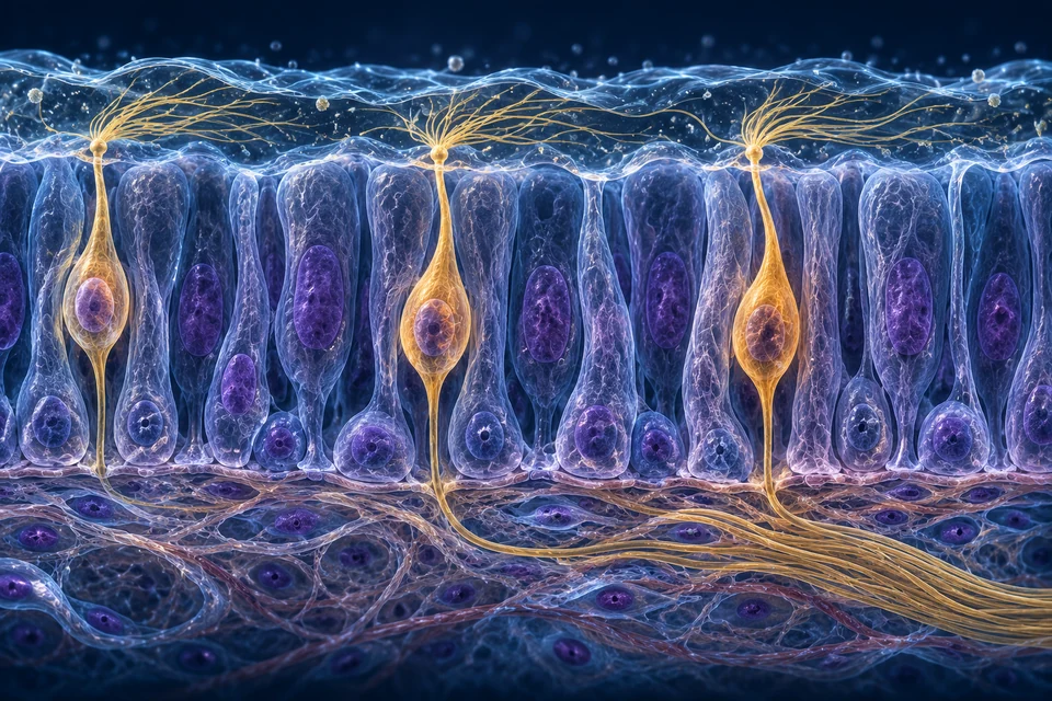
An olfactory sensory neuron places an apical process and sensory cilia toward the mucus interface. Its cell body remains within the epithelial setting, and its own basal axon carries neural information away. Chemical access and axonal communication are different events along one living neuron.
A taste epithelial receptor cell uses a different organisation: its apical region approaches the taste pore, while separate afferent fibres communicate with its basal region. Calling both cells receptors does not mean they have the same shape or each sends an identical long axon to the brain.
The two tissue preparations make this comparison visible. They do not identify receptor molecules, exact cell counts or synaptic types from generated colour.
Source links: olfactoryCells · humanMucosa · tasteCells
Teaching references: NLM · Olfactory epithelium and sensory neurons · PubMed · Human olfactory-mucosa ultrastructure study
04 / SURFACE-ENVIRONMENT
Odorants must reach receptor-bearing membranes in a suitable surface environment. Supporting cells and subepithelial glandular tissue contribute to local maintenance; they are not interchangeable neurons.
Sensory cilia / mucus
Receptor-bearing membrane meets chemical input.
Olfactory sensory cilia are distinct from motile respiratory cilia.
Within and beneath the epithelium
Selected cells and secretions maintain the local interface.
The generated gland is not a verified duct-by-duct specimen.
Axons / nerve fascicles
Neural activity travels toward bulb circuits.
Odor molecules do not travel inside these axons.
Teaching references: NLM · Olfactory epithelium and sensory neurons · PubMed · Human olfactory-mucosa ultrastructure study
05 / OLFACTORY-TRANSDUCTION
Receptor activation can initiate intracellular signalling and ion-channel responses. A local electrical response is converted into propagating activity in the sensory neuron; the chemical itself does not become an electrical arrow.
Structure and function · select a teaching emphasis; all basic explanations stay visible.
The dashed outline locates a compartment for the selected explanation; it is not a moving odorant or a measured electrical trace. The neuron is continuous between its apical process, cell body and basal axon.
Odorant → mucus / receptor membrane
Delivery brings molecules to sensory cilia.
No molecular transfer into the axon is depicted.
Receptor → local signalling / membrane change
Selected receptor activation alters electrical responsiveness.
This is not an exact ion-current recording.
Sensory axon → bulb circuits
Activity propagates along the receptor neuron’s own axon.
The bulb has processing circuits, not an odor-filled chamber.
The supporting sequence deliberately separates chemical delivery, receptor-cell response and axonal output. It is a causal explanation, not a measured electrical recording or complete molecular diagram.
Teaching references: OpenStax · Sensory perception, checked critically · NLM · Olfactory epithelium and sensory neurons
06 / RECEPTOR-PATTERNS
An odorant can activate a combination of receptor populations. Bulb and later circuits process patterns; mixture, concentration and context influence what is perceived.
Structure and function · select a teaching emphasis; all basic explanations stay visible.
Several sensory neurons are shown, but this is not a receptor assay, unique smell cable or measured population-firing pattern. Read the three default-visible explanations before using the optional topology summary.
Return to the annotated anatomical figure →The left-to-right map organises the lesson; real population activity involves parallel and recurrent circuits, not exactly three serial cells.
Several receptor populations
One chemical can contribute to a combination of responses.
Letters and dots are invented teaching keys.
Convergence / local processing
Combinations of input differ across stimuli and conditions.
No one-to-one labelled smell cable is asserted.
Distributed interpretation
Context and experience contribute to recognition.
No actual receptor assay or intensity scale is shown.
Illustrative pattern keys have no measured receptor identity, intensity scale or fixed smell code.
Teaching references: NIDCD / NIH · Normal smell and two delivery routes · NIDCD / NIH · Sensory neurons to bulb and brain
07 / BULB-TO-BRAIN
Sensory axons make connections in bulb circuits. Output through the olfactory tract reaches connected brain regions; identification, associations and perception depend on distributed processing.
Receptor axons
Carries information from the mucosa.
Do not confuse these axons with the bulb output tract.
Glomerular and local processing
Convergence and local interactions help organise input.
The main cutaway does not show verified individual glomeruli.
Olfactory cortical and associated networks
Contribute to interpretation and learned context.
A memory is not stored in a single coloured point.
Teaching references: NCBI · Olfactory nerve and bulb relationships · NIDCD / NIH · Sensory neurons to bulb and brain
08 / TWO-DELIVERY-ROUTES
Orthonasal delivery begins at the nostrils. Retronasal delivery brings volatile food aromas from the mouth and upper throat toward nasal sensory tissue. These are delivery routes, not two different kinds of receptor neuron.
Structure and function · select a teaching emphasis; all basic explanations stay visible.
Gold: front-nasal access. Coral: access from the oral/pharyngeal side. The short arrows mark access directions; the cyan arrow identifies the shared upper sensory interface. Their gaps do not hide a duct or claim a continuous streamline around the nasal folds. Neither food particles nor axons follow these arrows. Swallowing protection is a different mechanism.
The two left branches compare parallel inputs or alternatives. Their convergence groups a shared destination; one branch does not turn into the other.
Environment → nostrils → upper nasal region
Airborne odorants can approach through the front route.
A functional access map, not computed airflow.
Food aroma → upper throat → nasal region
Volatile food chemicals can reach the same sensory interface from behind.
Taste cells do not send odorants through nerves.
Olfactory mucosa → neural information
Delivery routes converge on smell-sensing tissue.
No food particles entering the brain or exclusive new receptor type.
Volatile molecules can approach the upper nasal sensory region through the nostrils. During eating, aromas can also reach the nose from the oral and pharyngeal side. The names orthonasal and retronasal distinguish access routes, not two separate smell organs.
The oral route does not mean that food travels into the brain or that taste nerves transport odorant molecules. Both routes bring chemicals toward a sensory surface; electrical information then follows neural pathways. The illustrated curves are route guides, not a computed map of flow around every nasal fold.
Source links: smell · smellJourney
Teaching references: NIDCD / NIH · Normal smell and two delivery routes · NIDCD / NIH · Normal taste, flavour and non-zonal tongue
09 / CHANGING-RESPONSIVENESS
Repeated exposure can reduce perceived intensity. Peripheral adaptation and central habituation contribute; response change is not proof that the odorant vanished or that neurons became permanently inactive.
Repeated or sustained exposure
The input can continue while its salience changes.
Peripheral and central processes
Several levels can alter responsiveness.
No universal countdown or exact intensity curve is asserted.
Attention / context
A change may become more noticeable than a steady background.
This is not a diagnostic smell test.
Teaching references: PubMed · Human odour habituation and adaptation
10 / TONGUE-SURFACE
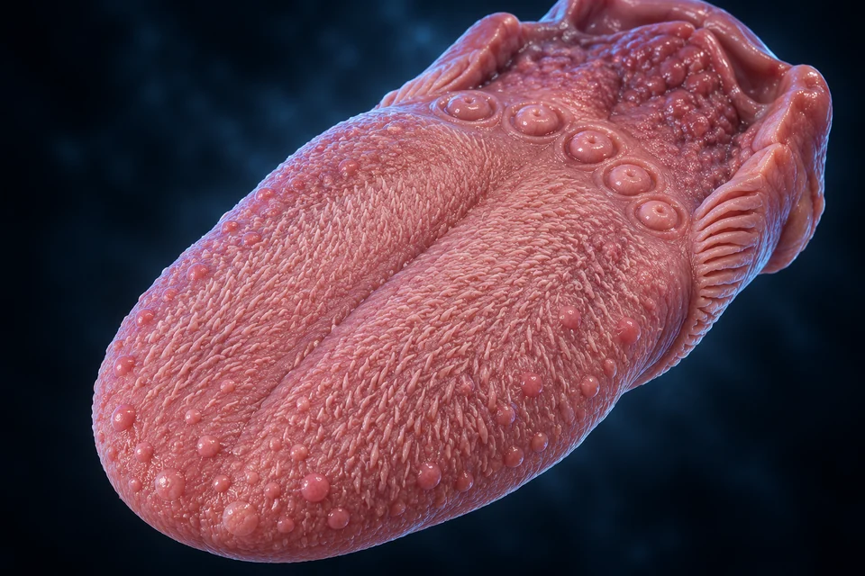
The muscular tongue has an epithelial surface with different projections. Some provide taste-bearing epithelial contexts; numerous filiform projections serve mechanical roles and lack taste buds.
Teaching references: NCBI · Tongue taste-bud organisation · University of Leeds · Tongue tissue relationships
11 / PAPILLA-COMPARISON
Gross projections are much larger than individual taste cells. Their distribution and prominence vary; no coloured region of the tongue exclusively owns a basic taste quality.
Scattered anterior surface
Rounded projections can contain taste buds.
Near the back of the oral tongue
Trench-wall epithelium contains buds; serous glands clear the clefts.
Posterolateral folds
Another taste-bearing epithelial setting.
Human prominence varies.
Much of the dorsal oral surface
Texture supports mechanical handling.
No taste buds.
Teaching references: NCBI · Tongue taste-bud organisation · University of Leeds · Tongue tissue relationships · NIDCD / NIH · Normal taste, flavour and non-zonal tongue
12 / TASTE-BUD
A taste bud is an epithelial unit with an apical pore and neural contacts near its base. Its specialised cells communicate with afferents; they do not each extend an axon to the brain.
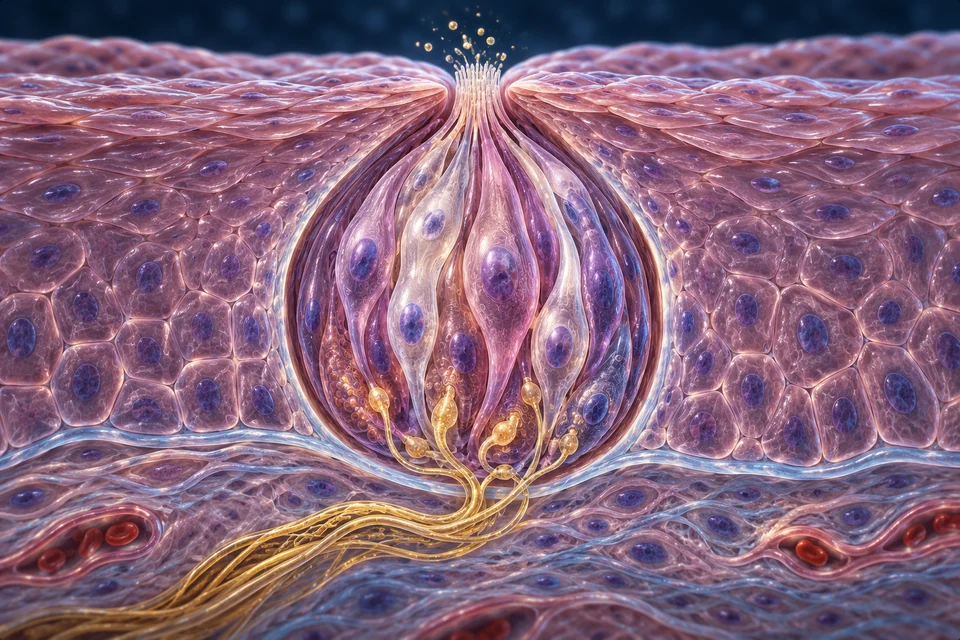
A papilla is a larger surface structure with its own epithelial and connective setting. A taste bud is a cluster of specialised epithelial cells located within an appropriate epithelial region. One papilla is therefore not one receptor cell, and a visible bump cannot be counted as a taste bud without the relevant tissue evidence.
The enlarged bud preparation places apical processes beside an opening and separate afferent contacts toward the base. The surrounding epithelium, basal boundary and deeper connective tissue answer different structural questions. Their positions help explain how surface chemicals and neural information meet without turning the epithelial cell into a sensory axon.
The local illustration is not a human biopsy, a fixed cell inventory or an identification of molecular subtypes by colour.
Source links: papillae · tongueTissue · tasteCells · histology
Teaching references: NCBI · Tongue taste-bud organisation · PubMed · Taste cells, signals and synapses · University of Leeds · Tongue tissue relationships
13 / CELL-JOBS
Taste buds contain receptor, supporting and renewing populations. Molecularly identified cell groups help explain transduction, but cell colour in an illustration cannot establish identity.
Sweet / bitter / umami signalling
Different receptor families can use related downstream signalling.
Not every taste mechanism is the same.
Sour-related signalling
Ion-sensitive responses and conventional synaptic specialisations differ from type-II channel signalling.
Fine generated cells are not assigned these types.
Local epithelial unit
Support and replacement help maintain the receptor environment.
This is not a fixed cell-count diagram.
Teaching references: PubMed · Taste cells, signals and synapses · PubMed · Taste transduction and channel synapses
14 / SALIVA-AND-CLEFTS
Saliva helps chemicals reach taste-bearing epithelial openings. Local serous secretions also clear papillary trenches. Oral fluid, mechanical food handling and chemical sensing cooperate without being the same process.
Teaching references: University of Leeds · Tongue tissue relationships · NIDCD / NIH · Normal taste, flavour and non-zonal tongue
15 / TASTE-QUALITIES
Sweet, sour, salty, bitter and umami are useful basic taste categories. They are not a map of separate exclusive territories, and a food commonly combines qualities rather than activating one category alone.
Selected receptor-cell signalling
Associated with sugars and other sweet stimuli.
Not a reliable measure of nutrient value.
Selected receptor-cell signalling
A family of bitter-sensitive receptors contributes.
Not a universal test of toxicity.
Amino-acid-related input
Glutamate contributes to savoury taste.
Flavour remains multisensory.
Acid-related input
Protons affect selected cellular signalling.
Temperature and irritation are distinct inputs.
Ionic input
Salt-related mechanisms depend on stimulus and context.
Do not equate every human salt response with one rodent channel.
The familiar qualities sweet, sour, salty, bitter and umami are not separate exclusive territories painted onto the tongue. Taste-bearing sites contain different cellular populations, and a food can combine qualities. Local differences do not justify a five-zone map.
Surface texture and tissue organisation answer another question. A papillary form can contribute mechanical or sensory context without representing one entire taste quality. Sweetness is not a measure of nutritional value, and bitterness is not a reliable toxicity test.
Source links: taste · papillae · tasteCells
Teaching references: NIDCD / NIH · Normal taste, flavour and non-zonal tongue · PubMed · Taste cells, signals and synapses
16 / TASTE-TRANSDUCTION
Sweet, bitter and umami include G-protein-coupled receptor pathways. Sour involves proton-sensitive cellular mechanisms. Transmitter release communicates with afferents; a simplified route should not hide these differences.
Structure and function · select a teaching emphasis; all basic explanations stay visible.
Both receptor-mediated and ion-sensitive examples concern epithelial cells; the nerve is separate. Generated colours do not assign a pictured cell to type II or III. The same broad cell region is retained for both chemistry examples, with basal nerve contact distinguished for communication.
The two left branches compare parallel inputs or alternatives. Their convergence groups a shared destination; one branch does not turn into the other.
Sweet / bitter / umami
Different GPCRs can recruit related downstream pathways.
Selected research principles; not identical receptors.
Sour / proton sensing
OTOP1-related mechanisms contribute to acid detection.
Not every salt mechanism is extrapolated from mice to humans.
Taste cell → transmitter → nerve
Type-II channel signalling and type-III conventional contacts differ.
Neither diagram assigns a generated cell subtype by colour.
OTOP1 and channel-synapse relationships come from molecular research. The supporting diagram shows selected principles, not a complete human salt model or every intermediate protein.
Teaching references: PubMed · Taste transduction and channel synapses · PubMed · Sour receptors, cells and circuits · PubMed · Taste cells, signals and synapses
17 / CRANIAL-DELIVERY
Taste afferents travel in selected facial, glossopharyngeal and vagal components. General oral sensation also involves trigeminal pathways; tongue movement uses motor supply. One tongue therefore has several neural jobs.
Selected anterior / palatal taste routes
The chorda tympani contributes anterior-tongue taste.
General tongue sensation is not all facial-nerve taste.
Posterior and circumvallate taste contexts
Supplies selected taste-bearing sites toward the back.
Circumvallate position does not imply facial supply.
Selected epiglottic / lower pharyngeal sites
Carries a smaller taste contribution from these regions.
The taste buds are not limited to the tongue.
Taste information, general sensation and movement are distinct tasks. Selected facial, glossopharyngeal and vagal components carry gustatory input; general oral touch and temperature also involve other sensory routes, while moving the tongue requires motor supply.
The chorda tympani contributes anterior-tongue taste, whereas circumvallate taste sites belong to glossopharyngeal contexts. Site position alone should not be converted into a simple left/right or front/back nerve-colour rule. Beyond the periphery, brainstem and ascending processing connect with insular, opercular and wider networks.
The chapter supplies a route to those roles, not verified individual fibres, a complete crossing diagram or a clinical cranial-nerve test.
Source links: papillae · tasteRoute · tasteNetwork
Teaching references: NCBI · Tongue taste-bud organisation · NCBI · Neural taste pathway
18 / CENTRAL-TASTE
Afferent information reaches the rostral solitary nucleus and ascending pathways involving thalamic and cortical processing. Gustatory insular and opercular regions contribute; integration extends beyond one isolated taste centre.
Structure and function · select a teaching emphasis; all basic explanations stay visible.
The basal contacts locate peripheral delivery. Brainstem, thalamic and cortical relays described below are onward processing stages, not structures drawn inside this taste bud. Continue to brain anatomy →
Return to the annotated anatomical figure →Selected VII / IX / X afferents
Taste-bud output enters distinct sensory components.
This is not tongue motor or general tactile supply.
Rostral solitary nucleus / ascending relays
Information reaches brainstem and thalamic processing routes.
Not a complete relay count or crossing map.
Insular / opercular and connected networks
Sensory interpretation connects with wider processing.
No single point contains all flavour or memory.
Educational relationship map, not a precise fibre trajectory, complete set of relays or universal crossing diagram. Human and experimental-animal routes are not treated as identical.
Teaching references: NCBI · Neural taste pathway · NLM · Central taste organisation
19 / FLAVOUR
Taste contributes chemical qualities. Retronasal aroma, texture, temperature and chemesthesis add other information. Together with context, they help distinguish foods that share a taste quality.
Structure and function · select a teaching emphasis; all basic explanations stay visible.
Oral/pharyngeal access lets volatile aromas contribute to smell. Taste, texture, temperature and chemical irritation add different information; the anatomy does not assign a fixed percentage to each part of flavour.
Return to the annotated anatomical figure →The two left branches compare parallel inputs or alternatives. Their convergence groups a shared destination; one branch does not turn into the other.
Taste + retronasal aroma
Two chemical interfaces contribute different input.
Sweetness alone cannot identify every food.
Texture + temperature + chemesthesis
Separate nerve endings contribute other sensations.
Mint coolness and chilli irritation are not tongue taste zones.
Input + context → flavour
The combined perception distinguishes complex food experiences.
Not a fixed numerical mixture or consumer taste test.
Taste qualities and retronasal smell contribute different chemical information. Texture, temperature and chemesthesis add other inputs; mint coolness and chilli irritation are not extra tongue taste zones. Their integration helps make a food experience recognisable.
This does not assign a universal percentage to smell or taste, and the static pictures cannot measure a person’s perception. Follow the nasal access and epithelial-cell lessons to keep chemical delivery, sensory signalling and interpretation distinct.
Source links: taste · smell · tasteCells
Teaching references: NIDCD / NIH · Normal taste, flavour and non-zonal tongue · NIDCD / NIH · Normal smell and two delivery routes
20 / CHEMESTHESIS
Menthol coolness and chilli-related irritation can involve somatosensory nerve endings. They differ from taste-bud detection even when experienced in the same mouth.
Taste epithelial cells
Chemical qualities reach gustatory afferents.
Olfactory sensory neurons
Volatile chemicals contribute through the nose.
Temperature / texture / irritation
Other nerve endings contribute distinct signals.
No at-home provocation or diagnostic test is proposed.
Teaching references: NIDCD / NIH · Normal taste, flavour and non-zonal tongue · NIDCD / NIH · Normal smell and two delivery routes
21 / RENEWAL
Olfactory and taste receptor populations can be renewed. Progenitors, differentiation and the local environment support normal maintenance; replacement does not imply universal recovery after disease or injury.
Structure and function · select a teaching emphasis; all basic explanations stay visible.
The epithelial unit is a structural context, not a measured lineage or regeneration sequence. Smell and taste maintenance involve different populations; a basal-looking generated cell is not molecularly identified as a progenitor.
Return to the annotated anatomical figure →Local basal / epithelial context
Renewal begins with appropriate local precursor populations.
Not every pictured basal cell is molecularly identified.
Specialised sensory / supporting roles
New cells acquire functions within their tissue environment.
No universal replacement time or identical lineage is asserted.
Ongoing interface function
Renewal and local support contribute to normal maintenance.
Not a promise of recovery, therapy or clinical prognosis.
Representative sequence only. No fixed replacement interval, treatment claim, patient prognosis or identical developmental lineage is asserted.
A functioning sensory interface depends on local support, appropriate precursor populations and differentiation. These processes operate within distinct olfactory and taste epithelial environments; their cellular lineages should not be treated as one identical mechanism.
A generated basal-looking cell does not establish a molecular identity, fixed replacement time or capacity to restore a damaged sense. This normal-maintenance lesson therefore preserves renewal as a biological process while making no promise of recovery or treatment.
Source links: renewal · olfactoryCells · tasteCells
Teaching references: NIDCD / NIH · Taste and smell research programme · PubMed · Human olfactory-mucosa ultrastructure study · PubMed · Taste cells, signals and synapses
22 / COMPARE-RECEPTORS
The olfactory receptor is a bipolar neuron. A taste receptor cell is specialised epithelium communicating with a separate afferent. Both are chemical interfaces, but their output anatomy cannot be swapped.
Apical cilia → neuronal soma → basal axon
One receptor neuron carries its own output toward bulb circuits.
Apical microvilli → epithelial cell → afferent contact
The nerve belongs to a different sensory neuron.
Access / transduction / neural delivery
Separate the physical stimulus from the signal and the perception.
An illustrated arrow is not a measured recording.
Teaching references: NLM · Olfactory epithelium and sensory neurons · PubMed · Taste cells, signals and synapses
23 / OBSERVATION
A gross tongue view cannot establish taste-cell identity. A tissue section depends on plane and sampling; a named specimen record does not guarantee that a taste bud is visible.
MHS266 · H&E · record checked
The provider explicitly says no taste buds can be seen in this specimen.
Pixels not acquired, copied or presented as verified taste-bud evidence.
Published TEM description · abstract-level access
Supports broad bipolar-receptor and supporting-cell organisation.
Full-paper images not acquired or licensed for reuse.
Original AI-assisted teaching illustrations
Broad source-supported relationships are labelled separately.
Fine texture, counts, subtypes and complete neural connections remain illustrative.
Teaching references: Histology Guide · MHS266 tongue specimen record · PubMed · Human olfactory-mucosa ultrastructure study · University of Leeds · Tongue tissue relationships
25 / SCOPE
This chapter teaches sensory access, regional anatomy, tissue organisation, cellular principles, delivery and integration. It is not a complete histological or cranial-nerve atlas, diagnostic tool or treatment chapter.
Precise receptor maps, licensed high-resolution human specimen pixels, full olfactory/gustatory fibre geometry, clinical imaging and disease comparisons remain separate staged work. Human medical review and current physical-phone acceptance have not been performed.
The private reference book informs scope, depth and visual teaching method only. Its prose, artwork and page layout are not copied.
Teaching references: NIDCD / NIH · Normal smell and two delivery routes · NIDCD / NIH · Normal taste, flavour and non-zonal tongue
EDITORIAL RECORD
Normal smell/taste access, regional anatomy, epithelial organisation, selected transduction principles and neural delivery. Current references are checked critically; historical numerical and oversimplified claims are not adopted.
AI-assisted original teaching content and five retained detailed text-free anatomical illustrations. Visible labelled broad relationships are source-supported at the stated scope. Generated fine cellular detail, cell counts, subtype colours, ciliary dimensions, precise filaments, synapses and vascular branches are illustrative / not independently verified. No specimen microscopy, physician approval or human medical review is claimed. Human medical review: not performed. Educational only.
Revision: smell-taste-eyes-context-2026-10-19-v1. Stable local key: ahb_ch_smell_taste_english_v1. No new CMS entity or publication state. Anatomy concept keys are separate from representation files; the chapter key is a local stable content identity, not a fabricated CMS entity. Text-free masters and editable English labels remain separate. New physical-phone acceptance: not performed.