Chapter overview film planned. No playable video is currently available.
Original intestinal exhibit · fine detail illustrative
01 / ORGAN AND NEIGHBORS
Three regions, one continuous route.
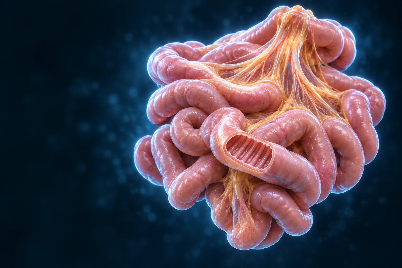
The small intestine links the stomach to the large bowel. Its loops occupy much of the central and lower abdomen. Their position changes; the regions are not three sealed containers.
New Golden-family expanded educational arrangement, not a surgical map. Person right is image left. Loop positions vary; hidden duodenal roots, regional loop identities and fine vessels remain illustrative.Structure notes · further context
Stomach
Its outlet leads into the duodenum; this overlapping view does not expose the entire connection.
Small-intestine loops
Duodenum, jejunum and ileum are continuous regions. Do not identify an individual overlapping loop by its color or this layout alone.
Mesentery
The mesentery helps suspend jejunal and ileal loops. This spread-out preparation is not the normal compact arrangement of all abdominal contents.
Colon
Its water-handling and microbial functions belong to a separate later lesson; it is not an extra small-intestine loop.
Cecum
The ileocecal relationship is shown broadly. Hidden junctions and individual vascular variants are not independently verified.
Duodenum
Receives gastric contents
Pancreatic enzymes and bicarbonate join bile to support digestion. Brunner glands help provide protective alkaline mucus.
Jejunum
Prominent folds and absorptive surface
A major absorptive region, not the exclusive site for every nutrient. Its wall and villi support contact with contents.
Ileum
Continues toward the cecum
Distal regions specialize in bile-acid recycling and intrinsic-factor–B12 uptake. Lymphoid tissue contributes to mucosal immune surveillance.
Source-supported route: stomach → duodenum → jejunum → ileum → cecum. This supporting order explains connections hidden by overlapping loops; it does not assign an exact region to every loop in the main image.
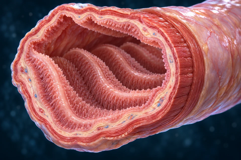
Three scales help create a useful absorption surface. Circular folds include mucosa and submucosa. Villi are mucosal projections. Microvilli belong to individual epithelial cells.
New Golden-family a jejunal teaching preparation. Folds, villi and microvilli are different scales; generated fine fibers and villus counts are illustrative, not quantitative microscopy.Structure notes · further context
Circular folds
They include mucosa and a submucosal core. They are not simply giant individual villi.
Villous surface
Villi contain connective tissue, blood vessels and lymphatic channels. Their form varies by region.
Lumen
Mixing keeps contents in contact with the surface. The lumen is separate from blood and lymphatic spaces.
Muscular wall
The muscularis externa has circular and longitudinal arrangements; the fine generated fiber pattern is not a measured specimen.
Outer covering
Many intraperitoneal segments have a serosa; some duodenal surfaces are attached rather than free. This segment is not every region.
Wall around the lumen
Mucosa faces the contents, followed by supporting submucosa, muscularis externa and an outer covering. Circular and longitudinal smooth-muscle arrangements mix and propel contents; local enteric nerve networks help coordinate them.
Regional differences matter
Duodenal submucosal glands, prominent jejunal folds and ileal lymphoid tissue provide useful distinctions. One opened segment cannot represent every regional microscopic feature.
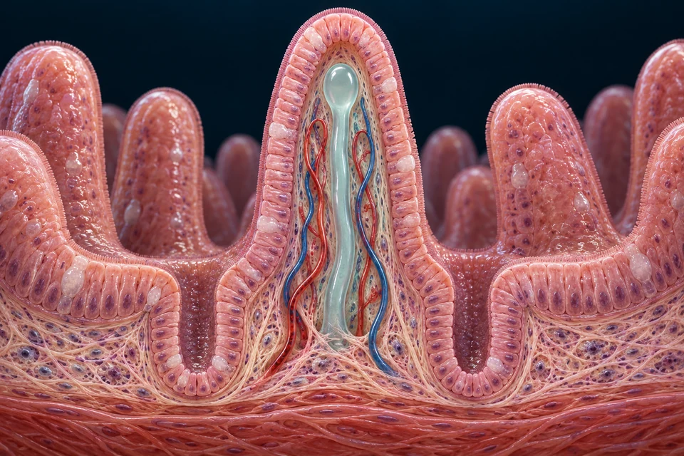
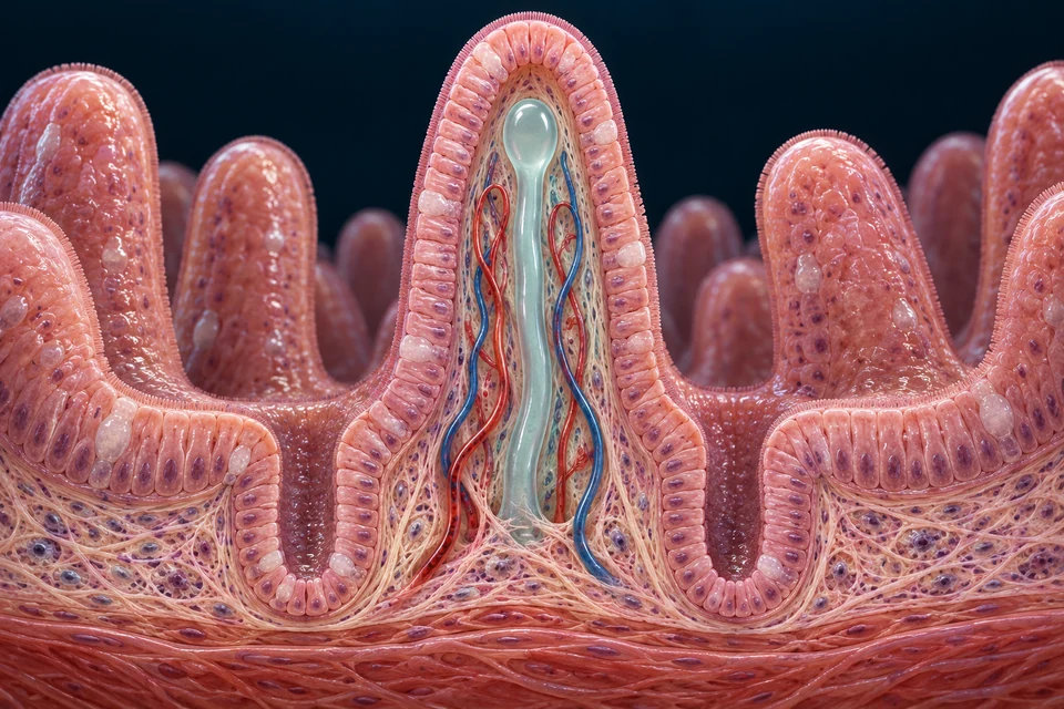
A villus points toward the digestive lumen. Crypts extend downward between villi. Inside a villus, connective tissue supports separate blood vessels and a lymphatic lacteal.
Different nutrients, different carriers and exits.
All three routes are visible. Choose one to focus the connection; selection does not reveal hidden basic information.
Across the epithelium, then into bloodSugars and protein products cross epithelial cells before reaching a blood supply within the villus. The onward portal route goes to the liver.
New Golden-family source-supported tissue relationships: lumen-facing villus, downward crypts and separate blood/lymph spaces. Individual generated cells, vascular branches and crypt details remain illustrative. The enlarged closed lacteal and vascular paths are selected teaching representations, not measured vessel diameters or an exact network.Structure notes · further context
Villus
Its core is supporting connective tissue, not an open digestive pipe.
Epithelial lining
Absorptive cells have apical microvilli. Junctions regulate passage between neighboring cells; the barrier is selective, not impermeable.
Lamina propria
It also contains immune and other supporting cells. A generated nucleus cannot establish a particular cell identity.
Blood vessels
The capillary network connects with larger vessels. Exact branch paths and red/blue vessel identities are illustrative.
Lacteal
Its tip is closed. It does not open into the lumen, and it does not carry every kind of absorbed nutrient.
Intestinal crypt
Crypts lie between villi. Stem-cell renewal and Paneth-cell roles are explained separately rather than guessed from generated cell colors.
Highlights identify anatomical compartments, not a measured route through cells or a specific transporter molecule. Blood and lymph remain separate from the digestive lumen.
Entry and processing. Glucose/galactose: apical SGLT1; fructose: apical GLUT5. Usual basolateral exit includes GLUT2.
Enterocyte → blood → portal circulation → liver
Protein products
Digestion. Protein → peptides → amino acids and small peptides
Entry and processing. Several carriers take up amino acids. PEPT1 takes up di- and tripeptides with a proton gradient; intracellular enzymes release amino acids.
Entry and processing. Products enter the cell; much long-chain lipid is reassembled and packaged into chylomicrons. The micelle itself is not swallowed as an intact package.
Educational route map, not measured transport anatomy. Many shorter-chain fatty acids can enter portal blood; the chylomicron route describes much long-chain dietary fat. It does not apply to every absorbed lipid.
Blood and lymph stay separate
The digestive lumen is outside the internal tissue spaces. Nutrients cross the epithelium before entering their onward routes; there is no open tube from food directly into a vessel.
A renewing surface
Crypt stem cells generate epithelial cells. Many descendants differentiate and move toward the villus surface, where older cells are shed. Different cell populations have different renewal patterns; no universal exact clock is asserted.
Barrier maintenance, secretion, signaling, defense and renewal work alongside nutrient uptake. These roles are source-supported; generated cell colors do not establish cell identity.
Enterocytes
Absorption and brush-border processing
Villus epithelium
Transfer nutrients through specialized membranes and help complete surface digestion.
Goblet cells
Mucus
Intestinal epithelium
Contribute mucus that supports lubrication and the mucosal barrier.
Paneth cells
Antimicrobial products and niche support
Crypt base
Help support local defense and the stem-cell environment.
Enteroendocrine cells
Digestive and metabolic signals
Scattered epithelial cells
Different populations release signals that coordinate secretion, movement and nutrient responses.
Stem cells
Epithelial renewal
Crypt region
Generate new epithelial cells; differentiation and migration maintain the surface.
Selected roles, not a complete cell census. Immune cells and lymphoid tissue provide additional surveillance within and beneath the epithelium.
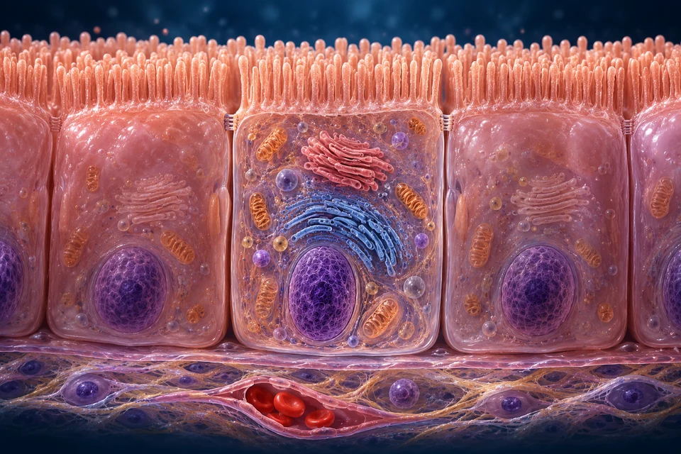
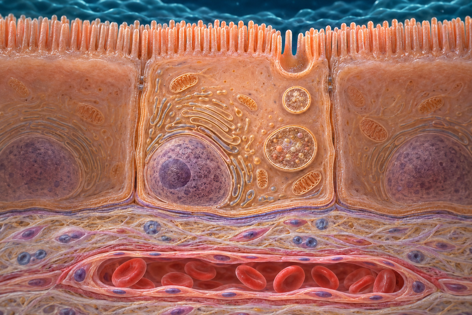
An enterocyte has an apical surface facing food and a basolateral surface facing internal tissue. The brush border combines surface area with enzymes and transport proteins.
A gradient connects two membranes.
The brush border faces the lumenSGLT1 couples glucose or galactose to sodium entry; GLUT5 handles fructose by facilitated diffusion. These protein names describe mechanisms at the apical side, not proteins identified in the generated texture.
New Golden-family the apical-to-basal orientation and brush-border relationship are educationally source-supported. Fine organelles, membranes and junction ultrastructure are illustrative / not independently verified. The small junctional marker is illustrative, not a verified molecular structure.Structure notes · further context
Brush border
These are projections of individual cells, not miniature whole villi. Membrane enzymes and transporters work at this surface.
Enterocyte
Its luminal and tissue-facing membranes have different transport roles. Organelle textures do not identify individual proteins.
Junctional region
This broad boundary is not a resolved molecular image of a tight junction. Both transcellular and regulated paracellular routes matter.
Basal side
Blood and lymph vessels lie beyond the epithelial cells in supporting tissue, not within the digestive lumen.
Nucleus
A typical absorptive epithelial cell has a basally positioned nucleus; this is an idealized educational view, not diagnostic histology.
Highlights identify anatomical compartments, not a measured route through cells or a specific transporter molecule. Blood and lymph remain separate from the digestive lumen.
Lumen → apical entry
Glucose / galactose → SGLT1 + Na⁺
Fructose → GLUT5
The luminal surface faces the digestive contents.
→
Enterocyte
Separate entry, processing and exit roles connect the two sides of the cell.
→
Basolateral side → internal tissue
Usual sugar exit: GLUT2
Separate gradient support: Na⁺/K⁺-ATPase + ATP
Transported sugars can enter the blood route beyond the epithelium.
Entry uses different mechanisms
SGLT1 couples glucose or galactose uptake to sodium moving down its gradient. GLUT5 transports fructose by facilitated diffusion. SGLT1 does not directly consume ATP as a pump.
The pump is on the other side
Basolateral Na⁺/K⁺-ATPase uses ATP to maintain the sodium gradient. Sugars ordinarily leave through tissue-facing transport pathways including GLUT2. Transporter regulation is more complex than this basic map.
Supporting membrane-route diagram; not a protein-scale image. A transporter name is a mechanism label, not an identification of a protein in the generated cell artwork.
Selective barrier
Junctions help regulate movement between cells. Substances can move through cells or, under regulated conditions, between them. A healthy epithelium is selective, not an impenetrable sheet.
What the artwork can show
The broad orientation, brush border and basal nuclear position are educational relationships. The fine generated organelles are illustrative; they do not reveal measured transporter positions or molecular interactions.
Deeper learning · when the absorptive surface is injured
The normal surface couples a selective epithelial barrier to digestion and transport. In celiac disease, a gluten-triggered immune disorder damages the small intestine and can impair nutrient absorption. This connects tissue injury with function; it does not mean that every digestive symptom has the same cause.
The normal preparations above do not show a diagnosed celiac biopsy. Recognizing an illness requires evidence beyond a teaching illustration; diagnosis and treatment are outside this lesson.
Pancreatic enzymes work in the lumen, while intestinal surface enzymes complete important steps at the brush border. Bicarbonate helps create suitable conditions after acidic contents arrive from the stomach.
FROM FOOD CHEMISTRY TO INTERNAL DELIVERY
Break the bonds. Cross the lining. Reach the blood.
Read the surface first: digestive lumen above, an absorptive cell in the lining, tissue and vessels beneath. Then follow the different chemical jobs along that route.
New Golden-family the apical-to-basal orientation and brush-border relationship are educationally source-supported. Fine organelles, membranes and junction ultrastructure are illustrative / not independently verified. The small junctional marker is illustrative, not a verified molecular structure. Selected lesson: Starch. Digestion reaches the exposed brush border. The upper surface faces the digestive lumen. Pancreatic amylase acts in that lumen; membrane-associated disaccharidases finish selected reactions at the microvillar surface. No specific enzyme molecule is visible in this illustration. Glucose uses apical SGLT1 with Na+. Gold outlines locate a compartment; blue arrows show a qualitative cross-boundary direction, not a measured flux or identified molecular pore.Structure notes · further context
Brush border
These are projections of individual cells, not miniature whole villi. Membrane enzymes and transporters work at this surface.
Enterocyte
Its luminal and tissue-facing membranes have different transport roles. Organelle textures do not identify individual proteins.
Junctional region
This broad boundary is not a resolved molecular image of a tight junction. Both transcellular and regulated paracellular routes matter.
Basal side
Blood and lymph vessels lie beyond the epithelial cells in supporting tissue, not within the digestive lumen.
Nucleus
A typical absorptive epithelial cell has a basally positioned nucleus; this is an idealized educational view, not diagnostic histology.
Digestion reaches the exposed brush border.
The upper surface faces the digestive lumen. Pancreatic amylase acts in that lumen; membrane-associated disaccharidases finish selected reactions at the microvillar surface. No specific enzyme molecule is visible in this illustration.
Original generated atlas, not microscopy. The same text-free images and anatomical endpoints are retained. Names and teaching cues are separate layers. Fine generated membrane proteins and vascular routes are not certified.
Digestive lumen → surfaceEnzymes make smaller sugar substrates.
Apical membrane → cellCarriers transfer selected monosaccharides.
Tissue side → bloodInternal delivery connects to the portal route.
Bond cleavagePancreatic alpha-amylase → smaller fragments; SI and MGAM at the brush border complete glucose release.
Absorbable productsGlucose
Apical entryGlucose uses apical SGLT1 with Na+.
A branched starch fragment is not a substrate that SGLT1 can carry intact. Hydrolysis changes chemical form before the usual monosaccharide entry route.
Chemical relationships beside the anatomy, not a meal simulation, measured transport speed or dietary recommendation.
Amylase opens chains; the surface finishes the job.
The chemical work is shared between soluble enzymes and the intestinal surface.
Starch contains glucose chains linked mainly through alpha-1,4 bonds, with alpha-1,6 branch points in amylopectin. Salivary and pancreatic alpha-amylases cut internal alpha-1,4 linkages. Their products include short chains and branched limit dextrins, rather than a complete stream of free glucose.
At the brush border, maltase-glucoamylase (MGAM) and sucrase-isomaltase (SI) provide overlapping alpha-glucosidase activities. Isomaltase helps handle alpha-1,6 linkages. It is misleading to assign all starch fragments to one exclusive enzyme or to depict pancreatic amylase as cutting every branch.
The reaction cleaves glycosidic bonds by hydrolysis; it does not pump the resulting sugar through the membrane. Conversely, a sugar carrier cannot make an intact starch fragment absorbable simply by binding it.
Why not predict a digestion speed from the enzyme name?
Purified enzyme experiments separate catalytic capabilities under chosen conditions. Meal structure, processing, accessible surface, mixing and transit also affect digestion in an intestine. Recombinant mammalian enzyme assays support overlapping activities, but their rates cannot be copied into a universal human meal animation.
Different starting sugars lead to different products.
Location and substrate identity matter as much as the word “digestion”.
Lactase acts at the intestinal brush border, yielding glucose and galactose from lactose. Sucrase yields glucose and fructose from sucrose. These products enter different combinations of carriers, so bond cleavage and epithelial entry should be followed as two connected questions.
A free monosaccharide does not first need to become a disaccharide. It can join the appropriate uptake route without that hydrolysis step. The substrate comparison shows normal biochemical relationships, not a ranking of foods or a prediction of blood glucose.
A shared surface, not one universal enzyme
Microvilli expand the apical surface bearing several digestive proteins and carriers. Their proximity helps connect terminal digestion with uptake, but they remain different proteins with different functions. The cell image locates the surface; it does not resolve enzyme complexes within its generated membrane texture.
Same epithelium; different transport relationships.
Glucose and fructose need not cross the apical membrane by the same mechanism.
SGLT1 couples glucose or galactose entry to Na+. GLUT5 facilitates fructose movement without that Na+ cotransport step. The separate basolateral Na+/K+-ATPase spends ATP to support ion gradients; neither lactase nor GLUT5 is an ATP-powered sugar pump.
The usual teaching route puts GLUT2 at the tissue-facing membrane. Transporter abundance and localisation can change with conditions, and findings combine different species and methods. This course therefore does not claim that one fixed arrangement predicts all sugar transfer in every human meal.
What established the fructose preference?
Experiments expressing human GLUT5 in Xenopus oocytes tested transport function independently of its name or appearance. This supports fructose selectivity, but a frog-oocyte assay is not a direct measurement of how much a person absorbs. The original study is linked separately from the broader physiological review.
An epithelial exit, tissue transfer and vascular transport are separate parts of delivery.
The tissue-facing side meets the villus interior. After epithelial transfer, soluble products encounter interstitial and vascular interfaces before venous drainage carries them toward the liver. This differs from long-chain lipid packaging and the initial lymphatic route through a lacteal.
The intestinal wall also uses and transforms nutrients. Mouse isotope-tracing experiments found substantial intestinal conversion of fructose under the tested conditions. That demonstrates a metabolic role, but it does not establish a universal human clearance percentage, safe dose or nutritional recommendation.
Portal carbon need not have the same chemical name as the meal
A labelled carbon atom can be followed into glucose, lactate or other metabolites. Detecting such products is different from showing that all fructose passes unchanged to the liver. Experimental species, dose, feeding state and analytical method constrain the interpretation.
Not every carbohydrate becomes a small-intestinal sugar.
Chemical bonds and physical access limit what human digestive enzymes can do.
Humans do not make cellulase to digest cellulose into glucose. Other carbohydrate may also escape small-intestinal digestion because of substrate structure or access. Material reaching the colon is therefore not all equivalent to sugar that should already have entered an enterocyte.
Do not generalise this to “humans cannot split any beta-linked sugar”: lactase hydrolyses lactose. Nor does one normal pathway diagnose a person with enzyme deficiency or malabsorption. The existing large-intestinal course follows onward microbial processing and remaining absorption.
Preserve the distinction between an enzyme and a clinical conclusion
A teaching comparison can identify what a normal enzyme acts on. Individual symptoms, inherited variation and test interpretation require different evidence. No elimination diet, supplement, diagnostic threshold or treatment follows from these interactive states.
No. Terminal digestion produces the monosaccharide substrate; transport does not replace hydrolysis.
Does sucrose produce two sugars with the same apical carrier?
No. Its glucose can use SGLT1; its fructose uses GLUT5.
Is a lacteal the direct sugar route to the liver?
No. The usual monosaccharide route is associated with blood and portal drainage; the lacteal serves a different lipid pathway.
Does a mouse fructose experiment prescribe a human dietary dose?
No. It establishes findings under the tested animal conditions, not a universal human threshold.
How the explanation was checked
Directed normal-physiology source check:22 October2026. Existing FB118 architecture and FB126 nutrient themes guide coverage. Qualified villus and cell art, existing protein uptake and water/electrolyte courses remain intact; no source figure or book wording is reproduced.
This is AI-assisted educational checking, not physician review or individual medical advice. Mouse tracing, recombinant enzyme assays and a human transporter expressed in frog oocytes are identified as different evidence. No physical-phone, fine generated histology or new WebGL acceptance is claimed.
Carbohydrates
Pancreatic amylase breaks starch into smaller carbohydrates. Brush-border enzymes produce absorbable monosaccharides; starch does not pass intact through a sugar transporter.
Proteins
Pancreatic proteases and intestinal peptidases make smaller peptides and amino acids. Enteropeptidase activates trypsinogen; trypsin helps activate other protease precursors.
Fats
Bile salts help disperse fat and support micellar delivery of lipid digestion products. Pancreatic lipase helps break down triglycerides. Bile is not a digestive enzyme.
Sugars and amino acids largely reach portal blood. Much long-chain dietary fat is repackaged into chylomicrons that first enter lymph. These routes should not be collapsed into one “everything goes directly to the liver” arrow.
PEPT1 couples di- and tripeptide uptake to a proton gradient. Intracellular processing releases amino acids for onward transport. Protein digestion need not produce only free amino acids before any uptake can occur.
A micelle is not a chylomicron
Micelles help deliver lipid products to the luminal surface. Inside the enterocyte, long-chain lipids are reassembled and packaged into lipoprotein particles. Those chylomicrons enter lacteals and lymph before reaching systemic blood.
Break a bond. Cross a membrane. Join an onward route.
Follow protein products from the digestive lumen into an absorptive cell, then toward villus blood. Peptide cleavage, membrane transport and vascular delivery are different jobs.
New Golden-family the apical-to-basal orientation and brush-border relationship are educationally source-supported. Fine organelles, membranes and junction ultrastructure are illustrative / not independently verified. The small junctional marker is illustrative, not a verified molecular structure.Structure notes · further context
Brush border
These are projections of individual cells, not miniature whole villi. Membrane enzymes and transporters work at this surface.
Enterocyte
Its luminal and tissue-facing membranes have different transport roles. Organelle textures do not identify individual proteins.
Junctional region
This broad boundary is not a resolved molecular image of a tight junction. Both transcellular and regulated paracellular routes matter.
Basal side
Blood and lymph vessels lie beyond the epithelial cells in supporting tissue, not within the digestive lumen.
Nucleus
A typical absorptive epithelial cell has a basally positioned nucleus; this is an idealized educational view, not diagnostic histology.
Gold locates a compartment on unchanged anatomy. No enzyme or transporter molecule is identified from the generated texture; it is not a measured uptake rate or a traced vessel.
Free amino acids
Apical amino-acid carriers → intracellular amino-acid pool.
Di- and tripeptides
PEPT1 + H+ → cytosolic hydrolysis → amino-acid pool.
Both connect to tissue-side transport and predominantly portal blood. Ion-gradient support is separate from transporting the nutrient itself.
Enterocyte · two membrane faces · Cross the brush border
Free amino acids use several carrier systems. PEPT1 carries di- and tripeptides with H+ across the apical membrane. NHE3 helps maintain the proton-driving conditions; basolateral Na+/K+-ATPase separately uses ATP to support ion gradients. No transporter molecule is identified by this artwork.
Digestion is shared across the lumen, surface and cell.
Gastric acid helps unfold proteins and provides conditions for pepsin. In the small intestine, pancreatic proteases cut different peptide bonds, and surface peptidases continue the work. A mixture of free amino acids and small peptides reaches the absorptive surface.
The existing pancreatic lesson explains enteropeptidase/trypsin activation and why not every digestive enzyme follows that switch. Here the next question is how the resulting products cross an epithelium; this is distinct from producing digestive enzymes.
PEPT1 couples peptide entry to H+, not directly to ATP.
PEPT1, encoded by SLC15A1, is an apical proton-coupled transporter with broad recognition of di- and tripeptides. The electrochemical H+ gradient supplies the driving force. Large intact dietary proteins do not simply pass through this small-peptide route.
H+ coupling and peptide selectivity describe function, not a visible pore in the generated brush border. Different substrates and experimental conditions can give different transport behaviour, so the drawing assigns no universal H+-to-peptide ratio or transport speed.
A sodium-dependent support system can drive H+-coupled entry.
NHE3 exchanges intracellular H+ for luminal Na+ at the apical surface. This helps handle the acid load associated with proton-coupled uptake and maintain the local driving conditions. On the other side, Na+/K+-ATPase consumes ATP while moving Na+ outward and K+ inward.
PEPT1 does not have to carry Na+ as its cotransported ion for its overall physiological support to depend indirectly on sodium gradients. The brush-border microenvironment is also not a requirement for the entire small intestine to remain as acidic as the stomach.
Different amino-acid transporters recognise different substrate classes and use different coupling relationships. B0AT1, encoded by SLC6A19, is an important apical Na+-dependent neutral amino-acid transporter. Other systems help handle charged amino acids and additional substrates.
A neutral amino acid and a dipeptide are different transport substrates. The two entry routes can contribute to the same intracellular amino-acid pool after peptides are hydrolysed; neither route means all dietary protein must first take exactly one chemical form.
A peptide entering the cell can be broken down inside it.
Cytosolic peptidases hydrolyse many absorbed small peptides to free amino acids. This intracellular step complements luminal and brush-border digestion. The nutritional route therefore cannot be described as either “all bonds are cut before absorption” or “peptides all stay intact”.
The opened cell shows a cytoplasmic compartment. It does not identify a specific peptidase in a Golgi stack, and ordinary peptide uptake is not a requirement to package food peptides into secretory vesicles.
The tissue-side membrane uses a cooperating transporter network.
Basolateral amino-acid movement involves several transporters rather than one universal exit pump. Some support exchange of one amino acid for another; others contribute to facilitated movement. Their combined activity and concentration conditions help determine net delivery.
LAT2 with its 4F2hc partner is one basolateral neutral amino-acid exchange system. Localisation studies help establish sidedness, but the presence of an exchanger alone does not establish the direction or amount of whole-tissue amino-acid transfer.
After crossing the tissue-facing membrane, amino acids enter the interstitial compartment and can be carried away by villus blood. Venous drainage joins the portal system before the liver. The digestive lumen and blood remain separated by tissue interfaces.
The lacteal is not an alternative label for the blood capillary. Long-chain lipid packaging and lymphatic delivery have their own existing lesson; normal protein products should not be shown entering chylomicrons as a universal meal pathway.
Enterocytes use amino acids for their own proteins and metabolism. First-pass handling is particularly important for selected amino acids, including glutamate and glutamine. The molecules reaching portal blood need not reproduce the ingested mixture in unchanged proportions.
Metabolic findings vary with substrate, developmental state, species and study design. No fixed percentage is assigned to every human meal, and this explanation is not a protein-intake or supplement recommendation.
Functional transport evidence is different from a cell picture.
Expression of a human intestinal transporter cDNA in experimental cells established H+-dependent peptide transport. Later human-derived Caco-2 monolayer work connected peptide uptake with NHE3-supported driving conditions. These are controlled functional studies, not measurements taken from this generated cell.
A transformed-cell monolayer, a cloned transporter system and a living human intestine each have limits. The normal account combines several kinds of evidence while leaving exact transporter positions, counts, kinetics and fine generated junctions uncertified.
Does all protein digestion have to finish before anything enters the cell?
No. Free amino acids and small peptides can enter by different routes; many absorbed peptides are hydrolysed inside enterocytes.
Does PEPT1 directly spend ATP or carry Na+ with every peptide?
No. PEPT1 is H+-coupled. NHE3 and the separate Na+/K+-ATPase help support the ion-driving conditions indirectly.
Is LAT2 a universal one-way amino-acid pump?
No. It is part of a cooperating basolateral transport network and supports amino-acid exchange.
Does the lacteal carry every absorbed meal product straight to the liver?
No. Protein products largely use portal blood, whereas packaged long-chain lipid initially follows lymph.
Normal physiology, not nutritional dosing, an allergy test or a treatment guide. Sources checked 21 October 2026. Original generated anatomy remains distinct from acquired tissue images; fine generated proteins/junctions, physician review and physical-phone acceptance are not certified.
The small bowel absorbs much of the water supplied by drinks, food and digestive secretions. Water movement is coupled to osmotic gradients and ion transport; it is not a separate nutrient pump.
Move solute. Create a gradient. Let water respond.
Follow the epithelial boundary from tissue scale to a cell. Absorption and secretion use different coordinated membrane functions; neither requires a separate pump that carries water into or out of the intestine.
Solute entry helps establish conditions for water absorption.
Locate the continuous epithelial boundary before following material toward internal tissue. Nutrient and electrolyte absorption helps favour net water uptake. Local vascular exchange carries away absorbed fluid; the lacteal remains a distinct lymphatic structure, not the only exit for intestinal water.
New Golden-family source-supported tissue relationships: lumen-facing villus, downward crypts and separate blood/lymph spaces. Individual generated cells, vascular branches and crypt details remain illustrative. The enlarged closed lacteal and vascular paths are selected teaching representations, not measured vessel diameters or an exact network.Structure notes · further context
Villus
Its core is supporting connective tissue, not an open digestive pipe.
Epithelial lining
Absorptive cells have apical microvilli. Junctions regulate passage between neighboring cells; the barrier is selective, not impermeable.
Lamina propria
It also contains immune and other supporting cells. A generated nucleus cannot establish a particular cell identity.
Blood vessels
The capillary network connects with larger vessels. Exact branch paths and red/blue vessel identities are illustrative.
Lacteal
Its tip is closed. It does not open into the lumen, and it does not carry every kind of absorbed nutrient.
Intestinal crypt
Crypts lie between villi. Stem-cell renewal and Paneth-cell roles are explained separately rather than guessed from generated cell colors.
Gold rings locate selected anatomical domains. In the cell view, cyan shows a local absorptive direction for sodium/nutrient or salt contexts, and the opposite anion-secretory direction for the secretion context. Counter-ion exchange is explained below. These cues are not one continuous carrier, measured water flow or identified molecular channels. The retained cell reference establishes orientation; it is not a recorded time series or a transporter-expression map.
1
Luminal membrane
Na⁺ + glucose → cell
SGLT1 couples nutrient entry to sodium movement.
2
Tissue-facing membrane
Na⁺ → tissue; glucose → tissue
The sodium pump and sugar exit use different proteins.
3
Across the epithelium
Water follows the resulting conditions
Solute transport supports water movement; these routes do not depict a separate ATP-driven water pump.
How transport, permeability and regulation work together.
Solute transport and water movement are closely linked
The intestine receives fluid from ingested material and from digestive secretions. Absorption retrieves much of this fluid as contents move along the bowel. Solute transport changes the conditions on either side of the epithelial barrier, creating local osmotic driving forces for water movement.
Water can cross cells and regulated routes between them. Experimental work also shows water permeation through SGLT1. Osmotic coupling is a standard tissue-level explanation, while the interpretation of tightly coupled water movement at this transporter has been debated. The lesson assigns no universal fraction to the proposed mechanisms and does not turn a cell-model result into a whole-person fluid rate.
Nutrient uptake and fluid absorption can cooperate
SGLT1 couples sodium entry to glucose or galactose uptake at the apical membrane. The sodium gradient is maintained by a different protein, Na⁺/K⁺-ATPase, on the tissue-facing side. Glucose exits by other transport machinery; the sodium pump does not transport the sugar molecule.
Together with other absorptive processes, this transfers osmotically active material toward internal tissue and supports water recovery. It is a normal physiological relationship, not a recipe or instruction for replacing fluid. The existing membrane lesson distinguishes fructose uptake, sugar exit and the separate gradient-maintaining pump in more detail.
NHE3, encoded by SLC9A3, exchanges sodium and hydrogen ions across the apical membrane. It contributes to sodium absorption and to the control of intracellular pH. It does not carry chloride in the same transport step, and its dependence on a sodium gradient does not make it an ATP-driven sodium pump.
Human small-intestinal enteroids have measurable NHE3-associated exchange activity. Changes in intracellular pH in these controlled preparations help identify exchanger function. They do not directly measure how many litres of fluid a whole person absorbs, and should not be converted into a universal post-meal transport rate.
Chloride/bicarbonate exchange is a partner, not the same carrier
At an apical anion exchanger, chloride can enter while bicarbonate leaves. Coordinating this with sodium/hydrogen exchange supports the classical electroneutral NaCl-absorbing function. The proton and bicarbonate exchanges also connect salt transport with local acid–base chemistry.
SLC26-family exchangers include DRA (SLC26A3) and PAT-1 (SLC26A6), with regional, species and experimental differences in their roles. Functional coupling with NHE3 was tested in gene-targeted mouse intestine; that result must not be relabelled as a precise transporter census in every human intestinal cell. The lesson therefore shows the paired function without assigning one universal regional combination or stoichiometric diagram.
Apical entry is only part of transepithelial absorption. The basolateral sodium pump removes sodium from the cell while importing potassium. Other pathways allow absorbed anions and nutrients to leave toward internal tissue. Potassium conductance contributes to the electrical conditions in which these systems operate.
This polarity matters: simply drawing every transporter on the luminal brush border loses the mechanism. The retained image locates apical and basal domains. Its organelles and junction-like details do not identify individual sodium pumps, exchanger complexes or channel numbers.
In a chloride-secretory context, basolateral NKCC1 cotransports sodium, potassium and chloride into the epithelial cell. It uses the sodium gradient sustained by Na⁺/K⁺-ATPase. This helps make chloride available for exit across the opposite, apical membrane.
Basolateral potassium channels and sodium pumping support this coordinated process. Human duodenal enteroid experiments show contributions from these transport systems, while also showing that chloride and bicarbonate handling are not exhausted by a single loading route. NKCC1 is not the apical chloride exit channel.
Regulated apical CFTR conductance supports chloride secretion in the selected mechanism. Channel opening permits anions to move according to their electrochemical driving force. ATP participates in channel gating; this does not make anion passage an ATP-powered pumping step. The active work maintaining the driving conditions is distributed across the epithelial system, not a pump physically carrying water through the cell.
CFTR-related intestinal secretion also involves bicarbonate, and regional anion exchange and bicarbonate supply contribute. The simplified chloride route is one explanatory view of a wider process. It neither identifies a visible CFTR molecule in the illustration nor establishes that every cell uses the same balance of anions.
Between cells is a selective route, not an open tear
Tight-junction organisation helps determine which substances can pass between epithelial neighbours. During electrogenic anion secretion, the electrical conditions can favour accompanying sodium movement through the paracellular route; water responds to the resulting osmotic conditions. Transcellular and paracellular transport work together at tissue level.
A regulated route between cells is different from a broken epithelial barrier. The small junctional feature in the retained artwork is only a broad positional cue. Its appearance does not establish molecular pore structure, selectivity, electrical resistance or the share of water taking each route.
Crypt-associated secretion and villus-associated absorption are useful introductory tendencies. They are not absolute assignments. Experiments in human intestinal enteroids have found absorptive exchanger function and stimulated anion secretion across different states of differentiation.
A separate transporter-distribution study supplies experimental anatomical context, but species and preparation matter. The villus-and-crypt illustration is used to orient the surface and supporting tissue, not to colour-code every cell as exclusively absorptive or secretory. Neither a pink cell nor its position proves a particular transporter profile.
Neural and local hormonal inputs can change epithelial transport through intracellular signalling. Cyclic nucleotides such as cAMP can favour secretory conductance while reducing selected absorptive processes. The resulting fluid response depends on how multiple membrane functions change together, not on one universal “water valve”.
Human epithelial models support interactions between enteroendocrine signalling and ion transport. These experiments help separate cellular mechanisms from the intact organism, where nerves, blood flow, luminal contents and other tissues also contribute. The selector compares functional contexts; it does not claim that every cell switches in synchrony.
Material can move in both directions across the intestine while the overall result is net absorption. Secretions help maintain a hydrated environment for digestion and movement; absorption returns fluid and solutes to internal tissue. A net result is the difference between opposing movements, not proof that one movement has stopped.
Supporting tissue and circulation remove absorbed material and help maintain the gradients on which epithelial exchange depends. The blood and lymphatic channels in the tissue view remain separate. This lesson gives no fixed daily fluid total, individual balance target or test for disease; the existing kidney and colonic lessons continue wider body-fluid handling.
A current trace, a swelling assay and a tissue image answer different questions
Human enteroid studies have used intracellular pH recovery, epithelial electrical measurements and fluid-related swelling to test aspects of transport. Each assay has defined conditions and limitations. A change in current alone is not a direct measurement of whole-person water absorption, and different anions may contribute to an experimental signal.
The two retained atlas preparations are original generated illustrations, not the experimental specimens. Their broad structure and independent annotations provide orientation for source-supported physiology. Fine organelles, junctions, protein localisation, real physical-phone usability and physician review remain separate unverified areas.
Does passive water movement mean the entire epithelial process uses no energy?
No. ATP-dependent sodium pumping maintains gradients used by solute carriers. Water movement is coupled to these processes; passive permeation and the experimental question of direct SGLT1 water coupling are distinguished in the deeper explanation.
Is NKCC1 the same as the apical CFTR channel?
No. Basolateral NKCC1 can load chloride into the cell; apical CFTR provides a regulated anion conductance toward the lumen. The sodium pump and potassium pathways support the system.
Does a secretory crypt label prove that villus cells can never secrete?
No. The tendency is useful for orientation, but experimental evidence shows overlapping functions. The tissue drawing is not a complete expression map.
Are sodium/hydrogen and chloride/bicarbonate exchange a single NaCl transporter?
No. Distinct apical exchange systems can support a coordinated NaCl-absorbing function, with contributions that vary by region and experimental context.
Normal physiology sources checked 21 October 2026. Human enteroids, animal experiments and generated atlas references answer different questions. No individual fluid targets, replacement recipes or treatment decisions are provided. AI-assisted source checking is not physician review or certification of fine generated anatomy.
Coordinated sodium handling
Sodium-coupled nutrient uptake and other ion pathways contribute to net absorption. Transport and regulated epithelial permeability together shape movement through and between cells.
Absorption and secretion coexist
Crypt and villus regions contribute differently to fluid handling. Secretory pathways also help maintain luminal conditions. This lesson is normal physiology, not a treatment or fluid-replacement instruction.
Mixing gives enzymes and surfaces access to the contents. Coordinated propulsion moves the changing mixture toward the large bowel. Enteric circuits organize local activity within wider autonomic control.
Contact and onward movement.
Local redistribution ↔ surface contact
Segmentation alternates local contractions to mix contents and increase contact with the lining.
Coordinated contractions → downstream
Propulsive patterns move contents along the continuous bowel. Mixing and propulsion cooperate rather than occur as completely separate programs.
Focused process: Mix nearby contents
This is a supporting functional comparison, not a time-resolved recording of bowel movement.
What arrives in the intestine influences pancreatic secretion, biliary delivery, gastric emptying and metabolic signals. No single hormone explains the entire digestive response.
Secretin
Duodenal acid stimulates signals that promote pancreatic bicarbonate-rich secretion, helping protect the environment for intestinal digestion.
CCK
Fat and protein products contribute to CCK release. It helps coordinate pancreatic enzyme secretion, gallbladder contraction and gastric delivery. It is a hormone, not an enzyme.
Incretin signals
GIP and GLP-1 link nutrient arrival with glucose-dependent insulin responses. Their wider actions belong to a more detailed endocrine lesson; this is not a medication discussion.
Regional uptake closes a recycling loop and completes a nutrient journey that began elsewhere.
Bile-acid recycling
Many bile acids are reabsorbed in the terminal ileum and return in portal blood to the liver for reuse. This enterohepatic route differs from the lymphatic route of newly packaged chylomicrons.
After food-bound B12 is released and transferred through binding steps, the intrinsic-factor complex can be taken up in the distal ileum. The stomach supplies intrinsic factor; it is not the main absorption site for that complex.
ONE VITAMIN / CHANGING ESCORTS / DISTINCT BOUNDARIES
How does vitamin B12 get from food into your blood?
Digestion frees the vitamin. Different proteins escort it through different stages. At the end of the small intestine, a cell takes in an intrinsic-factor–B12 package, breaks down that escort and passes the vitamin onward toward blood delivery.
The stomach, pancreas and ileum cooperate, but they do different jobs. The vitamin continues; its earlier protein escorts do not simply turn into its next carrier.
This deepens the existing three-step gastric introduction. It describes normal food-bound B12 handling, not supplement selection, deficiency diagnosis or treatment.
3 MINUTES / FOLLOW CARGO OR ESCORT
Same vitamin. Different companions.
Start in the ileum, or choose the earlier steps from food. Both views follow the same journey; they change what you pay attention to.
Terminal ileum · food-facing surface
Recognition comes first. Membrane enclosure brings the package in.
Vitamin: B12 carried by intrinsic factor is recognized at the cell surface. The membrane then encloses the package in a vesicle.
Protein: Intrinsic factor accompanies B12 into the uptake vesicle. The cubilin–amnionless recognition pair is a receptor complex, not the escort that later carries B12 in blood.
The package enters inside a membrane-bounded space, not by slipping through a cubilin pore directly into cytosol.
Follow each protein separately. They do not turn into one another.
Haptocorrin (HC)
Digestive haptocorrin is broken down during the pancreatic-enzyme handoff.
Intrinsic factor (IF)
Carries B12 toward ileal uptake and into its uptake vesicle.
Transcobalamin (TC)
The later blood-delivery carrier, not the early digestive escort.
◆ B12: the followed vitamin○ IF: intrinsic factor〉 TC: transcobalaminBrackets: surface recognition pairShapes and letters are teaching symbols, not molecular scale.
Original illustrative cutaway with independent semantic overlays. The open pocket and closed vesicle are selected stages, not one continuous tube. Symbols are enlarged teaching signs, not identified granules or molecular structures. Arrows show selected boundary crossings, not measured paths, rates or an exact human carrier-loading site.
Intestinal lumen: outside the tissue, above the brush border.
Closed uptake vesicle: its inside is not the surrounding cytosol.
Processing compartment: a closed, illustrative breakdown space.
Tissue-facing membrane: the enterocyte’s outer boundary.
Intervening tissue: between epithelium and vessel.
Capillary blood: enclosed by its own endothelial wall.
Read the entire journey without changing the picture
1 · Release from food: Stomach and early digestive contents
Vitamin: Digestion releases food-bound B12 so it can associate with haptocorrin.
Protein: Food proteins and haptocorrin are different. Haptocorrin carries released B12 in the early digestive environment.
This preparation occurs before the ileal cell shown below. No uptake into that cell is shown at this stage.
2 · Change escort: Duodenum
Vitamin: B12 is released when pancreatic proteases break down haptocorrin. It can bind intrinsic factor.
Protein: Haptocorrin’s digestive journey ends. Intrinsic factor is a different protein made by gastric parietal cells, not a pancreatic enzyme.
The vitamin is still in digestive contents. Making intrinsic factor in the stomach is not the same as absorbing its cargo there.
3 · Recognize and enclose: Terminal ileum · food-facing surface
Vitamin: B12 carried by intrinsic factor is recognized at the cell surface. The membrane then encloses the package in a vesicle.
Protein: Intrinsic factor accompanies B12 into the uptake vesicle. The cubilin–amnionless recognition pair is a receptor complex, not the escort that later carries B12 in blood.
The package enters inside a membrane-bounded space, not by slipping through a cubilin pore directly into cytosol.
4 · Separate their fates: Inside the ileal cell
Vitamin: After intracellular processing, B12 leaves the breakdown compartment and becomes available for onward transport through the cell.
Protein: Intrinsic factor is broken down in the endocytic–lysosomal pathway. It does not remain intact as the circulating B12 delivery protein.
Leaving the processing compartment crosses its membrane. That is a separate step from crossing the outer, tissue-facing cell membrane.
Vitamin: B12 passes toward tissue and blood. Newly absorbed B12 is carried on transcobalamin for delivery.
Protein: Transcobalamin is a new carrier, not intrinsic factor renamed. The handoff zone is schematic: its precise place and timing in humans are not assigned to one pixel.
The enterocyte’s membrane and the capillary’s endothelial wall are different boundaries, with tissue between them.
6 · Store and deliver: Intestinal circulation → liver and other tissues
Vitamin: Some B12 is stored, especially in the liver. Some is delivered to tissue cells for use.
Protein: A representative tissue-cell receptor, CD320, recognizes transcobalamin carrying B12. This is a different recognition event from the ileal intrinsic-factor receptor.
Blood delivery and tissue-cell uptake follow the intestinal crossing. Detailed cofactor chemistry is beyond this lesson.
KEEP THE SPACES AND ROLES SEPARATE
Binding is not entry. Entry is not delivery.
A recognition pair, not a pore
Cubilin recognizes the intrinsic-factor package. Amnionless helps anchor and internalize this receptor complex, often called cubam. Membrane enclosure takes the package into a vesicle.
The adult-human ileal study detected cubilin and amnionless but not megalin; an obligatory kidney-style megalin partner is not drawn here.
Intrinsic factor is broken down in intracellular processing. B12 continues. Releasing the vitamin from a processing compartment is not the same as exporting it across the cell’s outer membrane.
The decorative granules in the illustration do not identify intrinsic factor, B12 or a particular enzyme. Only the separate symbols make those teaching distinctions.
The cell’s tissue-facing side meets internal tissue, not an open blood space. A nearby capillary has a separate endothelial wall. The drawing keeps those boundaries visible instead of showing a food-to-blood tunnel.
Our generic exit arrows do not identify a single exclusive human transporter or a measured path through the tissue.
Optional molecular detail: two different membrane exits
ABCD4 is involved in B12 movement across the lysosomal membrane; LMBD1 helps its targeting to that compartment. This evidence includes protein, biochemical and cell studies, not direct recording of the complete normal-human ileal journey.
MRP1/ABCC1 is implicated in cellular export by cell, membrane-vesicle and mouse experiments. That is a different membrane step. It should not be placed on the lysosome as though it were ABCD4, or presented as the only proven human intestinal exit route.
The main diagram therefore distinguishes compartments and directions without adding invented protein shapes or a precise transport clock. It does not require every absorbed B12 molecule to become a coenzyme before leaving the intestine.
After the epithelial crossing, follow the vascular route toward the liver.
Liver: a storage destination
Some B12 is stored, especially in the liver. This is not a claim that all absorbed B12 stays there.
Circulation → tissue-cell uptake
Transcobalamin carries B12 for delivery. A representative destination receptor, CD320, recognizes that carrier–vitamin package.
Circulating B12 also associates with haptocorrin. This does not mean the earlier digestive haptocorrin molecule survived digestion and became the same plasma protein.
How does a destination cell receive the package?
1 · A different package
Transcobalamin is the escort in this delivery package. B12 is the vitamin cargo.
2 · A different receptor
CD320 at the destination-cell membrane recognizes TC–B12. This is not the ileal cubam receptor recognizing IF–B12.
3 · Enclosed, not free in cytosol
Membrane enclosure brings the package into a closed uptake vesicle. Its inside is still distinct from the surrounding cytosol.
Staged teaching schematic, not measured anatomy or molecular structure. The continuous cell boundary and separate closed vesicle distinguish recognition from uptake. Later receptor recycling and intracellular vitamin processing are not shown.
Why is the carrier-loading zone drawn schematically?
Human meal-tracer evidence supports newly absorbed B12 appearing on transcobalamin in blood. It does not locate the exact binding event. Intestinal experiments in guinea pigs proposed a tissue-side handoff; that is not a directly filmed human sequence.
The lesson follows the established change in carrier and destination without inventing one mandatory intracellular loading station or one exact human extracellular binding point. Detailed B12 cofactor chemistry, transport at specialized barriers and clinical problems are outside this lesson.
After the uptake package is processed, which can continue toward blood?
Read all three questions and answers
After the uptake package is processed, which can continue toward blood?
B12, while intrinsic factor is broken down. Track the vitamin and escort separately. Intrinsic factor helps deliver B12 into the ileal cell, but it is not the new circulating delivery carrier.
Which package does the illustrated tissue-cell delivery receptor recognize?
Transcobalamin carrying B12. The followed blood-delivery pathway uses transcobalamin and the tissue-cell receptor CD320. Ileal cubam recognizes the intrinsic-factor package earlier in the journey.
Is the blood vessel simply the lower part of the absorptive cell?
No. Supporting tissue and a separate endothelial boundary lie between them. Keep the spaces distinct: cytosol, tissue-facing membrane, intervening tissue, endothelial wall, then blood. The drawing shows relationships, not an exact human carrier-loading location.
SOURCES / WHAT EACH KIND OF EVIDENCE CAN SHOW
Human tissue, cell experiments and structures are different evidence.
The physiological route is taught with source-linked limits. A protein structure, a cell assay and a human blood sample do not reveal the same scale or answer the same question.
Protein structures, biochemistry and cell imaging. Explains lysosomal transport and targeting; not direct adult-human ileal tracking or a basolateral membrane model.
Human meal-tracer study. Supports carriage of newly absorbed B12 on transcobalamin. The small historical study does not locate the binding event or supply a universal percentage.
Human receptor-domain and cell assays. Supports CD320 recognition/internalization of the new carrier complex, not an identical exclusive route in every tissue.
Official physiology synthesis. Used for liver storage and tissue distribution, not fixed storage-duration predictions, intake advice or diagnostic thresholds.
Source and image–text correspondence checked 22 October 2026 using AI-assisted review. Human medical review has not been performed. Fine generated organelles, exact receptor positions, molecular folds and dimensions remain illustrative. The new cutaway supplements, rather than replaces, the existing qualified intestinal and cross-organ teaching.
ONE MATERIAL / DIFFERENT JOBS / REGULATED RELEASE
How does your body absorb iron?
Getting into a gut cell is only the first crossing. Iron can be retained there or exported toward the blood. Your liver helps regulate that release.
The proximal small intestine, especially the duodenum, is a major site of dietary iron absorption. Entry, storage, export and blood carriage are different jobs. Follow a selected nonheme route without treating the whole body as one open pipe.
The intestinal surface sits beside internal transport routes. Iron delivery is not the chylomicron route into the lacteal.
EnterCross the food-facing membrane.
Retain or releaseCell storage and export need not happen to the same extent.
Carry onwardTransferrin carries iron in plasma.
Normal physiology only. This is not an anemia assessment, supplement guide, absorption calculator or treatment plan.
3 MINUTES / FOLLOW NONHEME IRON
A cell is a checkpoint, not just a doorway.
Start at entry, or choose the earlier preparation. The anatomical numbers stay fixed while the broad focus changes.
Food-facing membrane → cell interior
The first crossing brings iron into an enterocyte, not into blood.
DMT1 couples Fe²⁺ entry to proton movement at the apical membrane. The entered iron then joins cellular handling pathways.
This is not an intact heme molecule passing through DMT1, an ATP-burning iron pump, or the vesicle-based IF–B12 uptake sequence.
Look at: Brush-border side → Enterocyte interior.
Qualified anatomical artwork retained unchanged. Numbers locate broad spaces and surfaces, not individual transporters or ferritin. Optional coloured outlines are teaching highlights, not additional membranes. Fine generated organelles and vessel details remain illustrative.Read the six anatomical meanings
Digestive lumen
Food-derived material is still outside the tissue here.
Brush-border side
The food-facing membrane is the entry boundary for the selected uptake route.
Enterocyte interior
Cellular handling and protein-based storage occur after entry. The coloured organelles are not ferritin identifications.
Tissue-facing edge
Basolateral export crosses the cell boundary toward internal tissue. Individual ferroportin proteins are not resolved.
Supporting tissue
This space is not the digestive lumen or the inside of a blood vessel.
Capillary blood
The vessel has its own lining. Transferrin carriage is in plasma, not inside the red cells drawn here.
Digestive lumenFe³⁺ → Fe²⁺
Reduction prepares iron for the selected entry route.
DMT1 + proton movement →
Inside the enterocyteHandle, retain, or make available
Ferritin retention and export availability can coexist.
Ferroportin export →
Beyond the epithelial cellSupporting tissue
Epithelial export is not yet an open blood space.
Separate vessel lining →
Blood transport poolTransferrin in plasma
Carriage supplies onward delivery, including the established marrow pathway.
Oxidation supports transferrin carriage. This extracellular relationship is not placed at one exact loading point. The boxes explain compartments; they do not identify an endothelial transport protein or a measured path.
1 · Start with the forms: Heme and nonheme are not two names for the same iron ion.
Food → proximal small intestine
Foods provide heme and nonheme iron. Animal foods can supply both; plant and fortified foods are common nonheme sources. This selected trace follows a nonheme entry route.
Heme is an iron-containing molecular form. It is not simply a synonym for Fe²⁺. Its initial processing is different; no single definitive human heme-entry protein is assigned here.
2 · Prepare for entry: Reduction changes oxidation state, not the size of an iron atom.
Lumen-facing surface
For the selected DMT1 route, ferric iron (Fe³⁺) is reduced to ferrous iron (Fe²⁺). DCYTB at the brush border can contribute to that preparation.
DCYTB is a contributor, not the only possible reductant or a required gate for every form of iron uptake.
3 · Cross into the cell: The first crossing brings iron into an enterocyte, not into blood.
Food-facing membrane → cell interior
DMT1 couples Fe²⁺ entry to proton movement at the apical membrane. The entered iron then joins cellular handling pathways.
This is not an intact heme molecule passing through DMT1, an ATP-burning iron pump, or the vesicle-based IF–B12 uptake sequence.
4 · Hold or make available: Storage can restrain onward delivery without being a second membrane.
Inside the enterocyte
Ferritin can retain iron in a protein assembly. Retention and availability for export are distinct, potentially concurrent functions; entering the cell does not guarantee immediate delivery to blood.
Ferritin is not a lysosome or a membrane-bound storage sac. The decorative purple or gold organelles in the anatomical image are not labelled as ferritin.
5 · Cross the outer edge: Ferroportin is an exporter, not the blood carrier.
Tissue-facing membrane → internal tissue
Ferroportin supports Fe²⁺ export across the basolateral membrane. This crosses the enterocyte’s outer boundary toward the tissue side.
The supporting tissue and the capillary’s own lining remain between epithelial export and blood carriage. There is no open food-to-blood pipe.
6 · Join blood carriage: Export and transferrin carriage are connected but different jobs.
Extracellular handling → circulating transport pool
Ferroxidase activity, including hephaestin-associated activity, helps prepare exported iron for transferrin carriage. Transferrin carries ferric iron in plasma.
The schematic explains the extracellular relationship. It does not fix every oxidation or carrier-loading event to one point beside or inside the pictured capillary.
Why not give heme one definitive entry-protein label?
Heme supplies iron after additional processing, but the read evidence does not establish one universal adult-human apical route. HCP1 has a well-supported proton-coupled folate-transport role; macrophage HRG1 findings do not establish its location on an intestinal food-facing membrane.
The main trace is therefore explicitly a selected nonheme route. It does not draw intact dietary hemoglobin emerging into blood or invent a human heme vesicle/ER sequence.
Optional molecular detail: reduction, coupling and oxidation
For the selected entry route, reduction changes Fe³⁺ to Fe²⁺. DCYTB can contribute, but knockout findings warn against making it the only possible contributor. DMT1 couples ferrous-ion entry to proton movement; it is not directly an ATP-powered or sodium/iron pump.
On the tissue side, hephaestin-associated ferroxidase activity supports oxidation and transferrin carriage. Ceruloplasmin can also contribute; it is not drawn as a compulsory second station after hephaestin. Recombinant-protein and mouse studies do not map every event in a normal human villus.
The cell also has handling controls beyond one hormone. No fixed iron fraction, particle count or transit time is assigned here.
Change the release signal, not the identity of iron.
Hepcidin is made chiefly by the liver. It can inhibit ferroportin and promote removal of the exporter from the cell surface, reducing release to plasma.
Supporting relationship schematic. Dashed purple is regulation; solid gold is iron movement. Liver position, protein shape, arrow brightness and timing are not anatomical or quantitative measurements.
Release is more restricted
More hepcidin influence can inhibit ferroportin-mediated release and reduce surface availability of the exporter. With ongoing entry, iron may remain in the cell rather than leave immediately.
A qualitative comparison with other conditions held conceptually unchanged, not a high/low lab result or a zero/all absorption switch. The arrow does not measure a rate.
Read both signal comparisons without changing the figure
Release is more restricted
More hepcidin influence can inhibit ferroportin-mediated release and reduce surface availability of the exporter. With ongoing entry, iron may remain in the cell rather than leave immediately.
Release is less restrained
With less hepcidin influence, this restraint on ferroportin-mediated release is weaker. That does not guarantee unlimited export: available iron and other cellular controls still matter.
A qualitative comparison with other conditions held conceptually unchanged, not a high/low lab result or a zero/all absorption switch. The arrow does not measure a rate.
Different restrictions can produce less immediate delivery.
Less new iron reaches the cell in this comparison.
If the selected entry step is reduced while other conditions are held conceptually unchanged, less dietary iron becomes available inside this enterocyte.
That alone does not tell us all circulating iron or body stores: existing iron and recycling also matter.
These are bounded causal comparisons, not mutually exclusive real-life states. They do not diagnose why a person has a particular blood result.
Read all three comparisons
Less entry: Less new iron reaches the cell in this comparison.
If the selected entry step is reduced while other conditions are held conceptually unchanged, less dietary iron becomes available inside this enterocyte.
That alone does not tell us all circulating iron or body stores: existing iron and recycling also matter.
More retention: More can be held rather than immediately released.
Greater retention in ferritin can restrain onward delivery. Storage and export need not be mutually exclusive.
Ferritin has not become a membrane gate, a blood carrier or a permanently sealed organelle.
More export restraint: The tissue-facing release step is more restricted.
Stronger hepcidin influence can inhibit ferroportin and promote its removal. With ongoing entry, iron may remain in the cell rather than being released immediately.
This is not proof that DMT1 has closed or that every iron-absorption route in the body has stopped.
CHECK THE DISTINCTIONS
Can you separate entry, storage and delivery?
Fe²⁺ has entered an enterocyte. Is it necessarily in the blood now?
Read all four questions and answers
Fe²⁺ has entered an enterocyte. Is it necessarily in the blood now?
No. Cellular retention, export and further boundaries still matter. Entry into an epithelial cell and delivery into the blood supply are separate events.
Which set keeps the three jobs distinct?
Ferritin stores; ferroportin exports; transferrin carries in plasma. A storage protein assembly, a membrane exporter and an extracellular carrier are not interchangeable.
What does stronger hepcidin influence tell us in this bounded comparison?
Ferroportin-mediated release is more restricted, not necessarily all entry. Follow the regulatory signal separately from iron material. Do not turn a graded effect into a whole-body switch.
Does “heme” simply mean Fe²⁺?
No. Heme is an iron-containing molecular form, not just an oxidation-state label. Dietary form and oxidation state are different distinctions. The selected DMT1 route is not an intact-heme uptake diagram.
DEEPER READING / LIMITS OF THE PICTURE
A protein assay and an intact human intestine are not the same evidence.
The course combines official education with primary studies while keeping their experimental systems explicit. Animal knockouts and purified proteins help explain mechanisms; they do not reveal a universal human percentage or an exact molecular path through this generated preparation.
Open the source register and evidence-system limits
Mouse intestinal ferritin-H experiment. Supports intracellular restraint beyond a single hormone switch; no numerical human retention fraction is inferred.
Human protein structure and functional assays. Molecular inhibition evidence is not a body-wide on/off measurement or a personal iron-status prediction.
Tissue-culture mechanism study. Supports reduced ferroportin-mediated release, not simultaneous complete closure of all entry routes.
Independent source correspondence and actual visual/interaction review are separate checks. AI-assisted educational production; human medical review has not been performed. The original qualified anatomy is retained, not regenerated. This English lesson does not claim medical translation, physical-phone or fine-ultrastructure certification.
An illustration connects ideas. A section shows tissue.
Institutional microscopy adds real tissue context without pretending generated cellular textures are a specimen.
A villus and a microvillus belong to different scales.
Compare a real mucosal projection with a separate electron-microscope view of an epithelial cell’s apical border. The images are different specimens and are not displayed at a common scale.
RECORDED · LIGHT MICROSCOPY
Villus coreEpithelial rimLuminal space
Real human small intestine; bright-field microscopy. Stain and precise bowel segment are not specified by the retrieved source. Original 0.1 mm bar retained without independent calibration certification; the historical printed magnification is not screen magnification.
Josef Reischig, CSc. · exact source and rights · CC BY-SA 3.0. Full acquisition frame retained, proportionally encoded for display; no generated microscopy, recolouring, sharpening or artificial detail.
A villus contains more than one cell.
Read the recorded structure
Follow the epithelial rim around a tissue core, beside the pale lumen. Neighbouring profiles look different when they are cut at different angles.
Connect structure with normal function
A villus carries an epithelial covering over supporting tissue. Blood and lymphatic routes within villi receive different absorbed products; those routes need the retained anatomical explanation.
Keep the inference within the evidence
This field does not reliably trace individual lacteals, capillaries, tight junctions or nutrient flow.
Gastric epithelium combines protection with specialised secretion. Intestinal epithelium receives material from the lumen, while renal tubules selectively exchange material with their surroundings after filtration. Shared epithelial organisation does not make these organs interchangeable or their transport mechanisms identical.
Sources checked 20 October 2026. Source-photo rights are unchanged; new independent labels and explanations are offered under CC BY-SA 4.0. Source identities and normal biology checks are distinct from physician, fine-histology and original-calibration certification.
Jejunal microscopy
Histology Guide’s MHS 219 preparation provides a real tissue context for villi, crypts, epithelium and supporting wall. A histological section is a slice through three-dimensional tissue, not this idealized open preparation.
External reference only. No specimen pixels are copied: browser redistribution rights were not established for those images.
Compare regions
MH 118 compares duodenum, jejunum and ileum. Brunner glands and ileal lymphoid aggregates illustrate why a single mucosal drawing cannot stand for the entire bowel.
This normal-function lesson is not a clinical imaging, disease, diagnosis or treatment guide.
CONNECT THE SCALES AND DESTINATIONS
A surface is useful because of what crosses it.
Is a microvillus a tiny villus?
A circular fold, a mucosal villus and the projections on one cell belong to different structural scales. Moving closer changes which boundary and transport machinery can be explained.
Lipid digestion products reach the cell from the lumen. Packaging into chylomicrons happens inside the enterocyte; the lacteal receives that onward product. Sugar and amino-acid blood routes remain distinct.
Sodium-coupled entry and the ATP-using sodium pump are separate mechanisms on different membrane domains. This distinction connects cell polarity to absorption.
Gastric delivery, pancreatic enzymes and bicarbonate, liver bile and intestinal transport cooperate. Terminal ileal recycling and B12 uptake connect a local surface to longer journeys.
Broad route and regional roles, fold/villus/microvillus distinctions, mucosal orientation, selected cell roles, digestion versus absorption, membrane transport and blood/lymph destinations, movement and cross-organ regulation. Source checks support these claims, not every generated pixel.
Boundaries remain explicit
AI-assisted visual/content production. Fine generated cells, organelles, junctions, vessels, fibers, hidden connections and proportions remain illustrative / not independently verified. Human medical review: not performed. Educational purpose only; not an exhaustive intestinal or clinical guide.
Directed mechanism/source correspondence check: 17 October 2026. Accessible independent references and retained source records support these teaching relationships; this is not physician review or verification of every generated detail.
English revision: small-intestine-english-2026-10-17-connected-quality. Chapter identity: ahb_ch_small_intestine_english_v1. No canonical intestinal CMS entity or verified chapter-specific 3D mapping is asserted. Physical-phone acceptance has not been performed. The anatomical masters are text-free; concept labels and supporting diagrams remain editable English code.