Digestive / VISUAL EXPLAINER
Are Microvilli Just Tiny Villi?
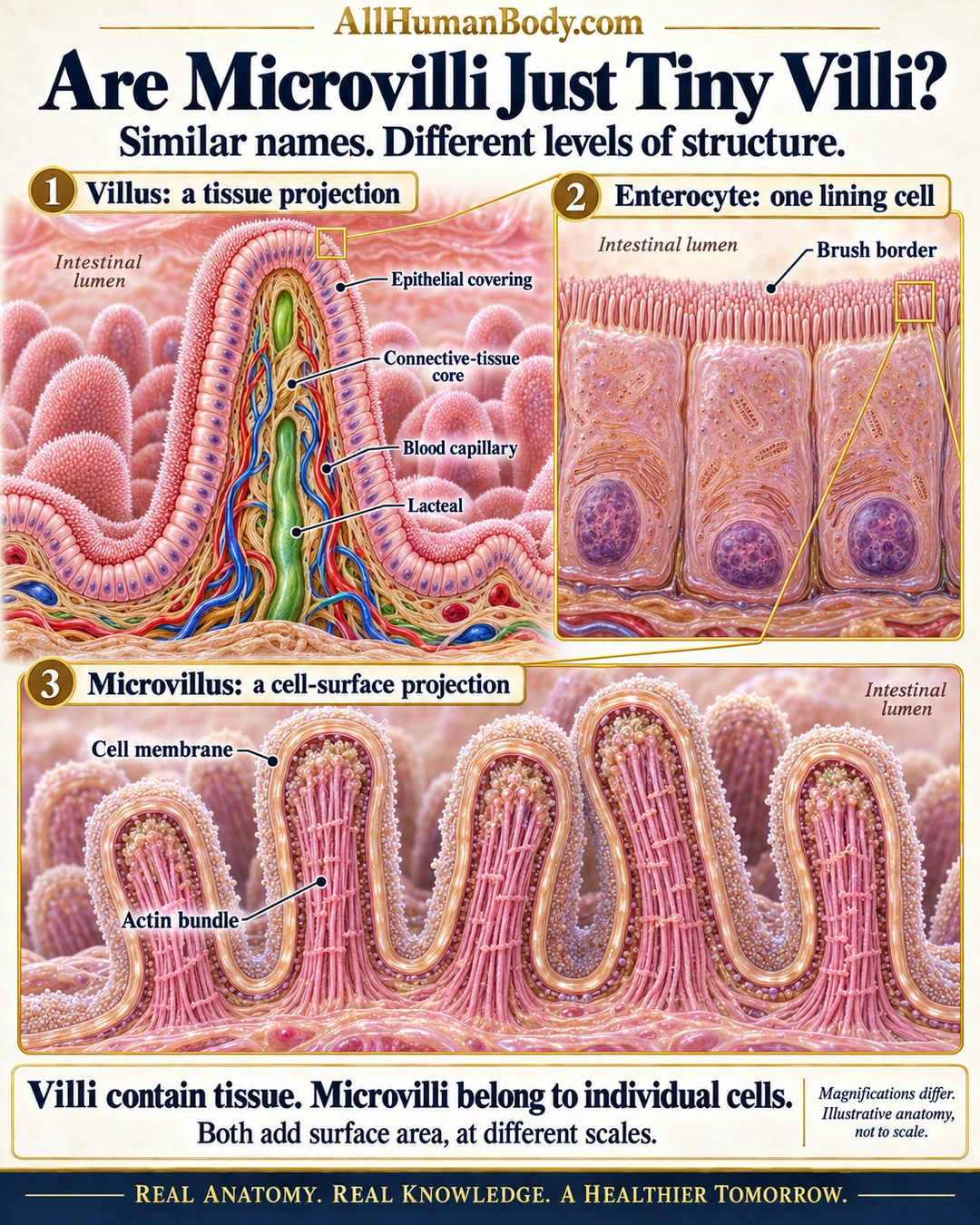
A villus is a multicellular mucosal projection with a connective-tissue core. Microvilli are much smaller projections of individual lining cells; both increase surface area, but they are different levels of organization.
READ THE IMAGE
What the illustration shows
English medical poster titled Are Microvilli Just Tiny Villi? Three connected magnifications show a cutaway villus with a single epithelial covering, tan connective-tissue core, red and blue capillaries and green lacteal; enlarged enterocytes with apical brush borders facing the intestinal lumen; and membrane-covered microvilli with internal actin bundles continuous with the parent cell. Gold connectors indicate magnification rather than transport. The takeaway says villi contain tissue, while microvilli belong to individual cells, and both add surface area at different scales. The image states that magnifications differ and anatomy is illustrative, not to scale. Gold AllHumanBody.com branding and the full project slogan appear.
How to interpret this poster
- Magnifications differ; no exact dimensions, molecular counts or vascular branching pattern.
- Microvilli are continuous with their parent cell and contain neither nuclei nor blood vessels.
- Gold connectors indicate magnification, not nutrient transport or cell migration.
- AI-assisted educational illustration; not physician certification, microscopy, a diagnosis or individualized medical advice.
- Fine annotations need enlargement; the full description provides an accessible text alternative.
- Display the full uncropped illustration; do not trim titles, legends or boundary annotations.
AI-assisted educational illustration, adapted into web-sized images without changing its composition. This is general education, not an individual diagnosis or treatment recommendation.
EVIDENCE / CONTEXT
Sources and what they support
- University of Leeds Histology Guide: Small Intestine ↗
Villus, columnar lining and vascular/lymphatic core.
- University of Leeds Histology Guide: Microvilli ↗
Cell-membrane projections with actin bundles at the luminal surface.