CHAPTER / NUTRIENTS, RESERVES AND CELLULAR WORK

Metabolism

Use. Build. Store. Mobilise.

Follow delivered nutrients into living reserves, changing tissue needs and the reactions that support a working body.

Watch chapter overviewFilm planned · not yet available
Approved current-family representative nucleated-cell Hero, general cellular work context rather than an adipocyte or microscope specimen.
Current Golden-family cellular context reused · fine detail illustrative

A circulating supply. A living reserve. A working cell.

Locate the tissue and its compartments, then distinguish delivered material, stored substrate, cellular work and heat.
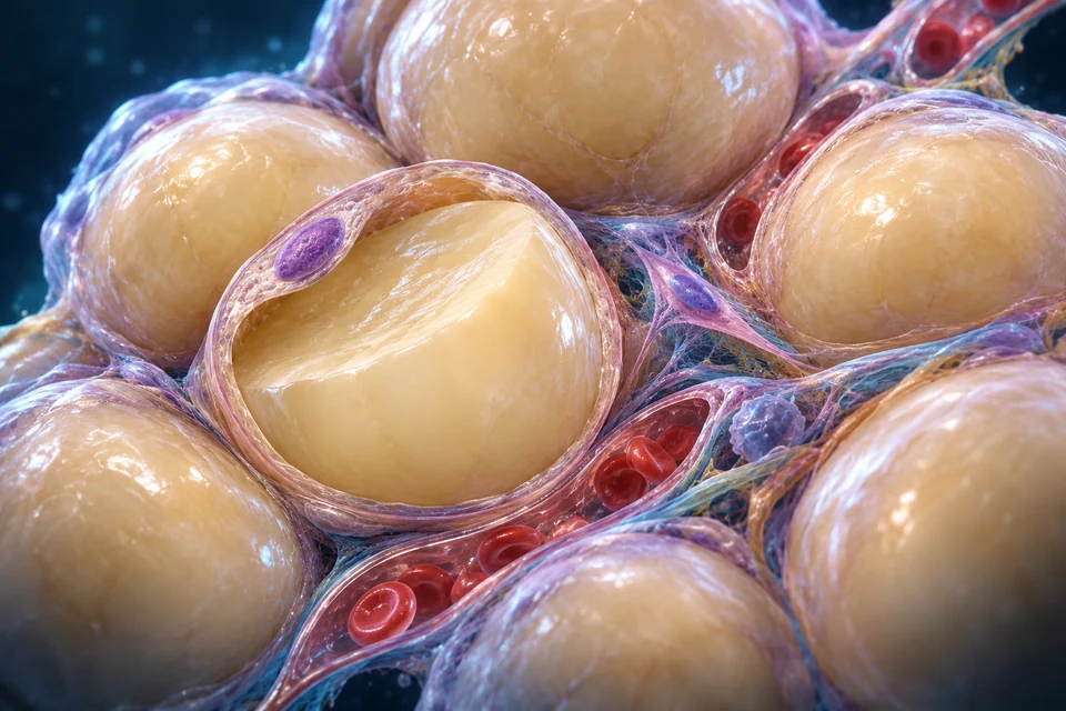
30-SECOND ORIENTATION / DELIVER · USE · BUILD · STORE · RELEASE

A reserve within a living tissue. Original Golden-family source-informed educational illustration; not observed histology or microscopy.
Retained original packed white-adipose teaching illustration. A local opening exposes a large lipid compartment and peripheral nucleus; interstitial vessels and supporting cells remain separate. Fine folds,cell proportions,counts and vascular topology illustrative,not measured histology. Human medical review: Not performed.
Structure notes · further context
White adipocyte
A living cell holds a lipid reserve.
Peripheral nucleus
Nuclear material lies toward the cell edge.
Microvascular context
Circulation connects the tissue with supply and release.
Supporting-cell context
Not every resident cell is a fat-storing adipocyte.
Connective support
Extracellular material links the tissue.

A reserve within a living tissue · atlas detail

A reserve within a living tissue. Original Golden-family source-informed educational illustration; not observed histology or microscopy.

Retained original packed white-adipose teaching illustration. A local opening exposes a large lipid compartment and peripheral nucleus; interstitial vessels and supporting cells remain separate. Fine folds,cell proportions,counts and vascular topology illustrative,not measured histology. Human medical review: Not performed.

Open full-resolution illustration ↗ · Image only; editable names and explanations remain in this lesson.

Circulation serves a living reserve.

Vessels remain outside the stored lipid. Cells receive material and signals; storing or releasing requires cellular reactions.

03 / ADIPOSE-CONTEXT

A storage tissue is made of living cells

Adipose includes fat-storing cells, vessels and supporting cells. Its organisation connects a reserve with circulating supply and signals. The original illustration shows selected relationships, not a stained specimen or a measured cell population.

A reserve within a living tissue. Original Golden-family source-informed educational illustration; not observed histology or microscopy.
Retained original packed white-adipose teaching illustration. A local opening exposes a large lipid compartment and peripheral nucleus; interstitial vessels and supporting cells remain separate. Fine folds,cell proportions,counts and vascular topology illustrative,not measured histology. Human medical review: Not performed.
Structure notes · further context
White adipocyte
A living cell holds a lipid reserve.
Peripheral nucleus
Nuclear material lies toward the cell edge.
Microvascular context
Circulation connects the tissue with supply and release.
Supporting-cell context
Not every resident cell is a fat-storing adipocyte.
Connective support
Extracellular material links the tissue.

A reserve within a living tissue · atlas detail

A reserve within a living tissue. Original Golden-family source-informed educational illustration; not observed histology or microscopy.

Retained original packed white-adipose teaching illustration. A local opening exposes a large lipid compartment and peripheral nucleus; interstitial vessels and supporting cells remain separate. Fine folds,cell proportions,counts and vascular topology illustrative,not measured histology. Human medical review: Not performed.

Open full-resolution illustration ↗ · Image only; editable names and explanations remain in this lesson.

Teaching references: NLM Endotext — Adipose physiology

01 / DESTINATIONS

Absorption starts a new set of decisions

Digestive surfaces deliver usable molecules; tissues can then use, transform or store them. Uptake is not the same event as conversion to ATP, and one nutrient does not have one unavoidable destination.

SOURCE-SUPPORTED RELATIONSHIP / SUPPORTING DIAGRAM

Delivery, fuel and material have different meanings
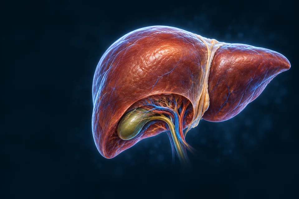
Retained detailed original organ illustration: Liver · blood supply. Gross context, not tissue-level measurement.

Liver · blood supply

Glycogen breakdown can support free glucose supply to blood.

Learn with this organ’s anatomy →
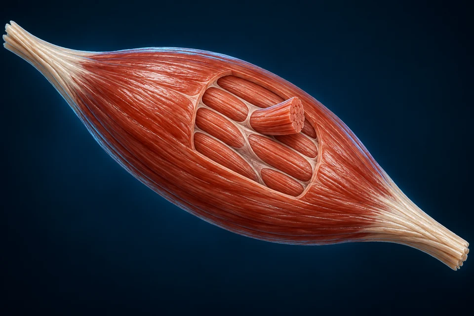
Retained detailed original organ illustration: Muscle · local work. Gross context, not tissue-level measurement.

Muscle · local work

Muscle glycogen supports metabolism within working muscle; it is not a direct free-glucose export store.

Learn with this organ’s anatomy →
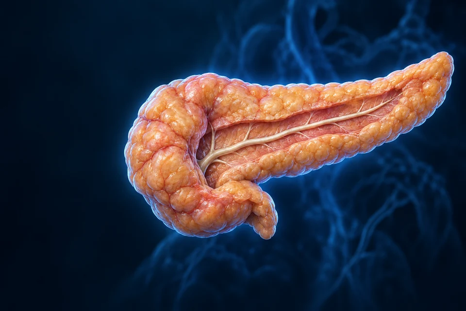
Retained detailed original organ illustration: Pancreas · coordination. Gross context, not tissue-level measurement.

Pancreas · coordination

Endocrine signals coordinate tissue responses; a hormone does not perform every tissue reaction.

Learn with this organ’s anatomy →

The named organs locate different jobs. Their layout is a functional comparison, not physical adjacency, a pipe between organs or measured flux. Glucose supply, tissue use and endocrine signals remain distinct.

Optional relationship summary · another scaleDelivery is distinct from use and storage — selected qualitative educational relationship, not measured histology or flux.Absorbed productsTissue deliveryUse / buildStoreSelected blood and lymph pathways.Not every nutrient enters one ATP route.

Retained editable diagram, with separate stable concept IDs, paths and text. Selected qualitative relations, not measured molecular thickness, tissue proportions or metabolic flux. Main illustrations teach cell/tissue organisation.

Read the relationships

Deliver: Absorbed products enter selected blood or lymph routes.

Use / build: Cells convert substrates for energy transfer and biosynthesis.

Store: Selected material enters glycogen or lipid reserves.

English First. Human medical review not performed.

Go deeper · Why does absorption not decide the final job?

A molecule entering circulation may be used, transformed or stored after it reaches a tissue. Follow the delivery route first, then ask which cellular reactions occur. The same delivered material can contribute to more than one destination; the organ cards locate jobs, not a single compulsory sequence.

Mechanism sources: reactions · states

Teaching references: OpenStax24.1 — Metabolic reaction roles · OpenStax24.5 — Metabolic states

02 / JOBS

A fuel molecule can also provide building material

Metabolism combines chemical breakdown with construction and maintenance. Energy transfer helps link these jobs, while enzymes and cellular compartments keep them organised.

STRUCTURE, CELLULAR WORK AND REGULATED RELATIONSHIPS

Breakdown and construction are coupled, not identical
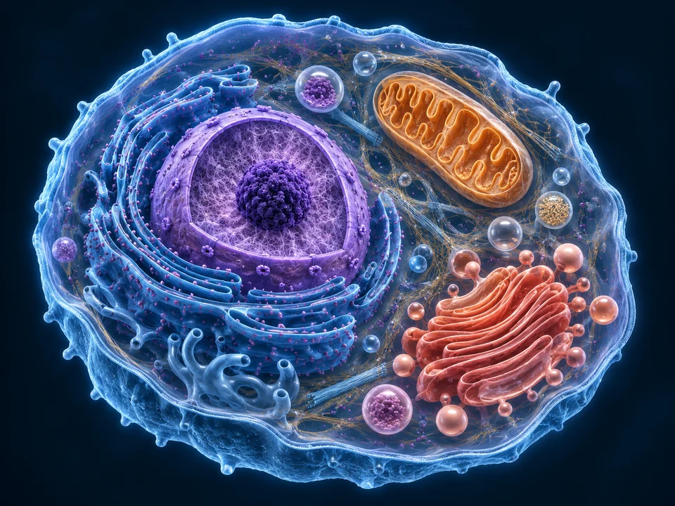
Retained detailed representative nucleated cell cutaway. Independent labels connect membrane, cytoplasm, nucleus, rough ER, mitochondrion and Golgi with their different jobs.
Original generated atlas, not acquired microscopy. A representative nucleated cell, not a universal human cell. Organelle positions and dimensions are composed for teaching; the image does not measure molecular topology or numbers.

One cell, connected kinds of work · atlas detail

Retained detailed representative nucleated cell cutaway. Independent labels connect membrane, cytoplasm, nucleus, rough ER, mitochondrion and Golgi with their different jobs.

Original generated atlas, not acquired microscopy. A representative nucleated cell, not a universal human cell. Organelle positions and dimensions are composed for teaching; the image does not measure molecular topology or numbers.

Open full-resolution image ↗ · Image only; editable labels remain in this atlas detail.

Optional process summary · qualitative relationshipsCoupled breakdown, transfer and construction — selected qualitative educational relationship, not measured histology or flux.Nutrient substratesEnergy / intermediatesWork / constructionSelected reaction roles.Not one enzyme or a fixed ATP yield.

Break down is emphasised; every essential explanation remains visible. Selected relationships, not exact reaction rates, a fixed ATP yield, an all-or-none clock or individual prediction.

Break down

Selected reactions release transferable energy and smaller substrates.

Transfer

ATP and other carriers link selected reactions with cellular work.

Build / maintain

Energy and substrates support biosynthesis and maintenance.

Teaching references: OpenStax24.1 — Metabolic reaction roles

04 / WHITE-CELL

One large lipid droplet changes the cell interior

In a typical mature white adipocyte, a large lipid droplet occupies much of the interior and the nucleus lies toward the edge. The opened illustration preserves this broad relationship; it exaggerates peripheral space for inspection.

A white adipocyte opened for inspection. Original Golden-family source-informed educational illustration; not observed histology or microscopy.
Retained original white-cell teaching cutaway. One dominant lipid droplet with a peripheral nucleus and remaining cytoplasm. Thin surface distinctions are qualitative; monolayer versus bilayer is explained in the separate editable diagram,not resolved as molecular detail in this art. Human medical review: Not performed.
Structure notes · further context
Large lipid droplet
Neutral lipid occupies much of this white-cell interior.
Peripheral nucleus
The nucleus remains outside the lipid core.
Cellular material
Working cytoplasm surrounds and connects compartments.
Cell boundary
The outer plasma membrane separates cell from environment.
Droplet surface
A monolayer-covered interface differs from the cell membrane.

A white adipocyte opened for inspection · atlas detail

A white adipocyte opened for inspection. Original Golden-family source-informed educational illustration; not observed histology or microscopy.

Retained original white-cell teaching cutaway. One dominant lipid droplet with a peripheral nucleus and remaining cytoplasm. Thin surface distinctions are qualitative; monolayer versus bilayer is explained in the separate editable diagram,not resolved as molecular detail in this art. Human medical review: Not performed.

Open full-resolution illustration ↗ · Image only; editable names and explanations remain in this lesson.

Teaching references: NLM Endotext — Adipose physiology · Zhang and Liu — Lipid droplet review

05 / BOUNDARIES

A droplet surface is not a second cell wall

The outer plasma membrane has a lipid bilayer. A lipid droplet has a neutral-lipid core and a phospholipid monolayer surface. Its surroundings remain cellular material, not an empty oil bag outside the body.

SOURCE-SUPPORTED RELATIONSHIP / SUPPORTING DIAGRAM

One boundary is a bilayer; another is a monolayer

A white adipocyte opened for inspection. Original Golden-family source-informed educational illustration; not observed histology or microscopy.
Retained original white-cell teaching cutaway. One dominant lipid droplet with a peripheral nucleus and remaining cytoplasm. Thin surface distinctions are qualitative; monolayer versus bilayer is explained in the separate editable diagram,not resolved as molecular detail in this art. Human medical review: Not performed.
Structure notes · further context
Large lipid droplet
Neutral lipid occupies much of this white-cell interior.
Peripheral nucleus
The nucleus remains outside the lipid core.
Cellular material
Working cytoplasm surrounds and connects compartments.
Cell boundary
The outer plasma membrane separates cell from environment.
Droplet surface
A monolayer-covered interface differs from the cell membrane.

A white adipocyte opened for inspection · atlas detail

A white adipocyte opened for inspection. Original Golden-family source-informed educational illustration; not observed histology or microscopy.

Retained original white-cell teaching cutaway. One dominant lipid droplet with a peripheral nucleus and remaining cytoplasm. Thin surface distinctions are qualitative; monolayer versus bilayer is explained in the separate editable diagram,not resolved as molecular detail in this art. Human medical review: Not performed.

Open full-resolution illustration ↗ · Image only; editable names and explanations remain in this lesson.

The detailed cell locates two different interfaces. Molecular leaflet organisation belongs to another scale; the optional chemistry summary compares a bilayer with a monolayer without claiming that individual leaflets are resolved in this artwork.

Optional relationship summary · another scalePlasma bilayer and droplet monolayer — selected qualitative educational relationship, not measured histology or flux.Plasma membrane / bilayerWater-facing surfaceWater-facing surfaceDroplet surface / monolayerCytoplasmNeutral lipid coreOrientation, not molecular thickness.

Retained editable diagram, with separate stable concept IDs, paths and text. Selected qualitative relations, not measured molecular thickness, tissue proportions or metabolic flux. Main illustrations teach cell/tissue organisation.

Read the relationships

Cell membrane: Two lipid leaflets face aqueous environments on both sides.

Droplet surface: One leaflet meets the neutral lipid core.

Different roles: This qualitative section does not show actual molecules or measured thickness.

English First. Human medical review not performed.

Go deeper · Why are the two lipid interfaces chemically different?

A cell membrane separates aqueous environments with a bilayer. A droplet has a neutral-lipid interior and a monolayer-facing cytoplasm. Locating the two surfaces in a cutaway is a spatial lesson; identifying their molecular arrangement requires a different scale of evidence. The pictured rim is not a measured membrane thickness.

Mechanism sources: droplet

Teaching references: Zhang and Liu — Lipid droplet review

06 / LIPID-RESERVE

Storing and releasing are regulated chemical changes

Triacylglycerol storage and lipolysis are different activities. Breakdown produces fatty acids and glycerol, with some material reused locally and some entering circulation. This is not dietary fat flowing unchanged out through a hole.

STRUCTURE, CELLULAR WORK AND REGULATED RELATIONSHIPS

A lipid reserve can be assembled or mobilised

A white adipocyte opened for inspection. Original Golden-family source-informed educational illustration; not observed histology or microscopy.
Retained original white-cell teaching cutaway. One dominant lipid droplet with a peripheral nucleus and remaining cytoplasm. Thin surface distinctions are qualitative; monolayer versus bilayer is explained in the separate editable diagram,not resolved as molecular detail in this art. Human medical review: Not performed.
Structure notes · further context
Large lipid droplet
Neutral lipid occupies much of this white-cell interior.
Peripheral nucleus
The nucleus remains outside the lipid core.
Cellular material
Working cytoplasm surrounds and connects compartments.
Cell boundary
The outer plasma membrane separates cell from environment.
Droplet surface
A monolayer-covered interface differs from the cell membrane.

A white adipocyte opened for inspection · atlas detail

A white adipocyte opened for inspection. Original Golden-family source-informed educational illustration; not observed histology or microscopy.

Retained original white-cell teaching cutaway. One dominant lipid droplet with a peripheral nucleus and remaining cytoplasm. Thin surface distinctions are qualitative; monolayer versus bilayer is explained in the separate editable diagram,not resolved as molecular detail in this art. Human medical review: Not performed.

Open full-resolution illustration ↗ · Image only; editable names and explanations remain in this lesson.

The outline locates a broad reserve, surface or outer-cell context for the selected explanation. It does not depict a pore, a reaction rate, a molecule moving through the cell, or direct dietary fat leaving unchanged.

Optional process summary · qualitative relationshipsAssembly and mobilisation of a lipid reserve — selected qualitative educational relationship, not measured histology or flux.Assembly substratesStored triacylglycerolFatty acids / glycerolSome products can be reused locally.Arrows do not measure reaction rates.

Store is emphasised; every essential explanation remains visible. Selected relationships, not exact reaction rates, a fixed ATP yield, an all-or-none clock or individual prediction.

Store

Fatty acids and glycerol-derived components contribute to triacylglycerol storage.

Mobilise

Lipolysis releases fatty acids and glycerol from stored triacylglycerol.

Reuse / supply

Products can be reused locally or carried to other tissues; not every released molecule must leave.

Teaching references: OpenStax24.3 — Lipid uses · NLM Endotext — Adipose physiology

07 / BLOOD-CARRIERS

Different forms require different carriers

Lipoprotein particles transport many lipids; released non-esterified fatty acids circulate largely associated with albumin. A particle, an albumin-bound fatty acid and an intracellular lipid droplet are different scales.

Go deeper · Which form is actually being transported?

Distinguish a lipoprotein particle from an albumin-associated non-esterified fatty acid and from a droplet inside a cell. They can participate in connected lipid handling without being the same object. A blood-vessel arrow cannot establish the carrier, cargo or transfer mechanism unless those are stated.

Follow existing digestive exits →

Mechanism sources: carriers

Teaching references: NLM Endotext — Lipid transport

08 / GLYCOGEN

Two glycogen stores serve different immediate needs

Liver glycogen supports glucose release into blood. Skeletal-muscle glycogen primarily supports work in that muscle; it is not a second direct glucose-export tank. Both are carbohydrate reserves, unlike the lipid droplets above.

SOURCE-SUPPORTED RELATIONSHIP / SUPPORTING DIAGRAM

Liver supply and muscle work are different destinations

Retained detailed original organ illustration: Liver · blood supply. Gross context, not tissue-level measurement.

Liver · blood supply

Glycogen breakdown can support free glucose supply to blood.

Learn with this organ’s anatomy →
Retained detailed original organ illustration: Muscle · local work. Gross context, not tissue-level measurement.

Muscle · local work

Muscle glycogen supports metabolism within working muscle; it is not a direct free-glucose export store.

Learn with this organ’s anatomy →

The named organs locate different jobs. Their layout is a functional comparison, not physical adjacency, a pipe between organs or measured flux. Glucose supply, tissue use and endocrine signals remain distinct.

Optional relationship summary · another scaleLiver supply and local skeletal-muscle work — selected qualitative educational relationship, not measured histology or flux.LiverSkeletal muscleGlycogenBlood glucoseGlycogenLocal workMuscle glycogen is not a directfree-glucose export tank.Other organs also produce glucose.

Retained editable diagram, with separate stable concept IDs, paths and text. Selected qualitative relations, not measured molecular thickness, tissue proportions or metabolic flux. Main illustrations teach cell/tissue organisation.

Read the relationships

Liver: Glycogen breakdown can support free glucose release into blood.

Skeletal muscle: Glycogen contributes locally to contraction-related metabolism.

Limits: No direct free-glucose export from muscle glycogen; other organs also contribute to glucose production.

English First. Human medical review not performed.

Go deeper · Why does the same polymer have different tissue roles?

The useful comparison is what each tissue can deliver: hepatic glycogen contributes to blood glucose supply, whereas muscle glycogen mainly supports local metabolism. This is not two identical tanks connected to one outlet. Other glucose-producing organs and substrates also matter; the two-organ comparison is deliberately bounded.

Continue hepatic regulation →

Mechanism sources: glycogen · gluconeogenesis

Teaching references: NLM — Glycogen tissue-specific roles

09 / CELL-ENERGY

A connected pathway is not one organelle

Glycolysis operates in cytosol. Mitochondrial reactions contribute to nutrient oxidation and ATP production in cells that possess mitochondria. Their contributions vary with fuels, demand and cell type.

Go deeper · What does a mitochondrion picture leave outside its border?

Glycolysis is cytosolic; mitochondrial reactions make another contribution in cells with mitochondria. A detailed organelle image therefore cannot contain the whole pathway. Fuel, cell type and demand change the balance, so one ATP number or one organelle cannot stand for all human energy metabolism.

Inspect the established mitochondrial lesson →

Mechanism sources: carbohydrate

Teaching references: OpenStax24.2 — Carbohydrate pathways

10 / CARBON

Similar destinations do not make substrates identical

Carbohydrate, lipid and amino-acid metabolism intersect through selected intermediates. Glycerol can contribute to glucose production; even-chain fatty acids do not provide net glucose by the ordinary human pathway. Nitrogen requires separate handling.

Carbohydrate

Glucose-derived pathways

Can support fuel use, glycogen and other synthesis.

Lipid

Fatty acids and glycerol

Different products follow different routes.

Amino acids

Carbon plus nitrogen

Carbon intermediates and nitrogen disposal have different jobs.

Teaching references: OpenStax24.3 — Lipid uses · OpenStax24.4 — Protein uses · NLM — Gluconeogenesis substrates

11 / PROTEINS

Building proteins is not filling a spare fuel tank

Amino acids contribute to enzymes, transporters, structure and tissue renewal. Protein turnover releases material for reuse or further metabolism. Body proteins are working tissues, not a dedicated inert reserve like a lipid store.

SOURCE-SUPPORTED RELATIONSHIP / SUPPORTING DIAGRAM

Carbon and nitrogen require separate handling

Retained detailed original organ illustration: Liver · nitrogen processing. Gross context, not tissue-level measurement.

Liver · nitrogen processing

Urea production handles much nitrogen separately from the carbon skeleton.

Learn with this organ’s anatomy →
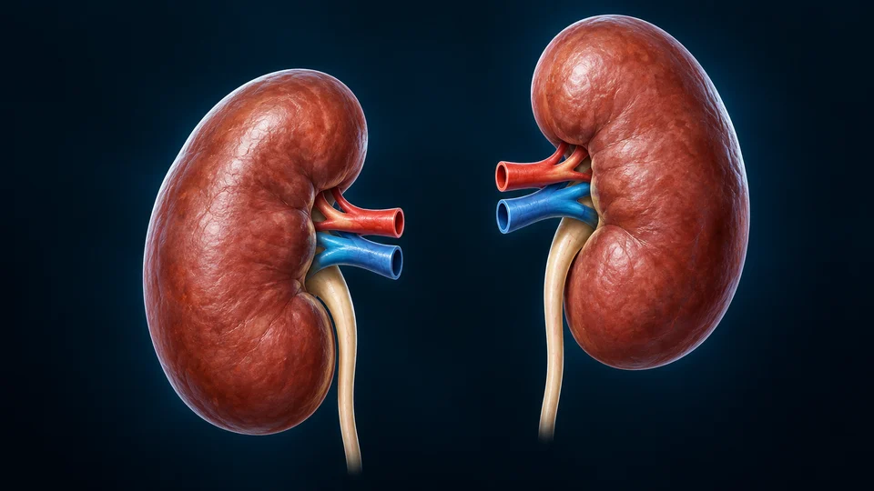
Retained detailed original organ illustration: Kidneys · excretion. Gross context, not tissue-level measurement.

Kidneys · excretion

Urinary excretion removes urea; these organs do not represent a complete molecular urea-cycle diagram.

Learn with this organ’s anatomy →

The named organs locate different jobs. Their layout is a functional comparison, not physical adjacency, a pipe between organs or measured flux. Glucose supply, tissue use and endocrine signals remain distinct.

Optional relationship summary · another scaleCarbon and nitrogen follow different roles — selected qualitative educational relationship, not measured histology or flux.Amino acidsCarbon skeletonNitrogenIntermediatesLiver / ureaKidneys / urineSelected handling, not a full urea cycle.No conversion of nitrogen into carbon.

Retained editable diagram, with separate stable concept IDs, paths and text. Selected qualitative relations, not measured molecular thickness, tissue proportions or metabolic flux. Main illustrations teach cell/tissue organisation.

Read the relationships

Carbon skeleton: Different amino acids contribute to different metabolic intermediates.

Nitrogen: Liver urea production processes much nitrogen for transport.

Kidneys: Urinary excretion handles urea; this is not a complete renal or molecular urea-cycle map.

English First. Human medical review not performed.

Go deeper · What happens to carbon and nitrogen when proteins turn over?

Protein synthesis and breakdown concern working tissues. Reused amino acids can support new proteins; further metabolism separates carbon handling from nitrogen handling. Liver urea production and renal excretion are linked but distinct processes. This comparison does not place a miniature urea cycle inside the kidney or make every amino acid a glucose precursor.

Liver recycling and nitrogen handling →

Mechanism sources: protein · gluconeogenesis

Teaching references: OpenStax24.4 — Protein uses

12 / STATES

Nutrient availability shifts several tissues together

After nutrient absorption, insulin favours selected storage processes. Between meals, supply from reserves and new glucose production contribute differently. These are overlapping regulated activities, not a timed switch that shuts all synthesis off.

STRUCTURE, CELLULAR WORK AND REGULATED RELATIONSHIPS

Availability changes the balance, not an all-or-none switch

Retained detailed original organ illustration: Liver · blood supply. Gross context, not tissue-level measurement.

Liver · blood supply

Glycogen breakdown can support free glucose supply to blood.

Learn with this organ’s anatomy →
Retained detailed original organ illustration: Muscle · local work. Gross context, not tissue-level measurement.

Muscle · local work

Muscle glycogen supports metabolism within working muscle; it is not a direct free-glucose export store.

Learn with this organ’s anatomy →
Retained detailed original organ illustration: Pancreas · coordination. Gross context, not tissue-level measurement.

Pancreas · coordination

Endocrine signals coordinate tissue responses; a hormone does not perform every tissue reaction.

Learn with this organ’s anatomy →

The named organs locate different jobs. Their layout is a functional comparison, not physical adjacency, a pipe between organs or measured flux. Glucose supply, tissue use and endocrine signals remain distinct.

Optional process summary · qualitative relationshipsOverlapping tissue needs and changing availability — selected qualitative educational relationship, not measured histology or flux.Nutrient uptakeReserves contributeWork changes demandThese activities overlap.Not a three-stage clock or fasting advice.

After uptake is emphasised; every essential explanation remains visible. Selected relationships, not exact reaction rates, a fixed ATP yield, an all-or-none clock or individual prediction.

After uptake

Insulin favours selected storage and reduces adipose lipolysis.

Between meals

Liver glucose supply and reserve mobilisation help meet ongoing needs.

During work

Local demand and neural/endocrine signals alter fuel handling. No fixed clock or prescribed fasting period.

Go deeper · Why is there no universal meal-to-fasting countdown?

Availability and tissue demand change continuously. Storage, use and endogenous supply can overlap rather than taking turns as exclusive programmes. The controls emphasise one teaching question at a time, not a prescribed fasting interval, a clinical glucose target or a claim that all organs switch fuel together.

Mechanism sources: states

Teaching references: OpenStax24.5 — Metabolic states · NLM Endotext — Glucagon physiology

13 / REGULATION

One hormone does not command every tissue identically

Pancreatic signals, sympathetic input and local cell conditions cooperate. Insulin suppresses adipose lipolysis; catecholamine input can favour mobilisation. Glucagon has a clear hepatic role, but direct physiological human adipose effects are less certain.

Go deeper · Why should a liver response not be copied onto a fat cell?

Hormones act through tissue-specific contexts. Glucagon has an established hepatic role; the extent of direct physiological adipose lipolysis in humans remains disputed. Keep insulin’s restraint on adipose lipolysis, catecholamine-related mobilisation and hepatic glucose supply distinguishable instead of using one universal hormone-to-all-tissues arrow.

See pancreatic glucose feedback →

Mechanism sources: glucagon · human-lipolysis

Teaching references: NLM Endotext — Glucagon physiology · Galsgaard et al. — Human glucagon/lipid scope

14 / BROWN-CELL

A different cell organisation can support heat production

Brown adipocytes contain several lipid droplets and many mitochondria. Their thermogenic machinery can dissipate part of a gradient as heat. This original view compares broad organisation, not an actual measured human sample.

Brown-cell organisation at another scale. Original Golden-family source-informed educational illustration; not observed histology or microscopy.
Retained original brown-cell teaching cutaway. Several smaller lipid droplets and mitochondrial context surround a separate nucleus. Fine organelle numbers,proportions,folds and membrane texture illustrative; no actual human sample,UCP1 molecules,depot distribution or thermogenic rate shown. Human medical review: Not performed.
Structure notes · further context
Multiple lipid droplets
Brown-cell storage is divided among smaller compartments.
Mitochondrial context
Numerous mitochondria support thermogenic organisation.
Nucleus
Genetic material remains in a separate compartment.
Cellular material
Working cytoplasm surrounds and connects compartments.

Brown-cell organisation at another scale · atlas detail

Brown-cell organisation at another scale. Original Golden-family source-informed educational illustration; not observed histology or microscopy.

Retained original brown-cell teaching cutaway. Several smaller lipid droplets and mitochondrial context surround a separate nucleus. Fine organelle numbers,proportions,folds and membrane texture illustrative; no actual human sample,UCP1 molecules,depot distribution or thermogenic rate shown. Human medical review: Not performed.

Open full-resolution illustration ↗ · Image only; editable names and explanations remain in this lesson.

Teaching references: NLM Endotext — Adipose physiology

15 / HEAT

A shared energy source can have different outputs

ATP supports cellular work; heat is another outcome of metabolism. Brown-fat thermogenesis differs from skeletal-muscle shivering. Adult humans can have active brown adipose, but its amount and activity vary.

Cellular work

ATP-dependent activity

Transport, movement and biosynthesis use energy.

Thermogenesis

Selected heat-producing activity

Brown-fat organisation supports one non-shivering contribution.

Variation

People and conditions differ

No universal depot size, temperature rise or calorie-burning estimate.

Go deeper · How can organisation help explain different energy outcomes?

Multiple lipid droplets and abundant mitochondrial context help distinguish a brown adipocyte from a typical white one. These are structural clues, not proof of a particular cell’s thermogenic rate. Non-shivering brown-fat activity and muscle shivering are different contributions; their roles vary among people and conditions.

Compare muscular work →

Mechanism sources: adipose

Teaching references: NLM Endotext — Adipose physiology

16 / COMMUNICATION

A reserve can also send messages

Adipose participates in endocrine communication. Leptin and adiponectin are examples, but a tissue secretion does not always originate in a fat-storing cell; other resident cells contribute.

Teaching references: NLM Endotext — Adipose physiology

17 / NONFUEL

Not every useful nutrient is a bulk energy source

Water, vitamins and minerals have different structural, transport and regulatory roles. Fibre also has distinct digestive and microbial relationships. These roles cannot be explained by directing every nutrient into one ATP arrow.

Teaching references: OpenStax24.1 — Metabolic reaction roles

18 / OBSERVATION

A constructed cell does not establish a specimen diagnosis

Sectioning and staining influence what a tissue sample shows. A bright empty-looking area in a prepared section can reflect processing, whereas this render exposes a deliberately illustrated lipid compartment. Neither is a live metabolic measurement.

Original model

Selected spatial relationships

Counts, textures and dimensions are illustrative.

Identified microscopy

Observed tissue with preparation limits

No specimen is imported or presented here.

Metabolic measurement

Another kind of evidence

No flux, patient glucose or energy-expenditure reading is claimed.

Teaching references: NLM Endotext — Adipose physiology · Zhang and Liu — Lipid droplet review

19 / VARIATION

Fuel choice changes without erasing the anatomy

Different cells use different combinations of fuels; changing demand does not mean every tissue adopts one universal program. Detailed enzyme maps, exact ATP yields, clinical imaging, disorders and dietary advice remain outside this normal teaching module.

Human medical review: not performed. Real physical-phone acceptance: not performed. Educational only. No new CMS entity, working database content or publication change.

Teaching references: OpenStax24.2 — Carbohydrate pathways · OpenStax24.5 — Metabolic states

20 / CONNECTIONS

Follow the same material across different systems

Digestion supplies; circulation carries; endocrine pathways coordinate; liver, adipose and muscle process; kidneys contribute to excretion and regulation. Existing deep organ chapters provide the corresponding anatomy, without repeating those plates here.

Teaching references: OpenStax24.1 — Metabolic reaction roles · OpenStax24.5 — Metabolic states

EDITORIAL RECORD

Sources & Editorial Status

Source-supported educational scope

Normal nutrient allocation, adipose organisation, reserves and qualitative metabolic relationships. Supporting pathways explain selected connections, not a complete metabolome.

Methods and limits

Three retained original high-detail text-free cell/tissue illustrations with separate semantic labels, plus the qualified Cells overview and organ context. Seven retained controlled SVG auxiliary summaries keep paths,stable concept IDs and typography separate. Current-family Cells Hero and Liver/Muscle/Pancreas cards reused exactly. No source artwork, microscopy or anatomy model was copied. Counts, dimensions, fine folds, texture and tissue topology remain illustrative. AI-assisted original content/code production. Human medical review: not performed. Educational only.

Revision: metabolism-eyes-context-2026-10-19-v1. Stable local identity: ahb_ch_metabolism_english_v1; not a new CMS entity. Original masters stay outside Git; labels and paths remain editable. New physical-phone acceptance: not performed. No patient diagnosis, surgical instruction, medication advice or physician approval claim. Observed microscopy and clinical disease remain separate work.