A route through a crowded thorax
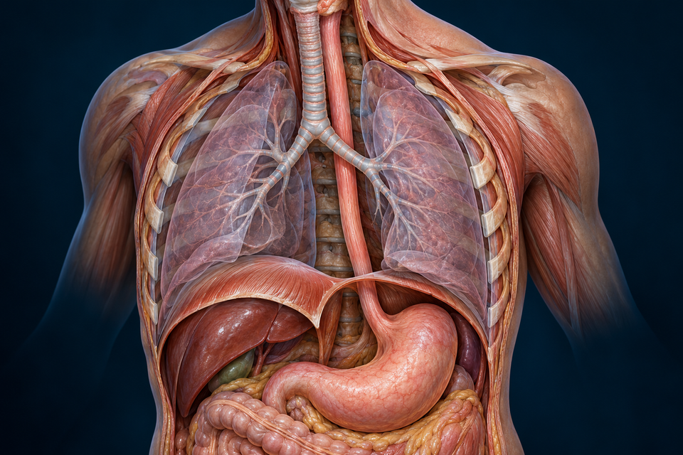
The esophagus begins at the lower pharynx in the neck, descends through the thorax and passes through the diaphragm before entering the stomach. Its cervical, thoracic and short abdominal portions are continuous regions of one organ, not separate tubes.
In the upper chest it lies behind the trachea and in front of the vertebral column. Farther down, its relationships include the heart anteriorly and major vessels nearby. A frontal opened view exposes structures that overlap in an intact body; transparency and tissue removal must not be mistaken for normal empty space.
The surrounding structures help explain changes in the passage’s contour. They do not make every narrowing a sphincter. The airway and alimentary passage have different destinations even where they are close together.
Mechanism references: Kenhub · Esophageal anatomy · Cleveland Clinic · Esophagus
Read from the lumen outward
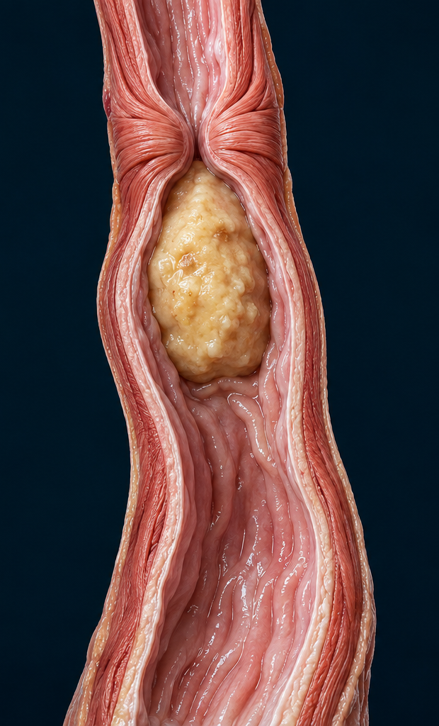
The mucosa comprises the epithelium, connective lamina propria and thin muscularis mucosae. Its surface is folded when the passage is not distended. Beneath it, the submucosa contains connective support, glands, vessels and nerves. Outside lies the thick muscularis externa, the major propulsive coat.
The main muscle has inner circular and outer longitudinal arrangements. Circular activity changes the calibre of the passage; longitudinal activity changes the length and geometry of a segment. Both contribute to coordinated transport. The much thinner muscularis mucosae belongs to a different anatomical layer and should not be labelled as the whole propulsive apparatus.
Most of the esophagus has an outer connective-tissue adventitia rather than the complete serosal covering familiar from freely mobile abdominal bowel. A small abdominal portion has a different peritoneal relationship; “the entire food tube has the same outer coat” is an unsafe generalisation.
Mechanism references: Histology Guide · Human esophagus MH109 · University of Leeds · Oesophageal histology · Kenhub · Esophageal anatomy
A surface suited to contact, not an absorptive forest
Nonkeratinised stratified squamous epithelium provides several cell layers between the lumen and underlying tissue. Basal cells replenish the sheet as more superficial cells are shed. The normal lining is not skin with a thick dry keratin layer, and it does not form the villi that enlarge small-intestinal absorptive surface.
The epithelial sheet itself lacks blood vessels. Supporting tissue underneath provides the vascular environment needed to maintain it. Mucus from esophageal glands, together with swallowed saliva, helps lubricate passage. A duct carries secretion from a small gland to the lumen; it is not a second route for the bolus.
At the stomach entrance, squamous mucosa gives way to gastric columnar mucosa with a different secretory organisation. The change is a tissue boundary. It is not a mechanical door, and the generated illustration cannot substitute for examination of a real section.
Mechanism references: Histology Guide · Human esophagus MH109 · University of Leeds · Oesophageal histology · Histology Guide · Gastroesophageal junction MH111a
The same direction requires different kinds of muscle control
Skeletal muscle predominates in the upper esophagus, smooth muscle in the lower part, with a mixed transition between. These are regional tendencies rather than three sharply separated compartments. An upper skeletal-muscle region can participate in an automatically coordinated swallow; skeletal does not mean that every later contraction is consciously chosen.
Brainstem swallowing coordination and vagal pathways link the pharyngeal transfer with esophageal activity. Within the smooth-muscle part, enteric circuits help organise relaxation and contraction. The myenteric plexus lies between the two main muscle layers; the submucosal neural network belongs nearer the mucosa and supporting tissue.
Sensory information about distension helps match clearing activity to contents. Neural control includes both excitation and inhibition: forward movement is not explained by turning every muscle cell on at the same time. This lesson shows the organisation, not the complete branching of each nerve or a patient-specific pressure pattern.
Mechanism references: Histology Guide · Human esophagus MH109 · Kenhub · Esophageal anatomy · Kenhub · Neurovascular and lymphatic context · Cleveland Clinic · Peristalsis
A swallow and a clearing response are not identical triggers
A primary peristaltic sequence follows swallowing. Secondary peristalsis can be triggered by esophageal distension, helping clear contents that remain. The two ideas describe how a response begins, not two separate anatomical channels.
The bolus and wall influence one another: the contents deform and distend the lumen, while coordinated muscle activity changes its shape. Liquids and a soft solid bolus do not produce identical transport conditions. A single cutaway cannot supply a universal transit time.
This makes the illustration useful for a question: where must the receiving region be relative to the contracting region? It cannot be read as measured motility, nor does the downward arrow mean that gravity alone performs the work.
Mechanism references: Cleveland Clinic · Peristalsis · NIDDK · Digestive transport
Opening, delivery and resistance to backward flow
The upper sphincter region helps coordinate transfer out of the pharynx. Lower down, the esophagogastric junction combines an intrinsic smooth-muscle pressure zone with surrounding anatomical support, including the crural diaphragm. The two ends have different tissue and control arrangements; they are not matching rubber rings.
During a coordinated swallow the distal junction relaxes so the bolus can enter the stomach. Between swallows, lower-junction tone helps resist return of gastric contents. Normal physiology also allows events such as venting gas; “permanently locked except when food arrives” is too absolute.
Reflux means gastric contents move back into the esophagus. GERD is not simply another name for any single reflux event. A transport problem, a lining problem and an antireflux-barrier problem are different concepts and cannot be diagnosed from this teaching image.
Mechanism references: NIDDK · Digestive transport · Kenhub · Esophageal anatomy · JCI · Human diaphragmatic contribution · NIDDK · Reflux and GERD
A long organ has regional supplies and connected drainage
Arterial contributors vary along the route: inferior thyroid branches serve the cervical region, branches of the thoracic aorta contribute in the chest, and left gastric and inferior phrenic contributions serve the distal region. These vessels supply the wall; they are not pathways for swallowed contents.
Venous return also changes by region. Cervical drainage communicates with inferior thyroid veins, thoracic drainage with the azygos system, and distal drainage with left gastric/portal pathways. Connections between systemic and portal territories have clinical importance, but a normal-route lesson is not a map for procedures.
Longitudinal lymphatic connections in the wall link drainage beyond a single short segment. Deep cervical, mediastinal and left gastric regional nodes are relevant along the route. A simple one-region–one-node cartoon would hide this continuity; the named regions here are an orientation, not an exhaustive drainage atlas.
Mechanism references: Kenhub · Neurovascular and lymphatic context
Use an illustration and a specimen for different questions
The original generated plates separate structures and expose otherwise hidden relationships. Their colour, selected openings and enlarged tissue window are teaching choices, not a record of one person’s anatomy. Cell texture and vessel profiles do not establish a diagnosis.
The linked human esophagus section lets you inspect folded mucosa, gland-bearing support and muscle in an actual stained preparation. The separate junction section shows a real tissue transition. A section is one plane through a specimen; an outer layer missing from the preparation does not prove it is normally absent everywhere.
No institutional slide pixels or copyrighted atlas figures are reproduced here. The external specimen links complement the original artwork. AI-assisted source checking and Owner visual approval are distinct from physician review, fine-histology certification and physical-phone testing.
Mechanism references: Histology Guide · Human esophagus MH109 · Histology Guide · Gastroesophageal junction MH111a