Follow the visible fibres and living cells, then connect their arrangement to support, exchange and maintenance. Existing tissue and organ lessons supply the specialised examples.
Outside a cell is not outside the tissue.
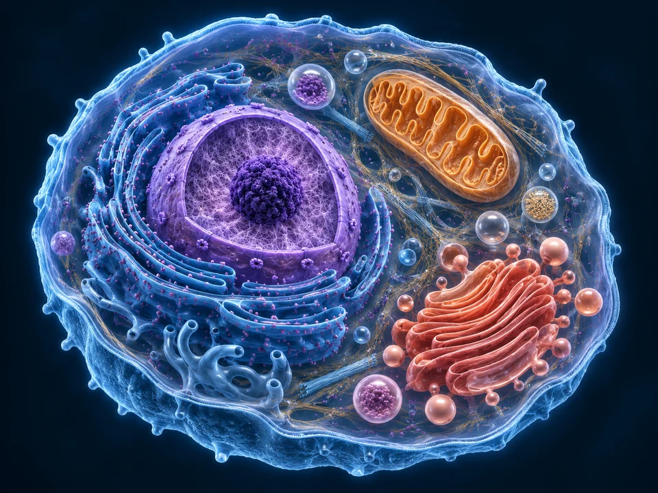
Cytoplasm belongs inside a cell. Extracellular matrix lies outside cells and includes fibrous proteins and hydrated ground substance. Interstitial fluid and dissolved substances occupy this environment; matrix is not the shared cytoplasm of a giant cell.
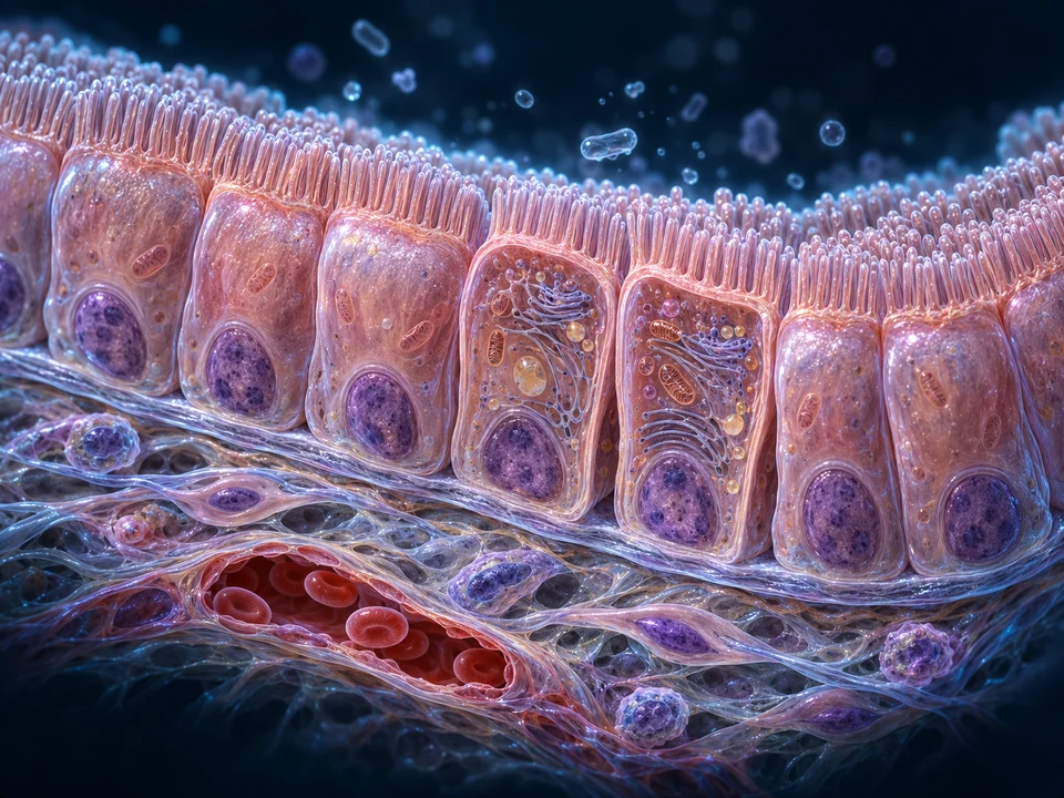
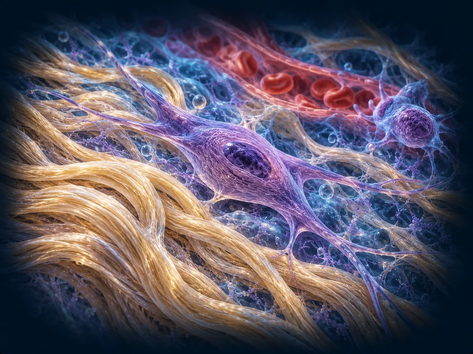
The retained loose-tissue image places a fibroblast beside collagen and a local vessel. Its generous spaces suit this selected arrangement. They must not be reused as the proportions of a dense tendon.
Structure and function: NCBI · Cells and the extracellular matrix · NCBI · Extracellular matrix organisation
Aligned fibres contribute to a larger tensile structure.
Collagen is organised at several scales. Small fibrils form larger fibrous structures; groups contribute to fascicles, and fascicles contribute to a tendon. Connective material separates and links neighbouring units. Terminology for the intermediate levels varies among descriptions.
The new cutaway contrasts a long-axis view with exposed cross-cut fascicular organisation. The little ends are illustrative texture, not counted collagen molecules or a calibrated fibril-size distribution.
Structure and function: Tendon hierarchical organisation · Tendon structure–function relationships
Resisting tension and recoiling are different properties.
Collagen-rich fibres contribute tensile support while remaining flexible. Elastin-rich structures contribute recoil. A reticular framework uses fine branching collagenous fibres to support cells in selected organs. One yellow line cannot establish the molecular identity of a fibre.
Many tissues combine these materials. The contrast is a set of structural roles, not a rule that tendon contains only collagen or that every ligament has the same elastic contribution.
Structure and function: NCBI · Cells and the extracellular matrix · NCBI · Extracellular matrix organisation
Water belongs in the mechanical setting.
Ground substance includes molecules that associate with water, including glycosaminoglycans and proteoglycans. This hydrated environment fills spaces around cells and fibres and contributes to how a tissue exchanges material and responds to loading.
A blue tint in the retained image only separates the surrounding material visually. It does not measure water content, pressure, chemical concentration or tissue stiffness. Follow cartilage for a different matrix specialised for regional support.
Structure and function: NCBI · Cells and the extracellular matrix · NCBI · Extracellular matrix organisation
Living cells maintain the material around them.
Fibroblasts synthesise and organise extracellular components. Tendon cells occupy a collagen-rich environment and participate in maintaining it. The elongated nuclear profiles in the dense illustration are cellular context, not dark collagen fibres.
Matrix is continually maintained and remodelled, but there is no single turnover clock for every protein, tissue or person. These normal relationships do not supply an injury-healing timetable.
Structure and function: NCBI · Cells and the extracellular matrix · Tendon structure–function relationships
A cell and its surroundings influence each other.
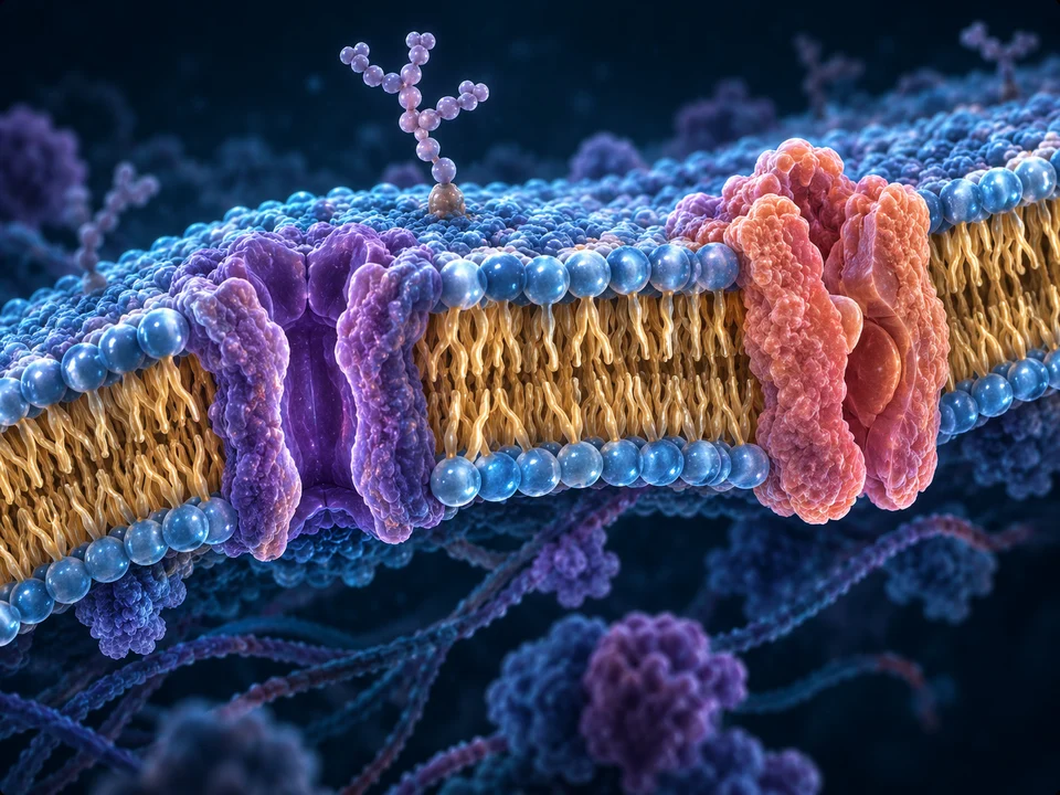
Connections between cell surfaces and extracellular components help link the cellular interior to the surrounding scaffold. The organisation and mechanical setting of matrix influence how cells behave; cells in turn produce and reorganise matrix.
The illustration locates cells and fibres. It does not resolve individual adhesion receptors, prove a specific signalling pathway or simulate a cell sensing a measured force.
Structure and function: NCBI · Extracellular matrix organisation · NCBI · Cells and the extracellular matrix
Density and direction answer different questions.
A dense arrangement contains abundant fibrous material. A regular arrangement has a strong preferred alignment; dense irregular tissue contains substantial bundles oriented in different directions. The dermis provides a familiar regional example of the latter.
This helps explain why aligned tendon is suited to transmitting tension along its principal axis. It does not imply perfect parallelism, identical behaviour across every tendon, or equal strength in all directions in an irregular tissue.
Structure and function: OpenStax · Connective tissue structure and function · Tendon structure–function relationships
Waviness is part of organisation, not a force meter.
Some tendon collagen structures show crimp or waviness. Recruitment, alignment and interactions among structural levels contribute to a tendon’s mechanical response. A simplified description of straightening does not explain every part of that response.
The image includes gentle waviness to convey living fibrous organisation. Neither its amplitude nor an on-screen arrow is a measured strain, a training target or a prediction of failure.
Structure and function: Tendon structure–function relationships · Load transfer in tendons and ligaments
Tendon and ligament are not interchangeable names.
Tendons commonly transmit muscle-generated force to bone. Ligaments connect bones and help guide or constrain joint movement. Their collagen-rich organisation can overlap, while attachments, regional structure and mechanical demands differ.
The tendon itself does not contract like a muscle fibre. Continue the established hand and foot lesson for tendon courses and attachments, rather than reading this generic tissue window as a named regional tendon.
Structure and function: OpenStax · Connective tissue structure and function · Load transfer in tendons and ligaments
Do not copy one vessel pattern onto every connective tissue.
The capillary in the loose-tissue image supports local exchange. Dense tendon has its own regional vascular and connective arrangements; absence of a drawn vessel in the new cutaway does not mean a living tendon is universally avascular.
Cartilage and mineralised bone solve supply and support differently again. Their existing lessons explain those arrangements; a capillary should not be added to an avascular cartilage matrix merely to reuse this picture.
Structure and function: Tendon structure–function relationships · OpenStax · Connective tissue structure and function
One family contains very different materials.
Adipose is cell-rich; blood has a fluid matrix; cartilage and bone have distinct supporting matrices. These are useful comparisons of cells, extracellular composition and organisation, not stages through which one loose tissue must pass.
Use the established organ and tissue lessons to see the appropriate structure at the right scale. The two images here are selected loose and dense examples, not a complete histological atlas.
Structure and function: OpenStax · Connective tissue structure and function · NCBI · Cells and the extracellular matrix
A section shows structure; a mechanical test asks another question.
Section orientation, preparation and staining affect the appearance of a real tissue image. A dark elongated profile may help locate a nucleus, but a routine field does not by itself identify every resident cell or collagen subtype.
The retained acquired tissue observations remain separate from these generated atlas illustrations. Neither an attractive render nor one section measures stiffness, three-dimensional fibre orientation or the full loading history.
Structure and function: Tendon structure–function relationships · NCBI · Extracellular matrix organisation
AI-assisted normal educational explanation and generated atlas. Human medical review and physical-phone acceptance have not been performed. Existing acquired observations retain their own identities and limits.