NORMAL REGIONAL ANATOMY / CONNECTION → POSITION → CONTROL
Shoulder Complex & Arm Elevation
A mobile socket sits on a moving base.
Locate the joints that connect the arm to the trunk. Open the glenohumeral joint, compare both faces of the cuff, and connect its tissue structure to coordinated elevation and control.
30 SECONDS / READ THE CONNECTED STRUCTURES
Clavicle connects. Scapula positions. Humerus moves.
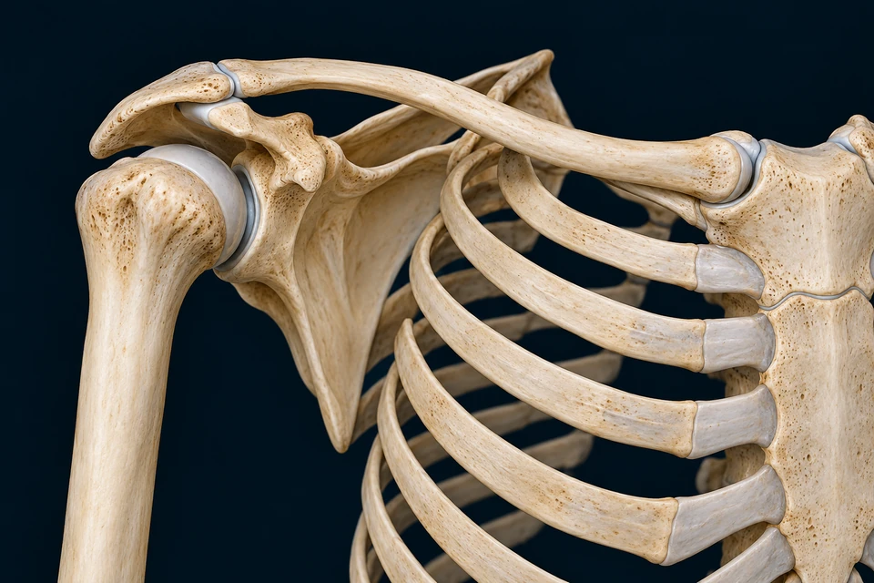
Structure notes · further context
- SC joint
- The medial clavicle articulates with the sternum. This is the girdle’s bony joint connection to the axial skeleton.
- Clavicle
- The curved strut bridges the sternum and scapula and moves during arm elevation.
- AC joint
- The lateral clavicle meets the acromion. It is separate from the humeral socket.
- GH joint
- The head of the humerus meets the shallow glenoid of the scapula.
- Coracoid
- An anterior scapular process projects below the clavicle. It is not the articular socket.
- Scapula
- A broad blade carries the socket. Its position changes relative to the thorax.
- Humerus
- The arm bone moves relative to the glenoid and to the moving shoulder girdle.
- Thoracic ribs
- The thorax supplies the curved surface around which the scapula is positioned; this is not another ordinary synovial joint.
- ConnectSternum → clavicle → scapula. SC and AC are different joints.
- PositionScapula moves on the thorax and carries the glenoid with it.
- Move and controlHumerus moves at the glenoid; cuff and larger muscles coordinate forces.
The clavicle connects the girdle to the axial skeleton.
Find the medial sternoclavicular joint, follow the clavicle and locate its lateral acromioclavicular connection. These are two separate joints.
Rings locate related structures. They do not show force, muscle activation, an exact motion axis or an extra rib joint.
Sources for this mechanism: OpenStax · The pectoral girdle · NCBI · Shoulder anatomy · NCBI · Rotator cuff anatomy
02 / ONE LINKED COMPLEX
The shoulder is more than one ball and socket.
Follow the clavicle from the sternum to the scapula, then locate the humeral head against the glenoid.
The sternoclavicular joint links clavicle to the manubrium; the acromioclavicular joint links clavicle to the acromion. The glenohumeral joint joins humerus and scapula. Together these connections let the upper limb move relative to the trunk.
The scapula also glides and rotates against the thoracic setting. This scapulothoracic articulation is a functional interface, not another ordinary synovial joint with a rib socket. The overview selectively exposes the bones; it is not a literal front-view X-ray.
Connection is different from contact
The clavicle provides the girdle’s bony connection to the axial skeleton. The scapula is also supported and positioned by muscles; its proximity to ribs does not mean that a new bony socket joins it to them.
Sources for this mechanism: OpenStax · The pectoral girdle · NCBI · Shoulder anatomy
03 / INSIDE THE JOINT
A large humeral head moves against a relatively shallow socket.
A selective oblique cutaway separates cut bone, smooth articular coverings, a peripheral rim and the enclosing capsule.

Structure notes · further context
- Humeral head
- Opened bone reveals a rounded head beneath its articular covering; the cut window is not a normal hole.
- Head cartilage
- Smooth hyaline articular cartilage covers the humeral joint surface.
- Glenoid surface
- The scapular articular surface faces the humeral head. Follow the curved opposing surface rather than the bone cut edge.
- Labral rim
- The peripheral fibrocartilaginous lip is distinct from the smooth central articular covering.
- Fibrous capsule
- An enclosing connective-tissue sleeve attaches around the articulation; its inferior part allows a recess.
- Inner capsule
- The opened inner capsular context faces the cavity. Synovial lining is associated with this inner surface, not spread over the articular cartilage.
Cartilage covers two bony articular surfaces.
Locate the smooth humeral covering and the glenoid surface. The neighbouring cut cancellous bone is a different structure.
The glenoid is the scapular articular region. Its relationship with the rounded humeral head permits movement about multiple axes. Stability depends on more than the depth of the bony socket.
The head remains normally aligned in this teaching preparation. A window is opened to reveal neighbouring surfaces, rather than showing a detached or dislocated ball. Visible separation and cut surfaces make structure readable; they do not measure an individual joint space.
A section can reveal a hidden relationship
A selective cutaway differs from an acquired MR slice. It can remove overlying tissue while preserving the intended gross connection; it cannot establish a patient-specific shape or the thickness of every membrane.
Sources for this mechanism: OpenStax · Selected synovial joints · NCBI · Glenohumeral joint
04 / SURFACE AND RIM
Smooth articular cartilage and a labral lip have different jobs.
Look at the covering of the humeral head, then at the margin of the glenoid.
Hyaline articular cartilage covers the opposing bony joint surfaces. It contributes to low-friction load transmission with the surrounding fluid environment. It is not the same structure as the fibrocartilaginous labrum at the socket’s edge.
The labrum augments the glenoid margin and participates in the capsulolabral complex. It is not a separate knee-like meniscus laid across the humeral head. Its detailed attachments and shape vary; this image teaches the broad rim rather than a surgical footprint.
A smooth surface still has living tissue biology
Cartilage structure, fluid and matrix properties matter for mechanical behaviour. Keep the detailed matrix lesson separate from claims about the thickness, material stiffness or wear of this generated joint.
Sources for this mechanism: OpenStax · Synovial joints · NCBI · Glenohumeral joint
05 / CAPSULE AND LINING
An enclosing sleeve can permit motion and constrain it.
The capsular envelope surrounds the articulation; its inner surface and the articular covering are separate tissue relationships.
The fibrous capsule connects around the glenoid and proximal humeral region. Capsular thickenings and ligaments contribute differently as joint position changes. A loose part of the envelope in one position is not evidence that it has no stabilising role.
Synovial lining lies along the inner capsular surface and contributes to the joint-fluid environment. It does not coat the articular cartilage as a continuous extra skin. The picture identifies an inner capsular context, without resolving every microscopic layer.
Variation limits one universal capsule drawing
Dissection and micro-CT studies show regional differences in attachments and thickness. A normal educational cutaway is a selected gross arrangement, not a millimetre-scale map of every capsular band or recess.
Sources for this mechanism: Whole shoulder-capsule attachment and thickness · anatomic study · Supporting layers of the glenohumeral joint · anatomic study · OpenStax · Synovial joints
06 / A COORDINATED CUFF
Four scapula-to-humerus muscles contribute to movement and centring.
Use the posterior plate for supraspinatus, infraspinatus and teres minor; use the separate anterior plate for subscapularis.
From behind · three cuff members
Right shoulder. Medial scapula is at image left; arm at image right.

Structure notes · further context
- Acromion
- The lateral continuation of the scapular spine arches over the cuff.
- Supraspinatus
- The superior posterior cuff member contributes to elevation and joint control.
- Scapular spine
- The bony ridge separates the supraspinous and infraspinous regions.
- Infraspinatus
- The broad muscle below the spine contributes to external rotation and stability.
- Teres minor
- The narrower lateral-border member is separate from the neighbouring teres major.
From in front · subscapularis
Right shoulder. Medial scapula is at image right; arm at image left.

Structure notes · further context
- Subscapularis
- The broad muscle occupies the anterior, costal scapular surface, not the posterior infraspinous fossa.
- Cuff tendon
- Muscle fibres converge toward the pale tendon at the proximal humerus.
- Humeral attachment
- The subscapularis reaches the anterior proximal humerus at the lesser-tubercle region.
- Coracoid
- The anterior scapular hook is above the subscapularis in this exposure.
- Medial border
- The medial border is at image right here, opposite to the posterior preparation.
- Humerus
- The arm bone is at image left in this anterior view.
A superior cuff contribution combines elevation and stability.
Read the muscle above the posterior scapular spine, then follow its tendon toward the humerus under the acromial region. Its contribution is not confined to one obligatory opening angle.
Compare two exposures of the same side, not two halves of one continuous picture. Selection changes teaching emphasis, not measured force or a simulated contraction.
The cuff tendons associate with the capsule and neighbouring tendons. Their coordinated force helps maintain humeral-head alignment while larger movers act. This does not mean that the cuff locks a normally moving joint in place.
Muscle names are useful location clues, but broad action labels are not rigid assignments. Joint position, load and recruitment change the contribution of each muscle and of its subregions. Supraspinatus and deltoid do not obey a universal hand-off at one fixed elevation angle.
A cuff is not four isolated ropes
Anatomical studies describe blending and interdigitation between adjacent tendon regions. The selected views expose the gross muscles and tendon transitions, not exact independent strips, measured footprints or individually certified fibre compartments.
Sources for this mechanism: NCBI · Rotator cuff anatomy · Rotator cuff subregions · anatomy and function review · Rotator cuff tendon insertion · anatomic study
07 / READ BOTH SIDES
The fourth cuff member lies on the front of the scapula.
A posterior view cannot reveal the full subscapularis origin-to-insertion relationship.
Subscapularis occupies the anterior, costal surface of the scapula and reaches the lesser-tubercle region of the humerus. Its contribution includes internal rotation and joint stability. The posterior image should not acquire an invented extra muscle to represent it.
Supraspinatus, infraspinatus and teres minor reach the greater-tubercle region from other scapular surfaces. The front and back preparations therefore reverse image-side relationships for the same right shoulder. Read each orientation note before comparing.
Compare the two oriented cuff exposures →
Teres major is a neighbour, not the fourth cuff member
Similar names do not establish membership. Teres major contributes to movements of the humerus but is not one of the four rotator-cuff muscles.
Sources for this mechanism: NCBI · Rotator cuff anatomy · NCBI · Shoulder muscles and innervation
08 / COORDINATED ELEVATION
Raising the arm changes both the humerus and its moving base.
Glenohumeral motion works with scapular and clavicular movement rather than exhausting one joint before starting another.
Structure notes · further context
- SC joint
- The medial clavicle articulates with the sternum. This is the girdle’s bony joint connection to the axial skeleton.
- Clavicle
- The curved strut bridges the sternum and scapula and moves during arm elevation.
- AC joint
- The lateral clavicle meets the acromion. It is separate from the humeral socket.
- GH joint
- The head of the humerus meets the shallow glenoid of the scapula.
- Coracoid
- An anterior scapular process projects below the clavicle. It is not the articular socket.
- Scapula
- A broad blade carries the socket. Its position changes relative to the thorax.
- Humerus
- The arm bone moves relative to the glenoid and to the moving shoulder girdle.
- Thoracic ribs
- The thorax supplies the curved surface around which the scapula is positioned; this is not another ordinary synovial joint.
The arm moves at the glenoid.
The gold cue indicates the broad raising direction of the humerus in this frontal anatomical context. Glenohumeral elevation is measured relative to the scapula, not the room or the thorax.
Gold arrow: a qualitative direction cue on the unchanged anatomical preparation. Rings locate the contributing structures. These movements overlap; this selector is not three successive phases, a measured axis, a fixed rhythm or a prediction of an individual range.
As the arm rises, scapular upward rotation and tilting help orient the glenoid. Clavicular motion at the sternoclavicular and acromioclavicular connections participates in this linked behaviour. The scapula does not simply slide straight upward.
Descriptions of scapulohumeral rhythm depend on the measured rotations, task, plane and individual. A familiar ratio is a teaching approximation rather than a universal rule at every instant. The static overview is not a calibrated animation of a person’s movement.
Locate the moving base, the clavicular link and the arm →
Be clear about the reference frame
Humerus relative to scapula differs from humerus relative to thorax. A change in the latter combines movements across the complex; a single external arm angle cannot independently quantify each contribution.
Sources for this mechanism: NCBI · Shoulder anatomy · Scapular kinematics variability · systematic review
09 / MOVING AND SUPPORTING
Scapular positioning and humeral movement are related tasks.
A muscle may help move one structure while helping stabilise another connection.
Serratus anterior and trapezius contribute to scapular positioning, with other regional muscles participating. Deltoid and the cuff act in the humeral setting; pectoralis major and latissimus dorsi connect the arm with wider trunk regions.
Holding an arm still can require active force. Adding a load changes the external moment and the muscular demand without necessarily changing visible joint position. The same pose therefore does not uniquely identify which muscles are active or how much force they produce.
Position is not a muscle-activation recording
The main pictures locate anatomy and attachments. They do not measure electromyography, force sharing or the activation of every muscle around the thorax. Full-body muscular atlas gaps remain separately recorded.
Sources for this mechanism: OpenStax · Pectoral girdle and upper-limb muscles · Rotator cuff subregions · anatomy and function review
10 / ADJACENT PATHWAYS
A tendon, a capsule and a bursa should not be merged into one object.
The long biceps tendon and the subacromial bursa have relationships to the shoulder without becoming additional cuff muscles.
The long head of biceps has a superior scapular/labral attachment and passes through the superior joint region toward the humeral groove. Its intracapsular segment is classically extrasynovial. It is not a free cord floating through an unrestricted fluid chamber.
The subacromial-subdeltoid bursa lies superficial to the supraspinatus tendon and normally remains separate from the glenohumeral cavity. Its job is a local gliding relationship. The new pictures omit a complete biceps-pulley and bursal reconstruction rather than drawing uncertain extra cords.
Omission does not establish absence
Selective exposure makes one relationship clearer while hiding others. A clean teaching picture cannot demonstrate a normal communication, tendon defect or inflamed bursa in a living person.
Sources for this mechanism: Long bicipital tendon · normal MR anatomy · NCBI · Glenohumeral joint
11 / FORCE INTO TISSUE
A muscle’s pull reaches bone through organised connective tissue.
Follow red muscle toward paler tendon, then separate attachment biology from a simple rope-to-hook analogy.
Tendons transmit force through collagen-rich tissue and their attachments to bone. A transition region can involve graded tissues; its material organisation differs from the contractile cell machinery that generates force.
The humeral attachment context shown here is broad anatomy. It is not an exact cuff-footprint measurement, collagen-fibre map or universal enthesis micrograph. The established tendon course supplies the deeper tissue and material lesson.
Connection does not fix the magnitude
Direction, moment arm, recruitment and tissue behaviour affect the mechanical result. The thickness or brightness of a drawn tendon cannot supply its load capacity or an individual safe exercise load.
Sources for this mechanism: Rotator cuff tendon insertion · anatomic study · Supporting layers of the glenohumeral joint · anatomic study
12 / REGIONAL SUPPLY
Muscle names and nerve names describe different parts of the relationship.
Trace supply in the regional knowledge route rather than guessing it from a coloured strand in a generated picture.
The suprascapular nerve supplies supraspinatus and infraspinatus. The axillary nerve supplies teres minor and deltoid. Upper and lower subscapular branches supply subscapularis. These are useful normal relationships, not a complete map of every individual’s branching variation.
Scapular and circumflex-humeral arterial networks support tissues around the region. Vessels, sensory pathways and motor supply have distinct courses. Their full hidden paths are intentionally not assigned to unverified details in these bone and muscle preparations.
Keep source identity and anatomical proof separate
A named mesh or generated line does not verify an exact nerve course, root contribution or arterial junction. The existing brachial-plexus teaching offers a source-bounded regional connection; no new WebGL or model-identity acceptance is implied.
Sources for this mechanism: NCBI · Shoulder muscles and innervation · NCBI · Rotator cuff anatomy
13 / FEEDBACK AND CONTROL
Movement depends on sensing, command and the mechanical setting.
Muscle, tendon, joint and cutaneous information contributes to coordinated movement; none works as a single universal shoulder-angle sensor.
Muscle activation is adjusted within wider neural control. Sensory feedback and the planned task influence coordination alongside the geometry and load of the shoulder complex. A static attachment picture does not represent the whole control loop.
The same cuff can contribute to rotation, elevation and stability in different combinations. This is why selected anatomical roles are shown as overlapping comparisons rather than a fixed serial chain of one muscle after another.
Control is not a personalised prescription
The lesson explains normal structural relationships and general function. It does not assess weakness, pain, instability, rehabilitation stage or return-to-sport readiness.
Sources for this mechanism: OpenStax · Pectoral girdle and upper-limb muscles · Rotator cuff subregions · anatomy and function review
14 / OBSERVE AND COMPARE
A specimen, scan, surface motion and muscle signal answer different questions.
Use the method and viewing direction to decide what a picture can support.
Anatomical dissections establish relationships and variation. Imaging reveals tissues under a specified acquisition and plane. Kinematic studies measure movement using their chosen coordinate definitions, while electromyography samples electrical activation rather than directly measuring tendon force.
These original atlas preparations are not acquired specimens or clinical scans. They explain selected normal anatomy and mechanisms with independent names and orientation notes. Fine generated tissue and a complete patient-specific joint remain outside their evidence.
Whole-chapter scope
This course retains the prior qualified cuff and tendon explanations while adding girdle, internal-joint and anterior-cuff teaching. It does not close the old full-Muscles trunk/head-neck artwork gaps, certify human medical review or teach a shoulder diagnostic test.
Sources for this mechanism: Whole shoulder-capsule attachment and thickness · anatomic study · Scapular kinematics variability · systematic review · Rotator cuff subregions · anatomy and function review
Check the structure before naming its job.
Does the scapula have a normal rib socket?
No. Scapulothoracic articulation describes a functional gliding/rotation relationship, unlike the ordinary synovial GH, AC and SC joints.
Is the labrum the same as the articular covering?
No. The fibrocartilaginous peripheral rim and hyaline articular surfaces have different positions and roles.
Can a posterior picture reveal all four cuff bellies?
No. Subscapularis occupies the anterior costal surface; compare the separately oriented anterior view.
Does one muscle stop at a universal elevation angle?
No. Recruitment, joint position and load affect overlapping contributions.
Does a visible arm angle measure GH motion alone?
No. Humerus-to-thorax movement includes the moving scapular/clavicular setting.
Does this atlas establish a safe exercise or diagnose pain?
No. It explains normal anatomy and general mechanics, not individual assessment or treatment.
Sources and image identity
Normal anatomy and mechanism source checks: 22 October 2026. Existing book-inventory entries FB017, FB028, FB032 and FB033 guide coverage; no book prose, source illustration or patient image is reproduced.
- OpenStax · The pectoral girdleReadable official text for clavicle/scapula landmarks and the axial connection. Source illustrations are not copied; generated selective exposure is not a radiograph.
- OpenStax · Selected synovial jointsOfficial educational shoulder text for shallow glenoid, labrum, capsule and cuff support. No clinical manoeuvres, treatment or size ratio adopted.
- OpenStax · Synovial jointsNormal cavity, articular-cartilage and capsule framework. The tissue illustration does not measure fluid thickness or identify a microscopic membrane.
- OpenStax · Pectoral girdle and upper-limb musclesReadable official regional framework; individual muscle roles additionally checked against independent cuff sources. Simplified image-description action/origin errors are not adopted.
- NCBI · Shoulder anatomyOfficial indexed anatomy for SC, AC, glenohumeral and functional scapulothoracic relationships. No universal motion range or shoulder test prescribed.
- NCBI · Glenohumeral jointOfficial indexed normal cartilage/labrum/capsule/bursa relationships. Direct request encountered an access challenge; no complete current full-text or fine-image certification claimed.
- NCBI · Rotator cuff anatomyOfficial indexed current anatomy for four members, attachments, stabilisation and suprascapular/axillary/subscapular supply. No fixed muscle hand-off angle or clinical injury claim.
- NCBI · Shoulder muscles and innervationOfficial indexed regional anatomy for named innervation and muscular roles. The new pictures do not trace hidden nerve or arterial branches.
- Supporting layers of the glenohumeral joint · anatomic studyOfficial indexed dissection abstract supports distinct overlying/supporting layers and deepest-layer variation. No copied dissection imagery or universal capsular thickness.
- Whole shoulder-capsule attachment and thickness · anatomic studyOfficial indexed primary abstract: thirteen cadaveric shoulders, including gross and micro-CT examination. No measured millimetres or specimen findings projected onto generated pixels.
- Rotator cuff tendon insertion · anatomic studyExisting qualified Muscle source check retained for blending/interdigitation. Fresh direct retrieval returned minimal content; no fresh full-methods read or exact footprint mapping claimed.
- Rotator cuff subregions · anatomy and function reviewReadable official abstract supports position-dependent and subregional function. Sports-treatment and return-to-play conclusions are not used as normal-person advice.
- Scapular kinematics variability · systematic reviewOfficial indexed abstract compares methods and participants with/without pain. Used to avoid a universal fixed rhythm; reported angle ranges and diagnostic implications are not applied.
- Long bicipital tendon · normal MR anatomyOfficial indexed normal-anatomy review and retained capsular anatomy distinguish intracapsular from extrasynovial. No imported MR image, complete tendon-pulley depiction or pathology diagnosis.
The girdle, joint cutaway and anterior cuff are new original generated teaching preparations. The qualified posterior cuff illustration is retained. Independent text, short names and anatomical positions are separate from the language-neutral bitmaps. These are not dissection photographs or clinical images.
Source checks, anatomical-image checks and teaching quality are distinct decisions. AI-assisted review is not physician certification. Fine generated fibre patterns, microscopic lining, hidden neurovascular routes and measured insertion footprints are not certified. This course does not diagnose shoulder pain or prescribe treatment, testing or rehabilitation.