Living support. Linked movement. Protected pathways.
Follow the column from regional curves to vertebral shape, disc tissue and neural spaces. Different scales reveal how bone, connective tissue and nerves share one framework.
▷
Watch chapter overviewFilm planned · not yet available
Source-guided bony preparation · discs omitted · fine detail illustrative
02 / COLUMN
A shared structure has different regional jobs.
Cervical, thoracic and lumbar regions transition into fused sacral support and a small coccygeal region. Differences in body size, joints and surrounding tissues matter.
Left lateral profile · anterior toward image left · bony-only view. Disc and neural tissues are shown in their own preparations below.
Original source-guided lateral bony-column preparation; anterior image left. Discs/ligaments/neural tissue are omitted from this bony-only view and taught separately. Regional form compared with licensed native atlas geometry; fine generated segmentation and individual numbering remain illustrative / not independently verified. Only empty surrounding background is cropped; the labelled detail retains this viewport; the full-resolution entry opens the uncropped source. Human medical review: Not performed.Structure notes · further context
Cervical region
The conventional seven cervical vertebrae include specialised atlas and axis. Cervical nerve pairs number eight; bone and nerve counts are different concepts.
Thoracic region
Twelve vertebrae are conventionally taught. Rib joints and surrounding tissue affect movement; not every generated level is a measured numbered specimen.
Lumbar region
Five lumbar vertebrae are conventional. Normal segmental variation exists; bone size does not prescribe one exact load capacity.
Sacrum
Five sacral segments commonly fuse during development. It is not a stack of freely moving lumbar discs.
Coccygeal region
Segment number and fusion vary. A representative tail cannot establish one universal count or posture.
The cervical region links head support with neck movement
Find the neck at the top of the profile. Atlas and axis specialise the first two joints; seven cervical bones and eight cervical nerve pairs are different counts. The bony view does not expose the nerves.
Gold emphasis identifies a teaching location, not a measured force, transport rate, movement or resolved nerve boundary.
Regional names do not imply identical repeated vertebrae.
Conventional counts aid orientation, but variation and fusion complicate the idea of a single adult bone count. Do not count generated pores or assume a numbered specimen.
Cervical
Conventionally7 vertebrae /8 nerve pairs
Specialised upper joints link head support to mobile neck.
Bone and nerve counts differ.
Thoracic
Conventionally12 vertebrae
Rib relationships contribute to the chest framework.
Movement differs across the region.
Lumbar
Conventionally5 vertebrae
Broad bodies and region-specific joints contribute to support.
Normal curvature is not a single compulsory posture.
Thoracic and sacral convexities relate to primary development; cervical and lumbar curves develop with later head and upright support. Curvature and positioning vary.
Anterior convexity
Cervical / lumbar regional context
The side profile bends toward the anterior side.
No universal angle from this image.
Posterior convexity
Thoracic / sacral regional context
Opposite regional curves connect within one column.
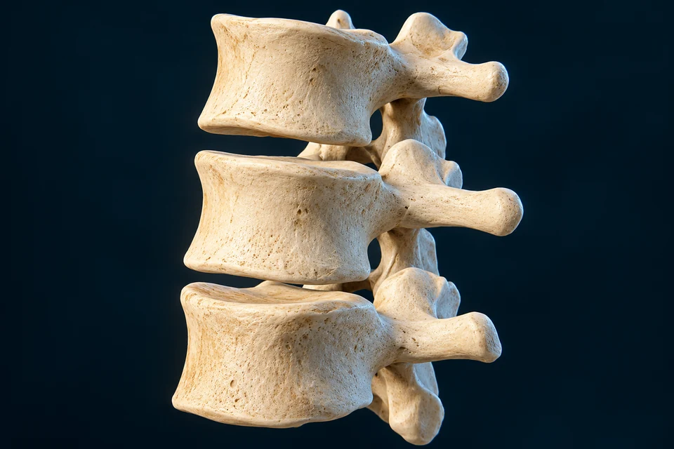
Each typical vertebra provides both support and space.
The body lies anterior to a neural opening; pedicles and laminae form an arch, with lateral and posterior projections for joints and attachments.
Original Golden-family text-free teaching preparation. Superior representative lumbar view; anterior top, posterior bottom. Broad body/arch/foramen/process relationships are source-supported. Fine generated pores and exact facet topology remain illustrative / not independently verified. This is not a named measured level or a cervical vertebra. Human medical review: Not performed.Structure notes · further context
Vertebral body
Cortical and trabecular bone coexist. It is distinct from the posterior neural canal and from a disc.
Vertebral foramen
Successive openings and surrounding tissues form the vertebral canal. A hole does not show every tissue occupying the canal.
Pedicle context
Neighbouring pedicles and other boundaries contribute to lateral intervertebral openings between levels.
Transverse process
Processes differ by spinal region. Paired cervical transverse foramina are not features of this lumbar plate.
Articular-process context
The complete facet joint requires the adjacent articular surface, capsule and lining; one isolated surface is not the whole joint.
Spinous process
Its shape differs between regions. This lumbar example is not the atlas, which lacks a conventional spinous process.
The body lies in front of the neural passage
In this superior lumbar view, anterior is toward image top. The broad body transmits load through the disc and endplate interfaces of neighbouring levels; this isolated bone has no disc tissue attached.
Gold emphasis identifies a teaching location, not a measured force, transport rate, movement or resolved nerve boundary.
The vertebral foramen is the opening in one vertebra. The vertebral canal is the connected space formed through the column and surrounding structures. Lateral intervertebral foramina are different passages between neighbouring levels.
Why are a foramen, a canal and a side opening different?
A vertebral foramen belongs to one bony level. The connected vertebral canal extends along the column, while intervertebral openings provide lateral passage between neighbouring levels. Neural tissues, coverings and vessels occupy these surroundings in life; an empty isolated-bone opening is not a complete nerve preparation.
Atlas has a ring and lateral masses. Axis has a body and dens. Skull–atlas and atlas–axis joints support linked motion, with no disc between skull/C1 or C1/C2.
Original Golden-family text-free teaching preparation. Anterior-superior regional relationship, not exact operative attachment geometry. Atlas ring, axis body/dens and broad paired contact relationships are source-supported. Fine generated facets, transverse-ligament attachments, pores and canal geometry remain illustrative / not independently verified. There is no intervertebral disc between C1 and C2. Human medical review: Not performed.Structure notes · further context
Atlas anterior arch
Atlas has arches and lateral masses rather than a vertebral body. This front arch is not an extra body.
Dens of axis
It relates to the anterior atlas arch and stabilising tissues. The drawing is not a measured rotation axis or injury model.
Axis body
Unlike the atlas, the axis does not lack its body. A posterior spinous process must not appear as an anterior body extension.
Atlas lateral mass
Superior articular regions receive skull-condyle context; inferior regions relate to the axis.
Space behind dens
A bony opening is not itself the spinal cord. Exact transverse-ligament attachments are not independently verified.
Lateral C1–C2 contact
The atlas–axis complex has specialised synovial joints rather than an intervertebral disc between the two bones.
The atlas is a ring rather than a typical body-bearing vertebra
Its anterior arch and lateral masses surround the upper passage. The skull rests on superior atlas surfaces in life. This regional view is not a complete ligament or vascular preparation.
Gold emphasis identifies a teaching location, not a measured force, transport rate, movement or resolved nerve boundary.
The atlas–axis complex allows rotation in relation to the dens and lateral contacts, supported by connective tissues. This is not the same as motion around a generic lumbar disc. No fixed angle or safe movement limit is prescribed.
Different materials share load and permit limited motion.
A hydrated nucleus, fibrous annulus and cartilage-to-bone endplate interfaces work together. The disc is not a freely floating jelly ball or a synovial cavity.
Original Golden-family text-free teaching preparation. Representative disc/endplate section, not a measured histology slide or exact named lumbar level. Broad nucleus/annulus/adjacent endplate/bone relationships are source-supported. Fine generated fibres, pores, cells and cartilage–bone interface thickness remain illustrative / not independently verified. The hydrated centre is not a blood vessel or sealed plastic balloon. Human medical review: Not performed.Structure notes · further context
Nucleus pulposus
Proteoglycans, water and cell activity contribute to behaviour. Its consistency and cell phenotype vary with age and species.
Annulus fibrosus
Collagen organisation changes across the tissue; this is not a literal set of hard concentric pipes.
Cartilage-interface context
Cartilaginous and bony endplates are distinct. Exact thickness, pores and fibre integration in generated texture are not independently verified.
Adjacent vertebral bone
Bone vascular context supports neighbouring nutrient supply. No vessel is shown delivering red blood cells directly into the nucleus.
Peripheral fibrous continuity
The illustration opens a teaching window; the disc is not normally an exposed cavity or separated stack.
A water-rich matrix resists compression
The nucleus contains a hydrated extracellular matrix maintained by cells. It is neither an empty cavity nor a free-floating fluid balloon; its behaviour depends on the surrounding annulus and endplate interfaces.
Gold emphasis identifies a teaching location, not a measured force, transport rate, movement or resolved nerve boundary.
The conventional adult mobile disc count is23, extending from C2/C3 to L5/S1. Atlas–axis lacks a disc; fused sacral levels are not ordinary mobile lumbar disc joints.
Posterior facet joints are distinct from the disc.
Adjacent articular processes meet through cartilage-covered surfaces, synovial lining and a capsule. Orientation varies across regions and helps constrain movement.
Disc articulation
Between adjacent vertebral bodies
Fibrous/cartilaginous materials share load.
Not a synovial cavity.
Facet articulation
Paired posterior articular contacts
Capsule, cartilage and synovial environment support joint motion.
Not the only synovial articulation in the wider spinal region.
Whole segment
Disc + paired facets + connective/active support
Interfaces interact rather than acting independently.
Compression, annular restraint and cartilage/bone interfaces interact. Forces vary with region, posture and activity; a simple comparison cannot supply measured biomechanics.
Compression
Body → disc / interfaces
Hydrated matrix and surrounding restraint share changing load.
Restraint
Annulus + facets + connective tissues
Different materials constrain relative motion.
Active support
Muscles + sensory control
Support is not provided by one passive tissue alone.
Supporting relationship map, not exact anatomical geometry or a measured time course. All steps are visible before highlighting.
Original Golden-family text-free teaching preparation. Representative disc/endplate section, not a measured histology slide or exact named lumbar level. Broad nucleus/annulus/adjacent endplate/bone relationships are source-supported. Fine generated fibres, pores, cells and cartilage–bone interface thickness remain illustrative / not independently verified. The hydrated centre is not a blood vessel or sealed plastic balloon. Human medical review: Not performed.Structure notes · further context
Nucleus pulposus
Proteoglycans, water and cell activity contribute to behaviour. Its consistency and cell phenotype vary with age and species.
Annulus fibrosus
Collagen organisation changes across the tissue; this is not a literal set of hard concentric pipes.
Cartilage-interface context
Cartilaginous and bony endplates are distinct. Exact thickness, pores and fibre integration in generated texture are not independently verified.
Adjacent vertebral bone
Bone vascular context supports neighbouring nutrient supply. No vessel is shown delivering red blood cells directly into the nucleus.
Peripheral fibrous continuity
The illustration opens a teaching window; the disc is not normally an exposed cavity or separated stack.
Load crosses neighbouring bodies and disc interfaces
The hydrated centre, annular matrix and endplate boundaries share a changing mechanical environment. Gold emphasis locates those materials; it is not a force arrow, pressure measurement or deformation simulation.
Gold emphasis identifies a teaching location, not a measured force, transport rate, movement or resolved nerve boundary.
Living disc cells need exchange beyond a central blood vessel.
The mature disc is largely avascular. Nutrients reach tissue through nearby vascular boundaries and matrix transport, particularly endplate context; waste moves outward.
Boundary supply
Nearby vessels → tissue boundaries
Blood is not flowing through a central nucleus vessel.
Matrix exchange
Small nutrients → diffusion through matrix
Endplate context is an important route, not a pump.
Cell / waste context
Use nutrients → exchange metabolites
Exact transport rates require measurements.
Supporting relationship map, not exact anatomical geometry or a measured time course. All steps are visible before highlighting.
Original Golden-family text-free teaching preparation. Representative disc/endplate section, not a measured histology slide or exact named lumbar level. Broad nucleus/annulus/adjacent endplate/bone relationships are source-supported. Fine generated fibres, pores, cells and cartilage–bone interface thickness remain illustrative / not independently verified. The hydrated centre is not a blood vessel or sealed plastic balloon. Human medical review: Not performed.Structure notes · further context
Nucleus pulposus
Proteoglycans, water and cell activity contribute to behaviour. Its consistency and cell phenotype vary with age and species.
Annulus fibrosus
Collagen organisation changes across the tissue; this is not a literal set of hard concentric pipes.
Cartilage-interface context
Cartilaginous and bony endplates are distinct. Exact thickness, pores and fibre integration in generated texture are not independently verified.
Adjacent vertebral bone
Bone vascular context supports neighbouring nutrient supply. No vessel is shown delivering red blood cells directly into the nucleus.
Peripheral fibrous continuity
The illustration opens a teaching window; the disc is not normally an exposed cavity or separated stack.
Blood supplies nearby boundaries rather than a central disc vessel
The largely avascular mature disc depends on exchange with vessels outside its centre. Vertebral endplate and peripheral contexts are relevant boundaries. This gross preparation locates them without pretending to resolve capillaries.
Gold emphasis identifies a teaching location, not a measured force, transport rate, movement or resolved nerve boundary.
Small-solute diffusion and fluid movement are not the same mechanism. The central nucleus is not filled with red blood cells. Exact exclusive transport fractions are not asserted.
Why is nutrient exchange different from squeezing water through a sponge?
Loading can change water distribution, but small nutrients also move down concentration gradients through matrix. Cells consume oxygen and glucose; exchange must balance that demand and remove metabolic products. The endplate and outer boundary supply contexts are not holes opening into a central blood vessel. A tissue image cannot provide an exact transport fraction or rate.
Similar-looking cells can have different regional roles.
Disc cells maintain distinct matrix environments. Adult phenotype, developmental lineage and animal models must not be treated as one universal microscopic portrait.
Anterior support and posterior neural enclosure coexist.
In a cord-bearing level, the cord lies behind bodies/discs. Side openings provide routes to local neural pathways, while posterior articular structures contribute to motion.
Original Golden-family text-free teaching preparation. Local cord-bearing upper-column context, not lower lumbar or sacral cord and not assigned an exact measured vertebral level. Broad anterior body/disc versus posterior canal and lateral neural passage relationships are source-supported. Fine generated rootlets, meninges, facet surfaces and attachments remain illustrative / not independently verified. No operative root map or precisely resolved ganglion is claimed. Human medical review: Not performed.Structure notes · further context
Spinal cord context
Cord is central nervous tissue, not a disc or a nerve passing through the body. The adult cord does not occupy the entire sacral canal.
Canal / covering context
Meningeal and CSF relationships require distinct layers; every fine membrane in this generated plate is not independently verified.
Lateral neural bundle
Rootlets join roots and local nerve pathways. This generated bundle is not assigned an exact sensory/motor branch or level.
Intervertebral opening context
It differs from the central vertebral canal. No exact foraminal dimensions or clinical compression measurement is shown.
Posterior articular context
Facet cartilage/capsule and disc articulation are different interfaces. Fine generated cartilage patches do not prove every joint boundary.
Body / disc interface
Neural tissue passes behind this interface, never through the centre of the disc or solid body.
Bodies and discs lie in front of the cord-bearing canal
Anterior is toward image left in this local cutaway. The canal occupies the space behind the bodies; posterior arch and articular regions contribute enclosure and movement. This is an upper, cord-bearing context.
Gold emphasis identifies a teaching location, not a measured force, transport rate, movement or resolved nerve boundary.
Roots and mixed spinal nerves have different roles.
Sensory information enters through dorsal-root pathways; motor output leaves through ventral-root pathways. After roots join, a local spinal nerve carries mixed fibres before further distribution.
Sensory input
Peripheral context → dorsal root → cord
A supporting direction, not a pictured exact sensory root.
Motor output
Cord → ventral root → peripheral context
Outward motor pathways differ from inward sensory pathways.
Mixed nerve
Joined roots → spinal nerve → distribution
Mixed fibres continue into regional branches and networks.
Supporting relationship map, not exact anatomical geometry or a measured time course. All steps are visible before highlighting.
Original Golden-family text-free teaching preparation. Local cord-bearing upper-column context, not lower lumbar or sacral cord and not assigned an exact measured vertebral level. Broad anterior body/disc versus posterior canal and lateral neural passage relationships are source-supported. Fine generated rootlets, meninges, facet surfaces and attachments remain illustrative / not independently verified. No operative root map or precisely resolved ganglion is claimed. Human medical review: Not performed.Structure notes · further context
Spinal cord context
Cord is central nervous tissue, not a disc or a nerve passing through the body. The adult cord does not occupy the entire sacral canal.
Canal / covering context
Meningeal and CSF relationships require distinct layers; every fine membrane in this generated plate is not independently verified.
Lateral neural bundle
Rootlets join roots and local nerve pathways. This generated bundle is not assigned an exact sensory/motor branch or level.
Intervertebral opening context
It differs from the central vertebral canal. No exact foraminal dimensions or clinical compression measurement is shown.
Posterior articular context
Facet cartilage/capsule and disc articulation are different interfaces. Fine generated cartilage patches do not prove every joint boundary.
Body / disc interface
Neural tissue passes behind this interface, never through the centre of the disc or solid body.
Sensory fibres enter the cord through dorsal-root pathways
The pale cord and lateral neural bundle locate central and peripheral surroundings. The displayed bundle does not resolve a particular sensory root. The direction statement below explains function without assigning an unverified tiny strand.
Gold emphasis identifies a teaching location, not a measured force, transport rate, movement or resolved nerve boundary.
Supporting direction guide, not exact generated root anatomy. The neural bundle in the primary plate is not individually classified as a resolved dorsal or ventral branch.
The tapered conus commonly lies around L1–L2, with individual variation. Descending roots continue below it as cauda-equina context before reaching lower exits.
Cord ending
Commonly upper lumbar vertebral context
Central neural tissue tapers before the entire canal ends.
No single fixed level for every person.
Descending roots
Below the conus within lower canal context
Roots travel farther to lower outlets.
Not a full cord continuing through the sacrum.
Levels / segments
Bony landmarks versus neural organisation
Relative positions differ along the column.
No operative access or diagnostic instruction.
Why can a vertebral level differ from a spinal-cord segment?
Column and cord growth do not preserve a one-to-one adult alignment at every level. Lower roots descend toward their exits after central cord tissue has tapered. The upper cord-bearing image therefore teaches enclosure and lateral passage; it must not be used as a picture of a continuous adult cord through the sacrum.
Muscle force, connective restraint and sensory feedback interact. Neural pathways carry information about body position and help coordinate movement with wider brain/cord circuits.
Head support, rib articulation, pelvic transfer, muscle control and neural connections coexist. Linked chapters provide deeper teaching without repeating the same main image.
A picture and a measured tissue sample answer different questions.
Bone specimens show surfaces; sections show interfaces; imaging shows living spatial relationships. These original source-informed plates do not become patient CT, MRI or microscopy.
Gross anatomy
Representative bony/soft-tissue relationships
Different views reveal different structures.
No exact named patient or level.
Microstructure
Matrix/cell/transport research
Sources support bounded normal tissue principles.
No acquired microscopy pixels or exact generated cell atlas.
Clinical imaging
Future rights/measurement work
Can add living material contrast when separately sourced.
A regional foundation keeps its remaining depth visible.
The column, vertebra, disc, canal/root context and upper-neck exception now have distinct primary views. Wider muscular, vascular, detailed nerve, developmental and clinical branches remain separate jobs.
Now taught
Region → structure → tissue → mechanism → related system
Five primary views and visible explanatory labels.
No fake coverage percentage.
Still staged
Full muscle/ligament/vascular maps, detailed neural levels and acquired specimens
Broad adult column/vertebral/disc/canal/upper-cervical relationships; selected normal matrix, transport, movement, neural, developmental and cross-system functions. Authoritative references are used critically rather than repeating every simplified or historical claim.
Explicit limits
AI-assisted visual/content production. Fine generated vascular, neural, facet, foraminal, matrix and attachment details remain illustrative / not independently verified. Human medical review: not performed. Educational only; not diagnostic, clinical or surgical material.
Revision: spine-english-2026-10-02-deep-v1. Stable local key: ahb_ch_spine_english_v1. No invented canonical CMS/atlas entity or 3D dataset. Six retained Golden-family text-free masters and twenty-eight original semantic targets retain independent names and explanations. Native anatomical teaching contexts and three source-linked deeper readings extend the normal foundation without replacing the qualified artwork. Fine generated facet, rootlet, vascular and matrix detail is illustrative / not independently verified. Bone source guidance does not establish exact individual numbering or surgical geometry. Migration physical-phone acceptance: Pending, non-blocking.
Bony structural guides and the regional nerve card derive from BodyParts3D (Database Center for Life Science, CC BY-SA 2.1 Japan) and Z-Anatomy (CC BY-SA 4.0; PC export by Lluís Vinent Juanico). New camera, lighting, materials and AI-assisted bony refinements by AllHumanBody; those derived views use CC BY-SA 4.0. Guide source: Z-Anatomy preserved version. Source geometry supports shape comparison, not proof of every generated pore, attachment or segment.