Musculoskeletal / VISUAL EXPLAINER
What Joins Your Pubic Bones at the Front of the Pelvis?
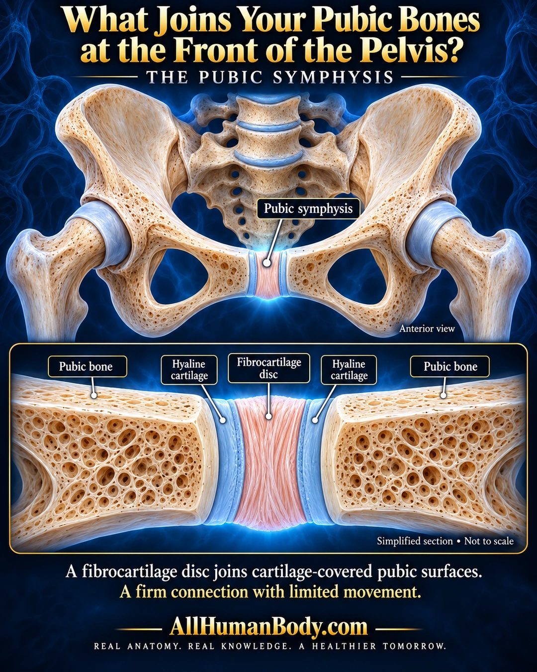
At the front of the pelvis, the pubic symphysis joins the two pubic bones. A fibrocartilage disc lies between their cartilage-covered surfaces, forming a firm connection with limited movement.
READ THE IMAGE
What the illustration shows
Navy and gold anatomy poster titled What Joins Your Pubic Bones at the Front of the Pelvis? An anterior pelvis in ivory shows a peach-and-blue pubic symphysis highlighted at the central junction above the pubic arch. Its label points to this junction, not either large obturator opening. A large lower inset shows pubic bone on each side, a blue hyaline cartilage layer at each facing surface, and a peach fibrous disc between them. All five regions are labeled. The inset says Simplified section, Not to scale. Text describes a firm connection with limited movement. Gold AllHumanBody.com and the full project slogan finish the poster.
How to interpret this poster
- Cartilage thickness and texture are exaggerated, not a normal-gap measurement or histology. Some discs contain a cleft; no pregnancy, birth-separation or diagnostic claim.
- AI-assisted educational illustration; not physician certification, microscopy, a diagnosis or individualized medical advice.
- Fine annotations need enlargement; the full description provides an accessible text alternative.
- Display the full uncropped illustration; do not trim titles, legends or boundary annotations.
AI-assisted educational illustration, adapted into web-sized images without changing its composition. This is general education, not an individual diagnosis or treatment recommendation.
EVIDENCE / CONTEXT
Sources and what they support
- Cleveland Clinic medically reviewed anatomy page ↗
Position, fibrocartilage disc, hyaline surface layers and limited flexibility.
- OpenStax anatomical textbook ↗
Pubic symphysis as a limited-mobility fibrocartilaginous connection.