Air travels through branching passages to delicate exchange surfaces. Chest movement renews that air, blood carries gases onward, and neural feedback adjusts breathing. The lung is an organ where airway, tissue, circulation and muscle mechanics must work together.
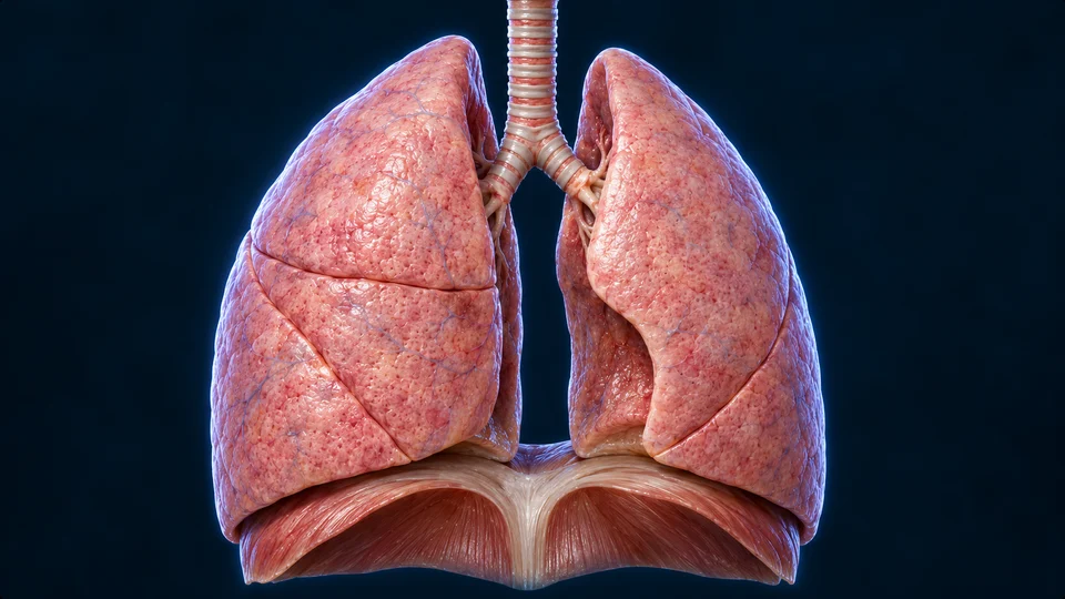
Generated anatomical atlas, not a lung photograph. Anterior view: the person’s right lung is image left.
Selected gross context. Fissure paths, surface vessels and diaphragm dimensions are illustrative; the main visible lobe relationship is not a map of every bronchopulmonary segment.
Chapter overview video is planned. No playable overview or invented duration is shown.
Upper passages → branches → respiratory region
Follow the route, then look through its wall.
The nose or mouth, pharynx and larynx lead to the trachea. Main bronchi distribute air into each lung; progressively smaller airways reach the exchange region.
Generated opened right-lung context. Selected branch ends are exposed by the illustrative dissection, not extra passages that spill into the chest. Fine segment connections, branch generations and vascular identities are not verified individually.
A passage changes into an exchange surface
The conducting region carries air without being the main site of gas exchange. Respiratory bronchioles lead into alveolar ducts and sacs. The change is regional: a ciliated conducting wall and an alveolar exchange wall are not interchangeable linings.
Conducting · Trachea, bronchi and terminal bronchioles. Transport and condition air before the exchange region.
Nasal surfaces help warm, moisten and filter incoming air. The shared pharynx leads toward the laryngeal airway and the separate food passage. The epiglottis and coordinated laryngeal closure help protect the airway during swallowing; the epiglottis is not a valve opening each alveolus.
A branch is not an alveolus
The sequence is trachea → main bronchi → smaller bronchi → bronchioles → respiratory bronchioles and alveolar region. Cartilage helps support the trachea and larger bronchi. Bronchioles lack the bronchial cartilage framework and their smooth muscle changes airway calibre. Conducting terminal bronchioles precede the gas-exchanging respiratory region.
Two lungs, unequal outlines
The right lung has three lobes; the left has two and a cardiac indentation. A hilum on each medial lung surface admits bronchi, vessels, nerves and lymphatic connections. A lobe contains many branches and exchange units; it is not one hollow balloon or one single airway.
Air and blood approach separately
Pulmonary arterial blood comes from the right ventricle toward exchange capillaries. Pulmonary veins return oxygenated blood toward the left atrium. Bronchial vessels also support conducting tissues. Red or blue artwork is a teaching colour code: actual blood is not bright blue, and colour alone cannot prove a branch’s identity.
Organ wall → epithelial cells → clearance
One sheet has several jobs.
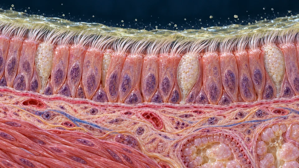
The airway lumen is above; connective support is below. The selected enlargement represents a proximal airway, not every bronchiole or alveolus.
Generated tissue atlas, not histology. Pseudostratified means one epithelial sheet with varied nuclear heights, not tall cells placed on a separate basal-cell floor. Counts, cell outlines, ciliary density and mucus thickness are illustrative.
A living lining meets incoming air
In a representative proximal airway, tall ciliated cells and secretory cells share a pseudostratified epithelial sheet. Nuclei occur at differing heights; this does not mean that the tall cells stand on a separate layer of basal cells. Small basal cells do not reach the lumen.
Capture and transport are different jobs
Mucus helps trap inhaled material. Apical cilia move the surface layer toward the upper airway, where material can be swallowed or cleared. Cilia are moving cell projections; mucus is the material they help transport. Neither is an oxygen-carrying blood vessel.
The smaller airways do not simply repeat the trachea
As airways narrow, their lining and supporting wall change. Distal bronchiolar secretory cells include club cells. Alveolar surfaces instead contain specialised thin and secretory epithelial populations. This proximal illustration is not a cell map of every airway generation.
A branching route becomes a large exchange surface
Many neighbouring spaces. Shared living walls.
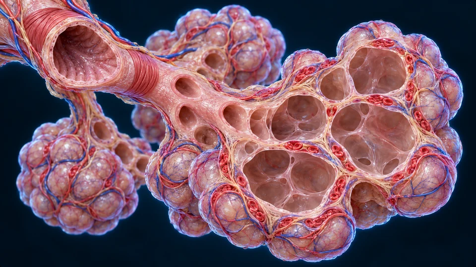
Opened air cavities make their relationship to the blood channels visible. The red cells remain inside those channels.
Generated selected respiratory context, not microscopy. Opened walls are a teaching cutaway; sizes, air-sac counts, alveolar duct geometry and capillary paths are not measured. Blood is not free inside alveolar air spaces.
Why so many small spaces?
The branched respiratory region brings a large tissue surface close to both air and blood. Thin septa, nearby capillaries and renewed alveolar gas support diffusion. Useful exchange depends on available surface, a short diffusion path and partial-pressure differences; a large air cavity without nearby blood would not do the same job.
Ventilation is not cellular respiration
Breathing moves air; lung exchange moves gases across a tissue boundary. Blood transport takes them between organs. Cellular metabolism uses oxygen in energy-related reactions and produces carbon dioxide. These are connected processes, not four names for the same movement.
Authentic light microscopy · compare with the atlas
Thin walls surround many air spaces.
A photographed tissue section shows a small airway among alveolar profiles. Compare its folded wall with the delicate partitions around it.
Authentic tissue photograph, not generated microscopy. Source title: Normal lung; source description identifies a bronchiole with surrounding alveoli. This is source attribution, not an independent patient diagnosis. Full frame retained; proportional WebP encoding only. No crop, recolouring, retouching, AI detail or invented scale. The image alone does not establish stain, magnification, species, individual cell types or the ultrastructural gas-exchange barrier.
The small airway provides a route for air. Around it, pale alveolar profiles are divided by much thinner tissue partitions. Capillaries normally lie within those septa; this field is useful for recognising the overall arrangement, not for assigning every dark nucleus to a specific cell or claiming each tiny blood channel is resolved.
A section is one slice through a three-dimensional organ
Tissue preparation and the cutting plane affect the apparent shape of the spaces and walls. A pale profile is not a live balloon whose volume can be read from the screen. This single field cannot trace every airway connection, identify the exact bronchiolar branch level, or show gas molecules moving. Use the separate barrier illustration below for a selected explanation of structures that this scale does not resolve.
Normal structural and preparation context: SIU respiratory histology guide. Original teaching explanations; no university image or prose reproduced. Source and AI-assisted image review do not constitute human medical review.
Surface film · epithelium · supporting interface · endothelium
Gases cross the wall. Blood cells do not.
Air lies above the opened septum; red cells lie within the capillary lumen below. Separation and some cell sizes are enlarged to show the relationship.
Original generated cellular atlas, not authentic microscopy. The supporting interval is expanded; it does not depict the thinnest fused interface at true scale. Fine membranes, organelles, nuclei and endothelial boundaries are illustrative. No macrophage is assigned to a non-resolved profile.
Air and blood meet across a wall, not in one space
This original opened septal illustration places alveolar air above a capillary blood space. Oxygen crosses the alveolar epithelial boundary, its supporting interface and capillary endothelium before entering blood. Carbon dioxide crosses in the opposite net direction during ordinary lung exchange.
Three cell roles should not become one generic lining
Thin type I alveolar epithelial cells provide much of the exchange surface. Type II cells produce surfactant and contribute to epithelial renewal. Alveolar macrophages participate in defence; they are not another continuous layer of the exchange wall.
Type I cell · Alveolar epithelial surface. A very thin cell body supports a short diffusion route.
Type II cell · Among alveolar epithelial cells. Produces surfactant and participates in repair.
Macrophage · Alveolar defence context. Engulfs material; omitted from this selected barrier view.
A surface film changes the mechanics of an air sac
Surfactant acts at the air–liquid interface and reduces surface tension. This helps stabilise air spaces and reduces the work needed to expand them. It is not the same surface material as the mucus-clearance layer in a conducting bronchus.
A septum contains exchange and support regions
The shortest exchange routes lie in thin regions where epithelial and endothelial supporting interfaces are closely apposed. Other septal regions contain more connective support. The illustration exaggerates separation to make boundaries visible; it does not claim that every part of every septum has one uniform thickness.
Direction follows partial pressure
Under ordinary lung conditions, oxygen has a higher partial pressure in alveolar gas than in arriving mixed-venous blood; its net movement is toward blood. Carbon dioxide has the opposite net movement. Gases do not travel because oxygen is always “pulled” by a red icon. At tissues, the local gradients and metabolic use support the corresponding delivery and removal.
A short route and a stable surface
Thin regions bring epithelial and endothelial support interfaces close together. Other septal areas carry connective support. Type II cells release surfactant, helping lower surface tension at the air–liquid interface. The whole lining cannot be replaced by one generic red cell; epithelial maintenance, defence and gas carriage belong to different populations.
STRUCTURE BEFORE SIMULATION
Connect the heart and lungs in source anatomy.
Use the existing adult male reference to locate the major receiving spaces, outflow vessels and paired lung context. The chapter preparations explain flow and exchange; the source surfaces do not simulate those processes.
Related learning, not a new approved anatomical representation. Existing source credits remain visible in the viewer. No new model, blood-flow measurement or patient treatment instruction is added.
Chest wall ↔ lung surface
Slide smoothly. Move together.
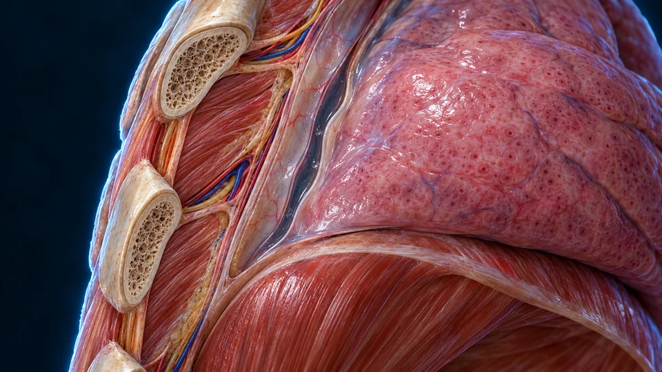
This section opens the chest-wall / lung relationship. The pleural interval is enlarged so its two facing surfaces can be distinguished.
Generated anatomical enlargement. The widened dark interface is not a normal air pocket or pneumothorax; lighting highlights are not bubbles to diagnose. Exact serosal thickness and the histological pleural boundaries are not independently resolved.
Two facing surfaces couple the lung to the chest
Visceral pleura covers the lung; parietal pleura lines the chest-wall side. A small fluid-filled interface allows sliding while helping transmit chest movement. The pleural space is not an airway or an open cavity full of breathing air.
Different pressures, different spaces
Alveolar pressure relates to air inside the lung; intrapleural pressure relates to the interface around it. Their difference helps keep the lung expanded against its elastic recoil. During ordinary inspiration, chest expansion makes intrapleural pressure more negative and expands the lung; airflow follows the resulting alveolar-to-ambient pressure difference.
Why the two pleural surfaces matter
Visceral pleura covers the lung and parietal pleura lines the thoracic boundary, including the diaphragmatic side. A small amount of fluid supports sliding without turning the lung into a loose organ disconnected from chest motion. The pleural interface does not join the bronchial tree or transport each inhaled breath.
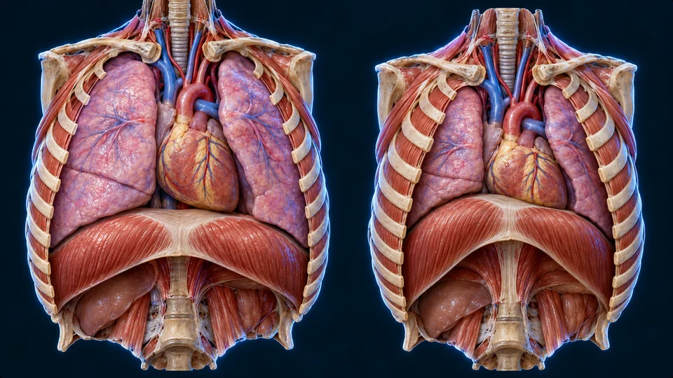
Left: an inspiratory context. Right: a quieter expiratory context. Both anterior preparations expose the diaphragm; neither is a measured time frame.
Generated paired anatomical contexts. Relative poses teach inspiration and quiet recoil, not calibrated chest volume, exact rib motion, a shared measured scale or pathology. Leaders exit their own preparation; the heart does not pump breathing air.
Air follows a pressure difference
When inspiratory muscles enlarge the chest, alveolar pressure falls relative to the outside air and air enters. During quiet expiration, relaxation and elastic recoil favour the reverse pressure difference. Airflow and chest movement are related but are not the same quantity.
Quiet expiration differs from active effort
Ordinary quiet expiration relies largely on relaxation and elastic recoil. Stronger expiration can recruit abdominal and other muscles. The paired anatomy and two-state comparison describe quiet breathing; it is not a model of every exercise, cough or forced breath.
A narrower route makes movement harder
Airway resistance opposes flow. Changes in airway radius strongly affect that resistance; the branching network and operating conditions also matter. This comparison explains a relationship, not an exact human airway calculation or a test for disease.
Moved air is not all fresh exchange-region air
Some inhaled air occupies conducting passages rather than reaching an exchange surface on that breath. Alveolar ventilation concerns fresh air delivered to the exchange region. Rate and breath size interact, so a faster visible chest rhythm alone cannot establish effective gas exchange.
Bulk movement · Air moved at the airway entrance. Includes conducting-passage space.
Alveolar delivery · Fresh air reaching exchange regions. Supports renewal of alveolar gas.
Volumes describe different parts of a breath
Tidal volume is air moved in one ordinary breath. Inspiratory and expiratory reserves describe additional air beyond that breath; residual volume remains after maximal expiration. Vital capacity combines the movable volumes, while total lung capacity also includes the residual component. These are definitions, not personalized targets or values inferred from the rendered chest.
Minute movement versus alveolar renewal
Total air movement depends on breath size and frequency, but some moved air fills conducting passages. Changing rate without considering depth and conducting-space ventilation can change effective alveolar renewal. Spirometry measures selected airflow and movable volumes; it cannot directly measure all residual-containing capacities by itself.
Lung exchange → blood → working tissues
Both sides of the boundary need a supply.
Air delivery and blood delivery work together
Ventilation supplies alveolar gas; perfusion supplies capillary blood. Useful exchange depends on both. A region receiving air without enough blood, or blood without enough fresh air, cannot contribute in the same way as a well-supplied region.
Crossing the lung wall begins a blood journey
Most oxygen in blood is carried by haemoglobin inside red cells; a smaller amount is dissolved. Carbon dioxide travels in several forms, including bicarbonate. Blood then connects lung exchange to tissues; this chapter links to the existing blood lesson instead of repeating its red-cell plate.
Supply is regional, not perfectly uniform
Posture, gravity and local airway / vessel conditions influence regional ventilation and perfusion. Low alveolar oxygen can constrict local pulmonary arterioles and redistribute flow; this differs from the usual systemic-tissue response to low oxygen. A mismatched region cannot be repaired just by drawing a thicker arrow across its wall.
Transport continues beyond diffusion
Oxygen enters plasma and binds haemoglobin in red cells. Much carbon dioxide is converted into bicarbonate, especially through red-cell chemistry, with additional dissolved and haemoglobin-associated forms. In the lungs, the chemistry and local partial-pressure conditions support carbon dioxide unloading; oxygenated blood returns to the left heart for systemic delivery.
Central chemoreception responds importantly to carbon-dioxide-related changes in brain-fluid pH. Peripheral chemoreceptors contribute information about oxygen as well as carbon dioxide and acidity. These signals interact with ongoing rhythm and context; a single low-oxygen on/off box would be misleading.
Automatic breathing still activates skeletal muscle
Brainstem networks generate and shape breathing activity. Motor commands to the diaphragm and intercostal muscles use somatic motor pathways. Automatic timing does not make those skeletal-muscle outputs wholly autonomic. Autonomic airway effects and respiratory muscle drive are distinct roles.
Breathing adjusts to activity and behaviour
Exercise, speech and deliberate changes interact with automatic breathing control and sensory feedback. No one sensor accounts for every change. The control explanation describes functional relationships, not a precise neuronal wiring map or measured timing sequence.
A distributed controller
Medullary and pontine networks generate and shape respiratory activity. Central sensing responds importantly to carbon-dioxide-related acidity in brain fluid. Carotid and aortic bodies provide peripheral chemical information, including arterial oxygen. Sensory input from lungs and higher-brain influences also contribute. This is not one isolated oxygen switch.
Commands reach muscles
Spinal somatic motor pathways activate respiratory skeletal muscles. The phrenic nerves supply the diaphragm; intercostal nerves supply intercostal muscles. Brainstem timing can be automatic even while its final muscle output is somatic. Autonomic effects on airway calibre are a related but different role.
Behaviour shares the same apparatus
Speech meters exhaled air, exercise changes ventilatory demand, and voluntary cortical control can alter breathing for a time. Chemical and mechanical feedback keep interacting with these behaviours. No illustration here maps every respiratory neuron or provides a breath-holding or treatment instruction.
An actual frame shows vocal folds around their central opening. It does not replace a full head-and-neck section.
Authentic acquired source, selected full frame at 30.000 seconds. Source describes normal female vocal folds; AllHumanBody has not made an independent diagnosis. Stroboscopy produces apparent sampled motion, not ordinary real-time motion. The still image cannot establish complete upper-airway orientation or airway function.
Air pressure from expiration supports vocal-fold vibration when the folds are brought into a suitable position. Laryngeal muscles alter their tension and position; the pharynx, mouth and nose shape resonance, while the tongue and lips contribute articulation. The lung supplies airflow but does not contain the vibrating vocal folds.
The airway must also stay protected
The larynx balances breathing, phonation and airway protection. Swallowing requires coordinated closure and movement, and coughing helps clear material. A sampled laryngoscopic frame answers a different question from a sagittal route map or a cellular barrier enlargement.
Evidence & limits
Different images answer different questions.
A rendering, a micrograph and a measured trace answer different questions
The main lung images teach gross anatomy. The new tissue plates are original source-bounded illustrations, not observed microscopy. Histology can reveal real tissue organisation; measured respiratory traces describe changing function. Clinical imaging and disease comparisons remain separate later work.
Gross anatomy, tissue relationships and mechanisms have been checked against independent sources; original explanations use the existing book-topic coverage as a learning benchmark. This is not human medical review or individual clinical advice. Generated fine cell dimensions, branch paths, serosal boundaries and receptor circuitry remain illustrative. Real laryngoscopy remains acquired imaging, never AI microscopy.
A verified full upper-airway cutaway, higher-resolution alveolar electron microscopy, paired measured chest imaging and quantitative spirometry are deeper visual gaps. They are not claimed complete by adding headings. Disease, emergency and treatment teaching belong in separate evidence-based topics.
Generated atlases are educational interpretations, not specimens, scans or histological measurements. Desktop and simulated narrow-screen checks are separate from physical-phone acceptance and human medical review.
 A living airway
 A different scale
 Moving together
 Why air moves


