Respiratory / VISUAL EXPLAINER
How Pleural Effusion Changes Lung Expansion
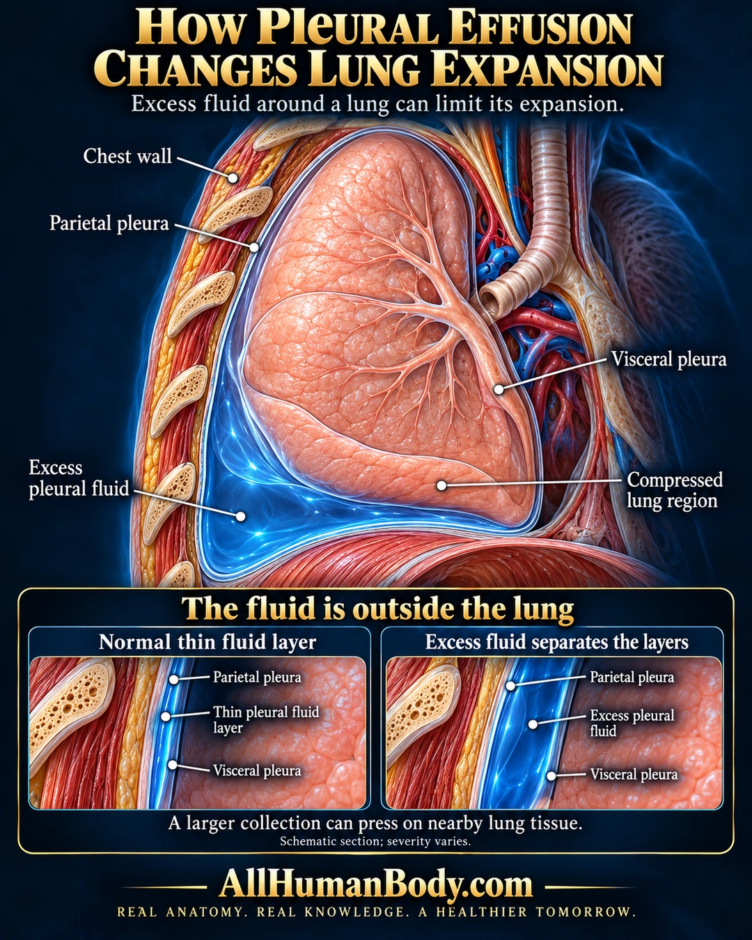
Pleural effusion is excess fluid between the lung-side and chest-wall pleural layers. A larger collection can press on nearby lung tissue and limit expansion. The fluid is outside the airways and air sacs.
READ THE IMAGE
What the illustration shows
English anatomy poster titled “How Pleural Effusion Changes Lung Expansion.” An upright chest cutaway shows a peach-colored lung attached at its medial root. A bright blue liquid collection lies mainly below and beside the lower lung, outside the lung and above the diaphragm. Direct labels identify chest wall, parietal pleura, visceral pleura, excess pleural fluid, and a compressed lower lung region. Two enlarged lower insets compare a normal thin fluid layer with excess fluid separating the pleural surfaces. The text states that a larger collection can press on nearby lung tissue, and notes that the section is schematic and severity varies. Gold AllHumanBody.com branding and the full slogan appear on a plain navy footer. Fine inset labels are best read enlarged.
How to interpret this poster
- Blue is coding, not actual fluid color. No volume threshold, cause, radiographic sign, complete-collapse guarantee or symptom/treatment guidance.
- AI-assisted educational illustration; not physician certification, a diagnosis or individualized medical advice.
- Fine annotations require enlargement; preserve the full uncropped illustration and its qualifiers.
AI-assisted educational illustration, adapted into web-sized images without changing its composition. This is general education, not an individual diagnosis or treatment recommendation.
EVIDENCE / CONTEXT
Sources and what they support
- MedlinePlus: Pleural effusion ↗
Excess external pleural liquid and normally small fluid amount.
- MedlinePlus: Atelectasis ↗
External fluid can compress lung.
- NHLBI: Pleural disorder types ↗
Pleural effusion compartment.