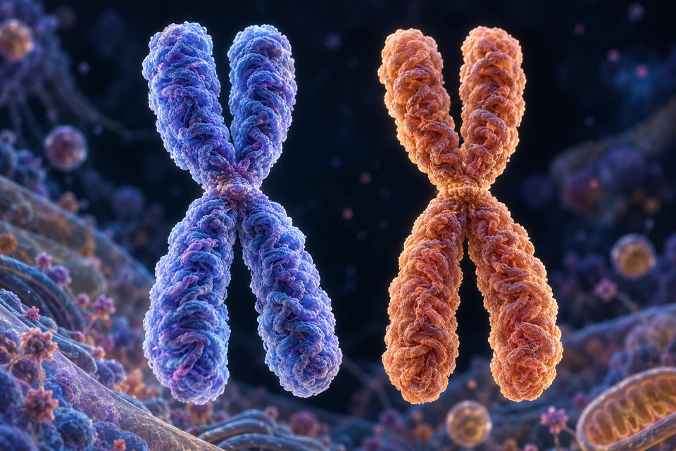
Original generated teaching illustration, not microscopy. Two selected replicated homologs, four chromatid bodies: not a complete human complement, synapsed meiotic bivalent or natural chromosome colour. Fine chromatin folding is illustrative.Structure notes · further context
Selected homolog A
One replicated member of an autosomal homolog pair. Its two chromatids belong to this same chromosome.
Selected homolog B
The corresponding parental homolog can carry different alleles at matching loci. It is not a copied sister of A.
One sister chromatid
One longitudinal DNA-containing body within the selected blue replicated chromosome.
Its sister chromatid
The other copy belongs to the same blue chromosome. Colour does not encode an allele sequence.
Centromeric region
The sisters remain associated through a constricted region. This is not a spherical bead or a resolved cohesion-protein model.
Condensed chromatin
DNA associated with proteins forms the illustrated chromosome material. Visible folds are not measured nucleosome organisation.
Two homologs are parental partners; sisters are copies within each replicated chromosome.
Follow each coloured chromosome as one homolog, then distinguish its two chromatids. Matching loci do not require matching alleles, and illustrative colour is not a genetic sequence.
DNA stores sequence information. Cell division distributes it; inheritance combines parental contributions. This chapter follows the missing connections between those ideas without repeating the molecular foundations.
Copy
Before ordinary division
Replication produces sister chromatids.
Distribute
Nuclear division
Mitosis and meiosis separate different partners.
Combine
Between generations
A gamete contributes one set, with a new mixture of alleles.
A typical autosomal homolog pair contains corresponding chromosomes from two parents. They have corresponding gene locations but can carry different sequence versions. A replicated chromosome has two sister chromatids.
Original generated teaching illustration, not microscopy. Two selected replicated homologs, four chromatid bodies: not a complete human complement, synapsed meiotic bivalent or natural chromosome colour. Fine chromatin folding is illustrative.Structure notes · further context
Selected homolog A
One replicated member of an autosomal homolog pair. Its two chromatids belong to this same chromosome.
Selected homolog B
The corresponding parental homolog can carry different alleles at matching loci. It is not a copied sister of A.
One sister chromatid
One longitudinal DNA-containing body within the selected blue replicated chromosome.
Its sister chromatid
The other copy belongs to the same blue chromosome. Colour does not encode an allele sequence.
Centromeric region
The sisters remain associated through a constricted region. This is not a spherical bead or a resolved cohesion-protein model.
Condensed chromatin
DNA associated with proteins forms the illustrated chromosome material. Visible folds are not measured nucleosome organisation.
Count-controlled source model · selected relationships
The authored model below preserves its explicit chromosome count and projected labels. It is a symbolic teaching reference, separate from the detailed cellular illustration; texture does not make it a measured specimen.
A homolog pair contains two replicated chromosomes
3
First sister chromatid
One replicated DNA-containing copy within the blue chromosome.
More about first sister chromatid
Fresh projection of authored model anchor; independently source-supported selected partner/count relationships. Fine chromatin, pole and membrane dimensions illustrative; not human medical review.
5
Centromeric context
A joining/attachment region in the model; not a measured molecular kinetochore structure.
More about centromeric context
Fresh projection of authored model anchor; independently source-supported selected partner/count relationships. Fine chromatin, pole and membrane dimensions illustrative; not human medical review.
1
Homolog A
One parental chromosome type; the other homolog has corresponding loci but can carry different alleles.
More about homolog a
Fresh projection of authored model anchor; independently source-supported selected partner/count relationships. Fine chromatin, pole and membrane dimensions illustrative; not human medical review.
123456
6
Chromatin surface context
A textured representation of condensed DNA/protein; fine granules are not microscopy.
More about chromatin surface context
Fresh projection of authored model anchor; independently source-supported selected partner/count relationships. Fine chromatin, pole and membrane dimensions illustrative; not human medical review.
4
Second sister chromatid
The replication partner belongs to the same blue chromosome, not the other parent.
More about second sister chromatid
Fresh projection of authored model anchor; independently source-supported selected partner/count relationships. Fine chromatin, pole and membrane dimensions illustrative; not human medical review.
2
Homolog B
The corresponding chromosome from the other parent; not a copied sister of Homolog A.
More about homolog b
Fresh projection of authored model anchor; independently source-supported selected partner/count relationships. Fine chromatin, pole and membrane dimensions illustrative; not human medical review.
A homolog pair contains two replicated chromosomes · names and basic functions are visible
One homolog pair after replication, not a full human karyotype. Colors identify model origin, not natural tissue or blood. Surface granules are illustrative chromatin texture, not resolved nucleosomes. Fresh Golden-family count-controlled rendering with2x supersampling, refined chromatin surface, warmkey/coolrim and nonmetallic materials. Geometry is original authored educational cytology, not measured human microscopy. Human medical review: Not performed. Leader lines or matching visible key numbers identify broad teaching locations, not measured borders or a surgical map.Which relationship is being compared?
A homolog pair compares corresponding chromosomes from the two genetic contributors. A sister pair compares the replicated copies within one chromosome. In the selected comparison, there are two homologs and four chromatid bodies; it is not a complete human chromosome display.
Matching loci do not require the same alleles. Blue and copper track the comparison, rather than measured sequence or natural colour. Recombination can also change relationships between chromatid sequences; parental origin must not be used as a guarantee that two sisters remain identical after meiosis proceeds.
During S phase, the DNA is copied. The copies remain associated as sisters until the appropriate separation. An X-shaped condensed representation is not the chromosome’s appearance throughout the cell cycle.
EDITABLE SUPPORTING RELATIONSHIP / NOT OBSERVED MICROSCOPY
Two homologs; each has two sisters
Original source-supported relationship; counts explicitly selected, shapes qualitative, no measured cytology or clinical genetic forecast. Separate text and arrows.
Read the relationships
Homolog pair: Corresponding parental chromosome types can carry different alleles.
Sister pair: Copies produced by replication belong to one chromosome.
Scope: These are different relationships, not four unrelated chromosomes.
Symbolic relationships, not personal genetic predictions.
Before sisters separate, a replicated chromosome still counts as one chromosome. The amount of DNA has doubled; the number of chromosomal sets has not simply doubled.
Before replication
Typical human nucleated diploid context
46 chromosomes; one DNA molecule per unreplicated chromosome.
After replication
Before sister separation
46 replicated chromosomes with 92 chromatids.
After ordinary mitosis
Each completed diploid daughter nucleus
Typically 46 chromosomes again; this is not a statement about every body cell.
Enucleated red blood cells and specialized cellular states are exceptions. The teaching models intentionally show selected subsets, not a full human count.
Two poles organize microtubules around condensed chromosomes. Sister kinetochore regions attach toward opposite poles; the chromosome arrangement lies across the spindle’s equatorial plane.
Original generated metaphase teaching preparation, not microscopy or a human chromosome count. Only selected exposed chromosomes are emphasised. The nuclear envelope has broken down; sister chromatids are still joined. Fine molecular attachment geometry is not a solved structural model.Structure notes · further context
Cell boundary
The plasma membrane encloses the cell. It must not be mistaken for a nuclear envelope around the spindle.
One spindle pole
Microtubules converge toward one centrosomal pole. The opposing pole lies across the same cell.
Opposing spindle pole
A bipolar spindle supports separation toward opposite poles; it is not a pipe carrying genetic material.
Joined sister chromatids
The textured replicated chromosome has two sister chromatids, still joined before anaphase. They are not a homologous pair.
Centromeric attachment context
Kinetochore protein assemblies at the centromeric region engage spindle microtubules. This expanded illustration locates the context, not individual binding molecules.
Spindle microtubules
Bundles connect the poles with centromeric attachment regions; other microtubules support spindle organisation. They do not copy DNA.
Count-controlled source model · selected relationships
The authored model below preserves its explicit chromosome count and projected labels. It is a symbolic teaching reference, separate from the detailed cellular illustration; texture does not make it a measured specimen.
Opposite poles organize a mitotic spindle
4
Kinetochore context
Spindle attachment belongs to a centromeric protein region; the molecular structure is not resolved.
More about kinetochore context
Fresh projection of authored model anchor; independently source-supported selected partner/count relationships. Fine chromatin, pole and membrane dimensions illustrative; not human medical review.
1
Left spindle pole
One microtubule-organizing region; the model depicts two opposing poles.
More about left spindle pole
Fresh projection of authored model anchor; independently source-supported selected partner/count relationships. Fine chromatin, pole and membrane dimensions illustrative; not human medical review.
5
Spindle microtubule guide
Selected fibers show attachment relationships, not every microtubule or all motor proteins.
More about spindle microtubule guide
Fresh projection of authored model anchor; independently source-supported selected partner/count relationships. Fine chromatin, pole and membrane dimensions illustrative; not human medical review.
1256A
3
Replicated chromosome
Two sister chromatids belong to one replicated chromosome; four such chromosomes are modeled.
More about replicated chromosome
Fresh projection of authored model anchor; independently source-supported selected partner/count relationships. Fine chromatin, pole and membrane dimensions illustrative; not human medical review.
2
Right spindle pole
The other pole faces the first across the cell.
More about right spindle pole
Fresh projection of authored model anchor; independently source-supported selected partner/count relationships. Fine chromatin, pole and membrane dimensions illustrative; not human medical review.
6
Cell boundary context
A transparent sectional shell locates the spindle within cellular space, not measured membrane thickness.
More about cell boundary context
Fresh projection of authored model anchor; independently source-supported selected partner/count relationships. Fine chromatin, pole and membrane dimensions illustrative; not human medical review.
A · Enlarged local detail
The outlined region is enlarged from the same artwork. Matching keys keep close teaching locations separate; no new anatomical detail is invented.
Opposite poles organize a mitotic spindle · names and basic functions are visible
Explicit teaching subset 2n=4,not a human cell with 46 chromosomes. Homologs are not paired as meiotic bivalents here. Fibers/pole/membrane shapes and abundance are illustrative; no complete microtubule inventory. Fresh Golden-family count-controlled rendering with2x supersampling, refined chromatin surface, warmkey/coolrim and nonmetallic materials. Geometry is original authored educational cytology, not measured human microscopy. Human medical review: Not performed. Leader lines or matching visible key numbers identify broad teaching locations, not measured borders or a surgical map.
G1 supports growth and activity; S copies DNA; G2 prepares for division. Mitosis redistributes nuclear material and cytokinesis divides cytoplasm. Some differentiated cells do not routinely re-enter this cycle.
CELLULAR CONTEXT / ALL ESSENTIAL STEPS VISIBLE
Replication precedes nuclear separation
G1 activity → S copying → G2 preparation and M division
Original generated medical illustration, not microscopy. The cutaway locates broad cellular structures; it does not measure membrane thickness, pore composition or native chromatin spacing.Structure notes · further context
Nucleolus
A non-membrane-bound region where ribosomal RNA production and ribosomal subunit assembly are organised. It is not a second nucleus.
Chromatin
DNA associated with proteins occupies the nucleus in a changing, heterogeneous organisation; not all genes are active at once.
Nuclear envelope
Two membranes separate nucleoplasm from cytoplasm. The outer membrane connects with the endoplasmic reticulum.
Nuclear pore
A protein-lined gateway regulates exchange. Selected RNAs and proteins cross by organised transport, rather than DNA escaping through an open hole.
Rough ER
Membrane surfaces in cytoplasm carry ribosomes making selected secreted and membrane proteins. Many other proteins are made on free ribosomes.
This interphase context locates nuclear DNA; it does not resolve replication forks or measure how much DNA has already been copied.
Selected focus: Prepare · A cell carries out functions and responds to signals.
Controlled symbolic guide · compare the relationship
Original editable mechanism. All basic relationships visible, colours and dimensions illustrative; not specimen microscopy or a personal genetic forecast.
Prepare
A cell carries out functions and responds to signals.
Many specialized cells do not routinely divide.
Copy
S phase replicates DNA into associated sister chromatids.
DNA amount changes before chromosome sets are reduced or distributed.
Distribute
Mitosis and cytokinesis redistribute nuclear material and cytoplasm.
Durations vary; this is not a timed human-cell recording.
The spindle checkpoint helps delay separation when chromosome attachments are not ready. Mitosis separates sisters; ordinary meiotic homolog pairing is a different arrangement.
Equatorial arrangement
Metaphase
Replicated chromosomes align across the middle of the spindle.
Attachment context
Kinetochore region
Opposite-sister attachments support distribution to opposite poles.
Checkpoint
Cell-cycle control
A signaling system assesses readiness, not a visible clock.
Original generated metaphase teaching preparation, not microscopy or a human chromosome count. Only selected exposed chromosomes are emphasised. The nuclear envelope has broken down; sister chromatids are still joined. Fine molecular attachment geometry is not a solved structural model.Structure notes · further context
Cell boundary
The plasma membrane encloses the cell. It must not be mistaken for a nuclear envelope around the spindle.
One spindle pole
Microtubules converge toward one centrosomal pole. The opposing pole lies across the same cell.
Opposing spindle pole
A bipolar spindle supports separation toward opposite poles; it is not a pipe carrying genetic material.
Joined sister chromatids
The textured replicated chromosome has two sister chromatids, still joined before anaphase. They are not a homologous pair.
Centromeric attachment context
Kinetochore protein assemblies at the centromeric region engage spindle microtubules. This expanded illustration locates the context, not individual binding molecules.
Spindle microtubules
Bundles connect the poles with centromeric attachment regions; other microtubules support spindle organisation. They do not copy DNA.
This preparation stays at metaphase while the cards describe alignment, later separation and rebuilding. A focus change is not a movie of those later stages.
Selected focus: Align · Opposing attachments organize replicated chromosomes near the equator.
Controlled symbolic guide · compare the relationship
Original editable mechanism. All basic relationships visible, colours and dimensions illustrative; not specimen microscopy or a personal genetic forecast.
Align
Opposing attachments organize replicated chromosomes near the equator.
Not meiotic homolog-pair synapsis.
Separate
Former sisters become chromosomes directed toward opposing poles.
Ordinary complement preservation is the teaching scope.
Rebuild
Nuclei reform and chromatin becomes less condensed.
Cytoplasmic division is related but distinct.
Ordinary mitosis aims to preserve a nuclear complement. This is not a guarantee that all daughter organelles, cell sizes or later gene activity are identical.
In an animal cell, a contractile system creates a cleavage furrow as the cell separates. Nuclear segregation and cytoplasmic separation are related processes, but they are not interchangeable words.
EDITABLE SUPPORTING RELATIONSHIP / NOT OBSERVED MICROSCOPY
Nuclear and cytoplasmic division differ
Original source-supported relationship; counts explicitly selected, shapes qualitative, no measured cytology or clinical genetic forecast. Separate text and arrows.
Meiosis occurs in the germ-cell lineage associated with egg and sperm formation. It differs from the divisions that grow and maintain many body tissues.
The first meiotic division separates homologs while sister chromatids remain associated. The second separates sisters. Compare the partners in the two selected models rather than assuming both stages do the same thing.
Original generated selected anaphase-I preparation. One illustrated homolog pair, not a full human chromosome count. Each moving homolog still has two associated sisters. No claim of four equal gametes, real microscopy or resolved kinetochore molecules.Structure notes · further context
Replicated homolog A
This selected homolog moves toward one pole while its sister chromatids remain associated.
Replicated homolog B
The corresponding homolog goes toward the opposite pole; the two parental homologs are being separated.
Sisters remain joined
Centromeric sister association is retained at this first division. DNA is not cut into two single strands.
One homolog faces one pole
The illustrated attachment context faces the right pole. Fine co-orientation proteins are not resolved here.
Left spindle pole
Microtubules organise the poleward route for the selected blue homolog. Abundance and speed are not measured.
Right spindle pole
The other pole organises the route for the selected copper homolog; this is one cell, not two products.
Count-controlled source model · selected relationships
The authored model below preserves its explicit chromosome count and projected labels. It is a symbolic teaching reference, separate from the detailed cellular illustration; texture does not make it a measured specimen.
Two divisions separate different partners
1
Meiosis I: one homolog
A replicated homolog moves toward one pole while its two sisters remain together.
More about meiosis i: one homolog
Fresh projection of authored model anchor; independently source-supported selected partner/count relationships. Fine chromatin, pole and membrane dimensions illustrative; not human medical review.
2
Meiosis I: other homolog
The corresponding homolog moves toward the other pole.
More about meiosis i: other homolog
Fresh projection of authored model anchor; independently source-supported selected partner/count relationships. Fine chromatin, pole and membrane dimensions illustrative; not human medical review.
3
Retained sisters
Sister chromatids remain associated during ordinary first-division homolog separation.
More about retained sisters
Fresh projection of authored model anchor; independently source-supported selected partner/count relationships. Fine chromatin, pole and membrane dimensions illustrative; not human medical review.
2456A
6
Second-division spindle
One selected cell divides again without a preceding second round of DNA replication.
More about second-division spindle
Fresh projection of authored model anchor; independently source-supported selected partner/count relationships. Fine chromatin, pole and membrane dimensions illustrative; not human medical review.
4
Meiosis II: first sister
A chromatid separates and is now a daughter chromosome.
More about meiosis ii: first sister
Fresh projection of authored model anchor; independently source-supported selected partner/count relationships. Fine chromatin, pole and membrane dimensions illustrative; not human medical review.
5
Meiosis II: other sister
Its former sister moves toward the opposite pole in this selected cell.
More about meiosis ii: other sister
Fresh projection of authored model anchor; independently source-supported selected partner/count relationships. Fine chromatin, pole and membrane dimensions illustrative; not human medical review.
A · Enlarged local detail
The outlined region is enlarged from the same artwork. Matching keys keep close teaching locations separate; no new anatomical detail is invented.
Two divisions separate different partners · names and basic functions are visible
Left: single homolog-pair subset in meiosis I. Right: one selected haploid-cell subset in meiosis II. These are comparative moments, not all daughter products or a complete human meiotic lineage. No intervening replication. Fresh Golden-family count-controlled rendering with2x supersampling, refined chromatin surface, warmkey/coolrim and nonmetallic materials. Geometry is original authored educational cytology, not measured human microscopy. Human medical review: Not performed. Leader lines or matching visible key numbers identify broad teaching locations, not measured borders or a surgical map.Which connections change in the first division?
Earlier meiotic pairing allows exchange between nonsister chromatids of homologous chromosomes. The partners can then retain chiasma-related connections until segregation. At the first anaphase, homologs part while centromeric sister association is protected.
The detailed image locates this selected separation: one replicated homolog travels toward each pole. It does not show the earlier molecular exchange reaction or each cohesion protein. The controlled guide separately explains counts, preventing a cropped illustrative pair from becoming a claimed whole human complement.
During prophase I, paired homologs can exchange corresponding DNA segments between nonsister chromatids. This recombination changes allele combinations along a chromosome; it does not create a new gene at every exchange.
EDITABLE SUPPORTING RELATIONSHIP / NOT OBSERVED MICROSCOPY
A selected reciprocal exchange involves two nonsisters
Original source-supported relationship; counts explicitly selected, shapes qualitative, no measured cytology or clinical genetic forecast. Separate text and arrows.
Read the relationships
Selected pair: One chromatid from each homolog participates in this example.
Exchange: Corresponding DNA segments change combinations.
Other sisters: The other chromatids are not participants in this selected crossover; no molecular resolution shown.
Symbolic relationships, not personal genetic predictions.
The supporting inset uses one idealized reciprocal exchange. It is not a measured hotspot, sequence map or guaranteed crossover count.
Pair orientation changes which set travels together
Different homolog pairs can orient independently at the first division. Their orientation influences which parental contributions enter a gamete; nearby loci on the same chromosome do not automatically assort independently.
Selected focus: Orient · Each homolog pair adopts an orientation at the first meiotic division.
Controlled symbolic guide · compare the relationship
Original editable mechanism. All basic relationships visible, colours and dimensions illustrative; not specimen microscopy or a personal genetic forecast.
Orient
Each homolog pair adopts an orientation at the first meiotic division.
This does not imply all loci are unlinked.
Separate
One homolog of each pair goes toward each pole.
Sisters normally remain together in this first division.
Combine
Different orientations produce different mixtures across a chromosome set.
Recombination adds further variation; no exhaustive human gamete count inferred.
After the first reduction division, the selected cells contain one member of each homolog pair, still replicated. Sisters separate during meiosis II without an intervening S phase.
Original generated selected anaphase-II preparation, not microscopy. Each shown moving body is one chromatid-derived chromosome with two arms, not a replicated X or severed DNA strand. This single cell is not the complete product set; shared colour does not certify sequence identity after recombination.Structure notes · further context
Separated chromosome
A former sister is now an independent chromosome moving toward the left pole. Its arms belong to one continuous chromatid body.
The other separated chromosome
The former sister moves toward the opposing pole. Separation distributes copies; it does not synthesize them again.
Leading centromeric region
The centromeric bend leads the leftward movement, with chromosome arms trailing inward.
Opposing attachment context
The rightward chromosome has its own attachment context. Individual molecular binding sites are not identified.
Spindle pole
A bipolar spindle acts within this selected second-division cell. There is no intervening S phase between divisions I and II.
Cell boundary
The plasma membrane surrounds cytoplasm. An intact nuclear envelope is not enclosing these separating chromosomes.
EDITABLE SUPPORTING RELATIONSHIP / NOT OBSERVED MICROSCOPY
Sets and DNA amount change at different steps
Original source-supported relationship; counts explicitly selected, shapes qualitative, no measured cytology or clinical genetic forecast. Separate text and arrows.
Read the relationships
Before meiosis: Typical diploid post-replication context: 2n, 4C.
After meiosis I: One homolog per pair, still replicated: n, 2C.
After meiosis II: One chromatid-derived chromosome per pair: n, 1C. C denotes DNA amount relative to an unreplicated haploid complement.
Symbolic relationships, not personal genetic predictions.
Haploid describes chromosome sets, not necessarily an unreplicated DNA amount at every meiotic stage.
How can a cell be haploid while its chromosomes are still copied?
Ploidy counts chromosome sets, not whether each chromosome has one or two chromatids. After meiosis I, a selected cell can have one member of each homolog pair while those chromosomes remain replicated.
The second division separates sisters without an intervening S phase. Each separated chromatid is then a chromosome. Our single-cell preparation selects this relationship; it does not imply four equal eggs or display all division products.
Oocyte divisions preserve most cytoplasm in one large cell and extrude small polar bodies. Spermatogenic divisions and oogenic divisions cannot be drawn as one universal four-equal-gamete outcome.
Oocyte
Asymmetric cytoplasm
The large cell retains substantial resources.
Polar body
Small division product
It carries a chromosomal complement without becoming an equivalent large egg.
Existing chronology
Pauses and completion
The reproductive chapter explains the normal division timing.
A typical human sperm and egg each contribute 23 chromosomes to a nucleated zygote. Their combination restores two sets; subsequent cell differentiation changes activity, and some specialized cells no longer retain that same nuclear state.
An allele is a sequence version at a genomic location. A diploid person generally has two alleles at an autosomal locus; the wider population can contain many versions at that same location.
Homozygous
Same locus, same versions
The two alleles match at the locus being considered.
Heterozygous
Same locus, different versions
The two alleles differ at that locus.
Population
Many people
More than two possible alleles can exist across the population.
A sequence combination is not the whole observed person
Genotype describes sequence or allele information. Phenotype describes observable characteristics that can involve gene activity, other genes, development and environment.
Dominance describes the relationship between alleles for a specified phenotype in a heterozygote. It does not say which allele is morally better, more frequent or necessarily the only allele whose RNA is made.
Specified phenotype
One observed feature
A dominance relationship can depend on what is measured.
Recessive contribution
Not automatically switched off
Molecular activity and observed phenotype are different levels.
Scope
Teaching model
Do not extend a fictional single-locus model to all human traits.
For a fictional autosomal locus, A/a × A/a with equal segregation gives possible genotypes AA, Aa, aA and aa. The middle two represent the same heterozygous genotype. Each modeled event has the same probabilities, not a promised sequence of four children.
CELLULAR CONTEXT / ALL ESSENTIAL STEPS VISIBLE
One fictional locus has four equiprobable grid cells
A or a gamete → combine two contributions → AA, Aa or aa
Selected focus: Segregate · Each A/a parent contributes A or a under the stated equal-segregation model.
Controlled symbolic guide · compare the relationship
Original editable mechanism. All basic relationships visible, colours and dimensions illustrative; not specimen microscopy or a personal genetic forecast.
Segregate
Each A/a parent contributes A or a under the stated equal-segregation model.
This is a fictional autosomal locus, not a family test.
Combine
The grid cells are AA, Aa, aA and aa.
Aa and aA represent the same heterozygous genotype.
Interpret
The model gives 25% AA, 50% Aa and 25% aa per event.
Not a quota of one AA, two Aa and one aa in each family.
EDITABLE SUPPORTING RELATIONSHIP / NOT OBSERVED MICROSCOPY
One model grid; probabilities are not quotas
Original source-supported relationship; counts explicitly selected, shapes qualitative, no measured cytology or clinical genetic forecast. Separate text and arrows.
Read the relationships
Assumptions: Fictional A/a × A/a autosomal locus with equal segregation.
Genotypes: AA: 25%; Aa: 50%; aa: 25%.
Boundary: No eye-color, disease, family or clinical prediction.
Symbolic relationships, not personal genetic predictions.
25% AA, 50% Aa, 25% aa are model genotype probabilities under stated assumptions. No disease, eye-color or personal reproductive prediction is calculated.
Why is a probability grid not a quota of children?
The fictional grid combines specified allele contributions under stated assumptions. Its outcomes are possibilities for a new event, rather than an order in which outcomes must occur. A previous outcome does not make the remaining boxes a family schedule.
Real inheritance depends on the locus, chromosome context and biological assumptions. The normal teaching model is not a personal estimate, diagnosis or recommendation for testing; individual questions need appropriate professional context.
At the ABO locus, IA and IB can both contribute to the AB phenotype; i supplies another common allele. This illustrates multiple population alleles and codominance, not one allele always erasing the other.
Population alleles
ABO example
IA, IB and i show more than two common versions.
Individual combination
IA with IB
Both contributions appear in the AB phenotype.
Boundary
Normal genetics
This is not a transfusion, compatibility or clinical blood-group service.
Loci close together on one chromosome can be inherited together more often than independently. Recombination can separate combinations; invented drawing distance is not a measured genetic map.
CELLULAR CONTEXT / ALL ESSENTIAL STEPS VISIBLE
Chromosome context limits independent-locus rules
Nearby loci → shared transmission → occasional recombination
Selected focus: Locate · Two loci can occupy the same chromosome.
Controlled symbolic guide · compare the relationship
Original editable mechanism. All basic relationships visible, colours and dimensions illustrative; not specimen microscopy or a personal genetic forecast.
Locate
Two loci can occupy the same chromosome.
Physical positions in a drawing are not measured genetic distances.
Travel
Nearby variants can be inherited together more often.
Independent assortment of chromosomes is not universal independence of loci.
Recombine
Exchange can change linked allele combinations.
No exact fraction or crossover hotspot is calculated here.
In a typicalXX/XY model, a non-pseudoautosomal X locus does not follow father-to-son X transmission: the sperm’s Y contribution forms that typicalXY combination. X-linked expression can vary with gene context and X-inactivation.
Non-PAR X model
TypicalXX/XY
Transmission differs from an autosomal two-copy grid.
Qualification
Chromosomal diversity
Actual human chromosomal and developmental variation is not reduced to this model.
Expression
X context
An X-linked allele does not guarantee identical expression in every person.
The diagrammatic model is about chromosome transmission, not a definition of gender or a prediction of every person’s anatomy.
Human mitochondrial DNA is ordinarily transmitted through the egg. Different proportions of mtDNA versions can occur through heteroplasmy; children need not have identical proportions or effects. Nuclear genes that support mitochondria follow nuclear inheritance patterns.
Variants can be inherited, arise in a gamete or early development, or occur in a somatic cell. Effects depend on location, function and context; many variants have no detectable adverse effect.
EDITABLE SUPPORTING RELATIONSHIP / NOT OBSERVED MICROSCOPY
Sequence variation has several possible outcomes
Original source-supported relationship; counts explicitly selected, shapes qualitative, no measured cytology or clinical genetic forecast. Separate text and arrows.
Read the relationships
Sequence: A DNA version differs at a location.
Molecular context: Effects may involve coding, regulation or no detectable functional change.
Observed feature: Other genes and environmental/developmental context can contribute; not every variant is harmful.
Symbolic relationships, not personal genetic predictions.
DNA damage and repair are not the same as a guarantee that every established inherited sequence variant is removed. Clinical variant interpretation belongs to a later, reviewed scope.
Whether a feature appears differs from how it appears
Penetrance concerns the proportion showing a specified feature among those with a relevant genotype. Expressivity concerns variation in the feature’s expression. These distinctions prevent inheritance diagrams from becoming certainty claims.
CELLULAR CONTEXT / ALL ESSENTIAL STEPS VISIBLE
Occurrence and expression are different questions
Relevant genotype → feature occurrence → range of expression
Selected focus: Genotype · A specified allele combination is present.
Controlled symbolic guide · compare the relationship
Original editable mechanism. All basic relationships visible, colours and dimensions illustrative; not specimen microscopy or a personal genetic forecast.
Genotype
A specified allele combination is present.
It does not specify every aspect of the person.
Occurrence
Penetrance concerns how often a specified feature appears.
No condition-specific risk estimate is provided.
Expression
Expressivity concerns variation in an expressed feature.
Other genes, development and context can contribute.
No named-condition probability or individualized family assessment is offered.
A sequence combination and an observed feature are different kinds of information
Penetrance describes how often a specified feature is observed among people with a relevant genotype. Expressivity describes variation in how that feature is expressed. Neither term turns one allele combination into a full description of a person.
Other genes, development and environment can influence an outcome. The selectable teaching states distinguish these questions without plotting a condition-specific probability or predicting an individual appearance.
Many traits involve many loci and other influences
Human eye color is influenced by multiple genes; it is not reliably explained by one brown-dominant and blue-recessive pair. Other complex traits also combine genetic and contextual contributions.
Many loci
Combined contributions
Single-locus grids explain one limited rule, not every trait.
Context
Development and environment
Observed outcomes can depend on more than sequence alone.
No ranking
People are not scores
No ancestry, ability, identity or worth is inferred.
Controlled models explain relationships, not specimens
These original offline Three.js models control chromosome counts and attachment/separation relationships. Condensed surface texture, membrane geometry and microtubule number are illustrative, not actual human microscopy.
The count-controlled references use authored geometry. The detailed cellular preparations are generated teaching illustrations; the separate molecular reference uses deposited experimental coordinates. These evidence types must not be mistaken for one another.
Experimental molecular reference · DNA packagingOriginal controlled rendering of unchanged CC0 PDB1KX5 coordinates. Xenopus histones and a deposited DNA construct; colours and display radii are illustrative. This is packaging evidence, not a complete human chromosome.
Model
Authored 3D geometry
Topological and count assertions can be tested.
Observed imaging
Separate evidence type
Actual microscopy requires source, license, scale and acquisition context.
Source verification
Stated teaching scope
Independent facts support relationships; they do not certify every surface detail.
Human medical review: not performed. New physical-phone acceptance: not performed. Rights-cleared observed microscopy and clinical genetics remain separate work.
Chromosome partners, mitotic and meiotic segregation, recombination, allele combinations and bounded normal inheritance. Fictional probability models are not personal genetic forecasts.
Methods and limits
Original authored count-controlled cytological references and their18 projected labels remain available as separate source models. New detailed original generated homolog and selected meiotic-cell preparations carry independent editable names and directions; qualified DNA cellular context is reused without assigning its exposed chromosomes the source model’s exact count. The retained PDB nucleosome reference illustrates experimental packaging at a different scale, not a full chromosome. All essential relationships visible by default. Separate labels and source-based supporting diagrams. Counts in the authored source models are controlled; generated cellular preparations identify only their selected chromosome relationships. Fine textures, proportions and microtubule abundance are illustrative, not microscopy. Human medical review: not performed. Educational only.
Edition: inheritance-cell-division-eyes-context-2026-10-19-v2. Original teaching illustrations and selected count-model references are distinguished from experimental molecular renderings. No family diagnosis, clinical genetic advice or physician approval claim. Rights-cleared observed microscopy and clinical interpretation remain separate work.