Explore the muscle behind the pump: its own blood supply, valve support, cell contacts and controlled activation.
▷
Watch chapter overviewFilm planned · not yet available
Current new-family Heart artwork reused · fine detail illustrative
30-SECOND ORIENTATION / SUPPLY · SUPPORT · CONNECT · CONTRACT
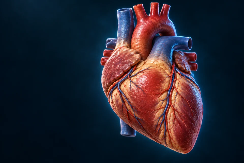
Original generated gross wall enlargement retained from the qualified Heart chapter. Thin surfaces and the fluid interval are expanded for teaching; this is not measured histology.Structure notes · further context
Myocardium
Working muscle occupies most of the wall and generates force.
Endocardial context
A thin inner lining faces the chamber cavity.
Epicardial context
Outer surface tissue supports vessels; epicardium is the visceral serous layer.
Pericardial context
The surrounding sac is outside the wall; its fluid interval is enlarged illustratively.
A muscular wall works within a supporting enclosure.
Locate the blood-facing lining, force-generating myocardium and outer covering before following their different jobs.
01 / SCOPE
Look beyond the chamber blood
The Heart chapter is the starting journey. This companion examines the tissues that generate force, their own blood routes and how separate cells coordinate. A cavity surface is a blood-space representation, not a muscle layer.
Endocardium faces chamber blood; myocardium provides most contractile tissue; epicardium covers the outside. Connective tissue, vessels and nerves support the muscle. These roles should not be inferred from the colour of an unlayered source mesh.
STRUCTURE TO FUNCTION / NORMAL TISSUE
Different tissues face different spaces
Original generated gross wall enlargement retained from the qualified Heart chapter. Thin surfaces and the fluid interval are expanded for teaching; this is not measured histology.Structure notes · further context
Myocardium
Working muscle occupies most of the wall and generates force.
Endocardial context
A thin inner lining faces the chamber cavity.
Epicardial context
Outer surface tissue supports vessels; epicardium is the visceral serous layer.
Pericardial context
The surrounding sac is outside the wall; its fluid interval is enlarged illustratively.
Start at the chamber-facing lining, cross the muscular middle and reach the outer epicardial context. The surrounding sac is outside the wall.
Optional qualitative process summary
Retained detailed organ or cell context comes first. The optional editable summary uses qualitative geometry; it is not histology, an electron micrograph or a clinical trace.
Read the relationships
Endocardium: Faces chamber blood with a lining and underlying support.
Myocardium: The contractile middle contains cells and supporting tissue.
English First. Human medical review not performed.
Endocardium
Inner lining
An endothelial surface and underlying connective tissue meet the blood space.
Myocardium
Muscular middle
Working cells generate force within a supporting network.
Epicardium
Outer surface
Visceral serous pericardium includes connective and vascular context.
Go deeper · Which side faces chamber blood?
Use the lining as the starting boundary, then locate contractile myocardium and the outer surface. The surrounding pericardial sac is a separate enclosure. A thick-looking rim in an enlarged teaching view is not a measurement of membrane thickness.
Start with the detailed anterior arterial and venous approaches. The optional source-native posterior preparation adds the coronary sinus context; these are different views, not one universal territorial map.
Original qualified Heart atlas illustration, not angiography. Anterior context cannot show the posterior coronary sinus outlet; fine branches and hidden origins are not traced here.Structure notes · further context
Right coronary approach
A major arterial approach follows the right atrioventricular groove.
Left coronary branching context
Visible arterial approaches extend toward the ventricular surface; the hidden origin is not mapped.
Anterior interventricular artery
The LAD follows the groove between the ventricles; fine branches vary between people.
Surface cardiac veins
Veins collect blood from myocardium. Most returns through the posterior coronary sinus.
Compare the preserved source-native positioning reference
Coronary arteries feed myocardial exchange beds; veins carry blood away from those beds. These routes are distinct from blood crossing the four chambers. This source-native posterior view shows named surface landmarks, not a universal territorial map.
Posterior supply and drainage
4
Great cardiac vein
A named venous route drains myocardial tissue.
More about great cardiac vein
Visible source-native mesh hit; source positions unchanged. Gross correspondence is not anatomical review. Fresh first-visible named source-surface raycast passed.
1
Circumflex branch
A left-coronary route follows the atrioventricular region.
More about circumflex branch
Visible source-native mesh hit; source positions unchanged. Gross correspondence is not anatomical review. Fresh first-visible named source-surface raycast passed.
5
Ventricular wall context
The source wall provides gross context, not individually resolved layers.
More about ventricular wall context
Visible source-native mesh hit; source positions unchanged. Gross correspondence is not anatomical review. Fresh first-visible named source-surface raycast passed.
12345
3
Coronary sinus
A posterior collector carries much venous drainage toward the right atrium.
More about coronary sinus
Visible source-native mesh hit; source positions unchanged. Gross correspondence is not anatomical review. Fresh first-visible named source-surface raycast passed.
2
Posterior interventricular branch
This source shows a posterior arterial route; origin patterns vary.
More about posterior interventricular branch
Visible source-native mesh hit; source positions unchanged. Gross correspondence is not anatomical review. Fresh first-visible named source-surface raycast passed.
Posterior supply and drainage · names and basic functions are visible
Existing adult male BodyParts3D CC-BY4.0 geometry. Camera/material/visibility only. Coarse surfaces and incomplete fine anatomy; human medical review not performed. New navy2x source-native rendering, separate warm key/cool rim and restrained ghost context. Source surfaces remain coarser than generated artwork; no missing branch, annulus or chordal topology is invented. Leader lines or matching visible key numbers identify broad teaching locations, not measured borders or a surgical map.
The coronary sinus lies on the posterior side and opens into the right atrium. It receives much of the heart’s venous drainage. Other cardiac veins can drain directly into a chamber, so an all-veins-through-the-sinus arrow would be misleading.
STRUCTURE TO FUNCTION / NORMAL TISSUE
The muscle’s route differs from the chamber route
Original qualified Heart atlas illustration, not angiography. Anterior context cannot show the posterior coronary sinus outlet; fine branches and hidden origins are not traced here.Structure notes · further context
Right coronary approach
A major arterial approach follows the right atrioventricular groove.
Left coronary branching context
Visible arterial approaches extend toward the ventricular surface; the hidden origin is not mapped.
Anterior interventricular artery
The LAD follows the groove between the ventricles; fine branches vary between people.
Surface cardiac veins
Veins collect blood from myocardium. Most returns through the posterior coronary sinus.
Compare surface arterial and venous approaches. The posterior collector and its outlet are outside this anterior view; the preserved native posterior preparation is available separately for positioning.
Optional qualitative process summary
Retained detailed organ or cell context comes first. The optional editable summary uses qualitative geometry; it is not histology, an electron micrograph or a clinical trace.
Collection: Many cardiac veins join the coronary sinus.
Exceptions: Other veins drain directly; this selected route is not exhaustive.
English First. Human medical review not performed.
Go deeper · Why is the coronary sinus not the only exit?
Surface veins and their collector belong to myocardial drainage. Some cardiac venous routes enter a chamber directly. Identify the large posterior collector without turning it into an obligatory route for every vein.
Ventricular contraction compresses intramyocardial vessels. Left-ventricular coronary flow is therefore mainly supported during diastole. Pressure, tissue compression and local vascular regulation all matter; not every coronary bed has the same flow pattern.
A surface artery is a route to tissue, not a direct opening from its lumen into the chamber. This illustration does not show individual myocardial capillaries.
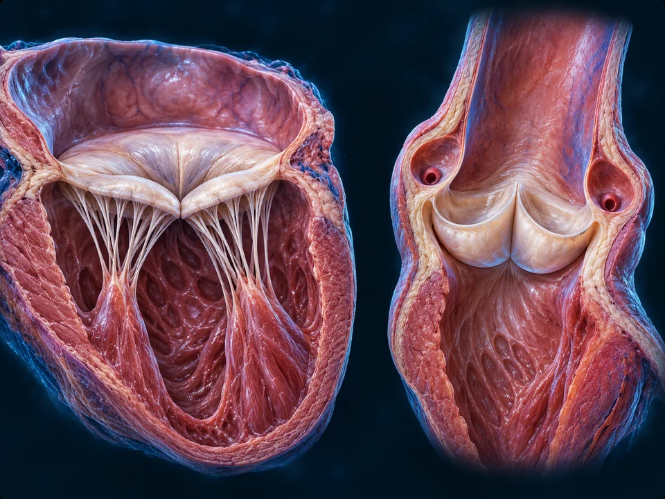
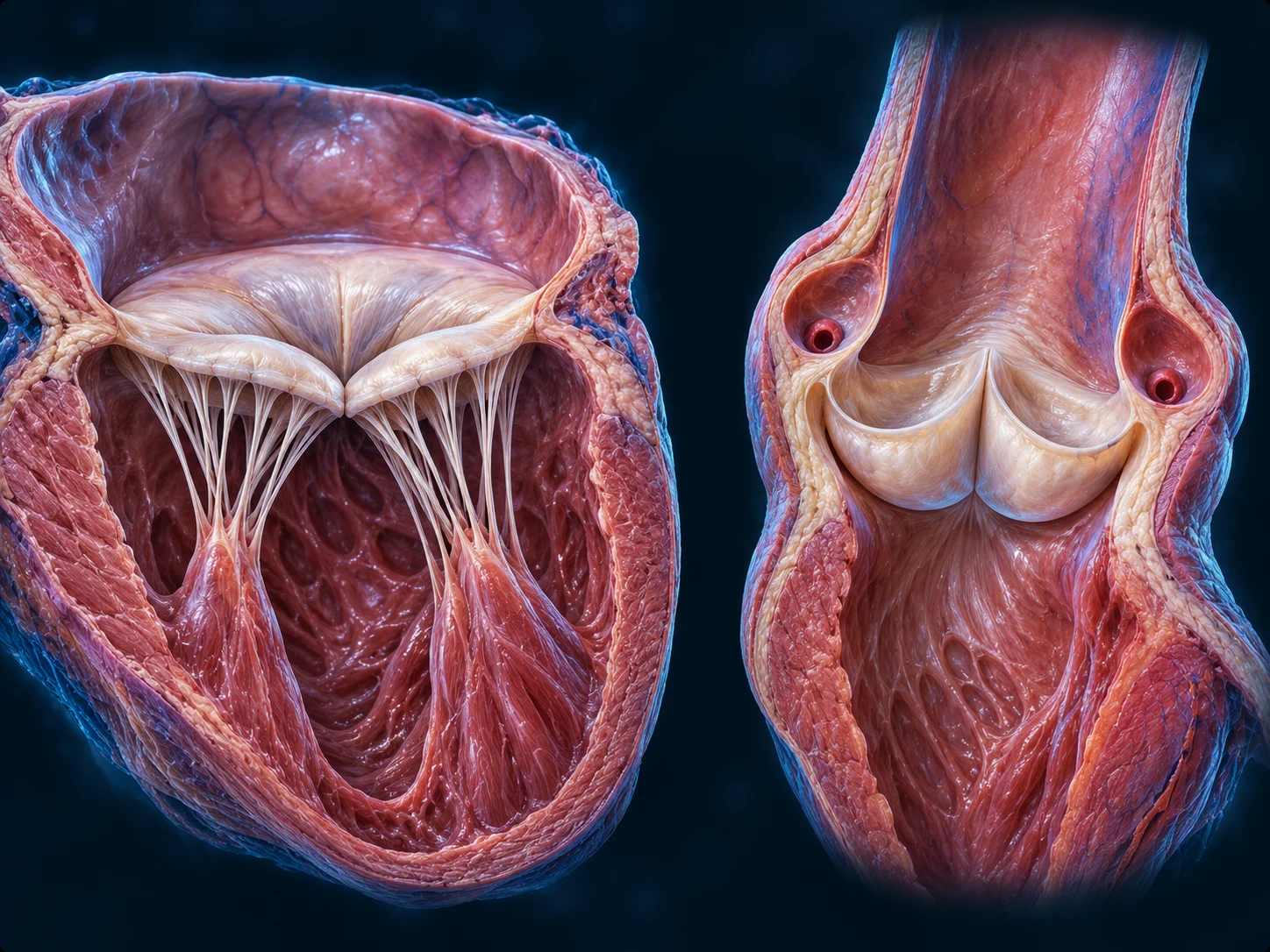
Compare the detailed mitral inlet with the separate aortic outlet. AV leaflets have a chordal-papillary support apparatus; semilunar cusps do not. The optional native preparation preserves its independently limited source geometry.
Original qualified Heart preparations, near closure and not one shared spatial scene. Chordae restrain AV leaflet displacement; semilunar cusps have no chordal apparatus. Fine attachments are illustrative.Structure notes · further context
Aortic root · right preparation
The artery begins beyond the left ventricular outlet; pocket cusps sit at its base.
Mitral leaflets · left preparation
The closed inlet has two main leaflets separating atrium from ventricle.
Aortic cusps
Three pocket-like cusps meet during closure. There are no chordal tethers.
Chordae tendineae
Tethers connect AV leaflets to papillary muscle; they restrain backward displacement.
Papillary muscle
A muscular projection helps maintain tether tension during ventricular contraction.
Original qualified Heart preparations, near closure and not one shared spatial scene. Chordae restrain AV leaflet displacement; semilunar cusps have no chordal apparatus. Fine attachments are illustrative. Same original, selected observation window; no anatomy regenerated or added.Structure notes · further context
Mitral leaflets · left preparation
The closed inlet has two main leaflets separating atrium from ventricle.
Chordae tendineae
Tethers connect AV leaflets to papillary muscle; they restrain backward displacement.
Papillary muscle
A muscular projection helps maintain tether tension during ventricular contraction.
Original qualified Heart preparations, near closure and not one shared spatial scene. Chordae restrain AV leaflet displacement; semilunar cusps have no chordal apparatus. Fine attachments are illustrative. Same original, selected observation window; no anatomy regenerated or added.Structure notes · further context
Aortic root · right preparation
The artery begins beyond the left ventricular outlet; pocket cusps sit at its base.
Aortic cusps
Three pocket-like cusps meet during closure. There are no chordal tethers.
Compare the preserved source-native positioning reference
The mitral leaflets occupy the atrioventricular opening. Papillary muscles project into the ventricular space and connect to leaflets through tendinous cords. The controlled source view removes surrounding walls to show available components; it is not a complete surgical dissection.
Selected mitral support in ventricular context
1
Anterior mitral leaflet
One leaflet guards the left atrioventricular opening.
More about anterior mitral leaflet
Visible source-native mesh hit; source positions unchanged. Gross correspondence is not anatomical review. Fresh first-visible named source-surface raycast passed.
3
Papillary head
A named head contributes to the ventricular support apparatus.
More about papillary head
Visible source-native mesh hit; source positions unchanged. Gross correspondence is not anatomical review. Fresh first-visible named source-surface raycast passed.
1234
2
Posterior mitral leaflet
The other leaflet complements closure at the same opening.
More about posterior mitral leaflet
Visible source-native mesh hit; source positions unchanged. Gross correspondence is not anatomical review. Fresh first-visible named source-surface raycast passed.
4
Papillary muscle context
Muscle projects into the chamber and tensions tendinous connections.
More about papillary muscle context
Visible source-native mesh hit; source positions unchanged. Gross correspondence is not anatomical review. Fresh first-visible named source-surface raycast passed.
Selected mitral support in ventricular context · names and basic functions are visible
Existing adult male BodyParts3D CC-BY4.0 geometry. Camera/material/visibility only. Coarse surfaces and incomplete fine anatomy; human medical review not performed. Surrounding wall hidden; ghost surface is a blood-space context, not muscle. Tendon-like leaflet extensions are not separately verified chordae. New navy2x source-native rendering, separate warm key/cool rim and restrained ghost context. Source surfaces remain coarser than generated artwork; no missing branch, annulus or chordal topology is invented. Leader lines or matching visible key numbers identify broad teaching locations, not measured borders or a surgical map.
Tendon-like extensions are fused into the coarse source leaflet meshes. They are not independently named or verified chordal attachments. No separately resolved annulus or precise chordal topology is claimed.
Support resists inversion; pressure moves the leaflet
Ventricular pressure rises as the wall contracts. Papillary contraction maintains chordal tension so an atrioventricular leaflet does not turn back into the atrium. Papillary muscles do not open a valve by pulling it down. Semilunar valves have no chordal-papillary apparatus.
STRUCTURE TO FUNCTION / NORMAL PHYSIOLOGY
Pressure closure needs a stable support apparatus
Original qualified Heart preparations, near closure and not one shared spatial scene. Chordae restrain AV leaflet displacement; semilunar cusps have no chordal apparatus. Fine attachments are illustrative.Structure notes · further context
Aortic root · right preparation
The artery begins beyond the left ventricular outlet; pocket cusps sit at its base.
Mitral leaflets · left preparation
The closed inlet has two main leaflets separating atrium from ventricle.
Aortic cusps
Three pocket-like cusps meet during closure. There are no chordal tethers.
Chordae tendineae
Tethers connect AV leaflets to papillary muscle; they restrain backward displacement.
Papillary muscle
A muscular projection helps maintain tether tension during ventricular contraction.
Original qualified Heart preparations, near closure and not one shared spatial scene. Chordae restrain AV leaflet displacement; semilunar cusps have no chordal apparatus. Fine attachments are illustrative. Same original, selected observation window; no anatomy regenerated or added.Structure notes · further context
Mitral leaflets · left preparation
The closed inlet has two main leaflets separating atrium from ventricle.
Chordae tendineae
Tethers connect AV leaflets to papillary muscle; they restrain backward displacement.
Papillary muscle
A muscular projection helps maintain tether tension during ventricular contraction.
Original qualified Heart preparations, near closure and not one shared spatial scene. Chordae restrain AV leaflet displacement; semilunar cusps have no chordal apparatus. Fine attachments are illustrative. Same original, selected observation window; no anatomy regenerated or added.Structure notes · further context
Aortic root · right preparation
The artery begins beyond the left ventricular outlet; pocket cusps sit at its base.
Aortic cusps
Three pocket-like cusps meet during closure. There are no chordal tethers.
Follow leaflet → chordae → papillary muscle in the mitral preparation. The separate aortic cusps have no chordal apparatus. The selected explanation concerns pressure or restraint, not a new image state.
Optional qualitative process summary
Selected normal relationships, not measured timing or a complete reaction network. Pressure is emphasised; every explanation remains visible. Pressure closure and papillary support are parallel roles, not a serial delay.
Pressure
A rising ventricular-to-atrial pressure difference favours AV closure.
Leaflet movement
Flow and pressure move the leaflet; papillary muscle does not pull the valve open.
Support
Papillary contraction tensions the chordal apparatus and helps prevent inversion into the atrium.
Go deeper · What changes pressure and what restrains a leaflet?
A pressure difference acts on the leaflet while its support apparatus resists displacement toward the atrium. The pictured connection from papillary muscle through chordae explains restraint; it does not represent a motor pulling an inlet open.
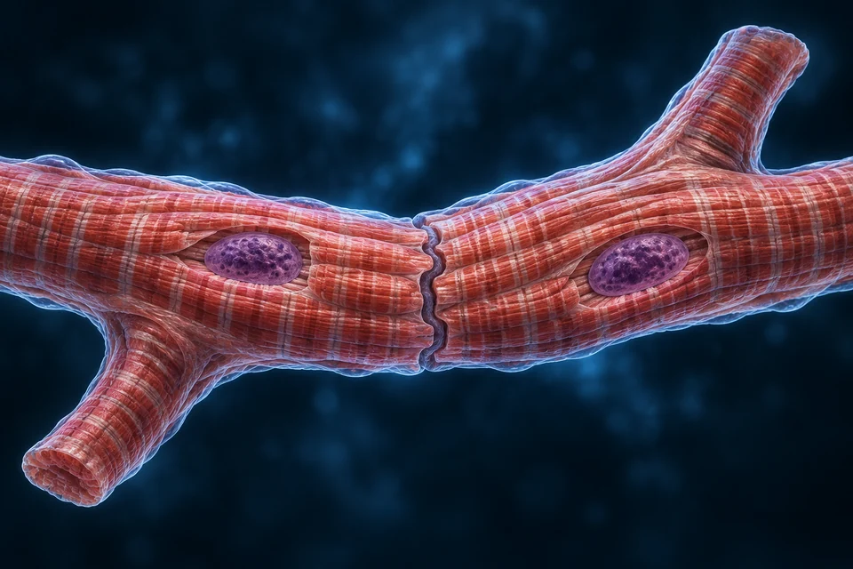
Working cardiomyocytes are elongated, branching and striated. Their nuclei are usually central; some cells contain more than one. The original tissue illustration exposes selected nuclei and boundaries, while keeping detailed membrane/junction nanostructure outside its teaching claim.
New original Golden-family educational illustration of two selected working cardiomyocytes. Local windows reveal central nuclei and the joining region. Striations/branching/separate cells are source-supported; fine folds, branch proportions, nuclear texture, cell-end crops and junction ultrastructure illustrative / not independently verified. This is not histology or microscopy. Human medical review not performed.Structure notes · further context
Branching cell form
Working cells connect within a tissue network.
Central nucleus
A nucleus lies within the elongated cell, not along a skeletal-fibre edge.
Cell-to-cell junction
A complex interface helps transfer load and electrical activation.
Striated organisation
Repeating filament units create a transverse pattern.
Separate cell boundary
Connected tissue does not mean one fused giant cell.
Adhesion and electrical coupling have different jobs
Intercalated discs organise contacts between neighbouring cells. Mechanical junctions transfer load and keep cells attached; gap junctions allow ionic current between them. The interface is variable and complex, not three identical blocks on every cell.
STRUCTURE TO FUNCTION / NORMAL TISSUE
A junction can transfer load and ionic current
New original Golden-family educational illustration of two selected working cardiomyocytes. Local windows reveal central nuclei and the joining region. Striations/branching/separate cells are source-supported; fine folds, branch proportions, nuclear texture, cell-end crops and junction ultrastructure illustrative / not independently verified. This is not histology or microscopy. Human medical review not performed.Structure notes · further context
Branching cell form
Working cells connect within a tissue network.
Central nucleus
A nucleus lies within the elongated cell, not along a skeletal-fibre edge.
Cell-to-cell junction
A complex interface helps transfer load and electrical activation.
Striated organisation
Repeating filament units create a transverse pattern.
Separate cell boundary
Connected tissue does not mean one fused giant cell.
The interface joins neighbouring cells without erasing their separate boundaries. The image locates the contact; mechanical junctions and gap-junction channels are not distinguished by these rendered colours.
Optional qualitative process summary
Retained detailed organ or cell context comes first. The optional editable summary uses qualitative geometry; it is not histology, an electron micrograph or a clinical trace.
Read the relationships
Mechanical link: Adherens junctions and desmosomes contribute to attachment and load transfer.
Electrical coupling: Gap junction channels permit current between neighbouring cells.
Complex interface: The detailed fold and molecular organisation vary; not three fixed universal compartments.
English First. Human medical review not performed.
Go deeper · How can tissue coordinate while cells stay separate?
Mechanical contacts transmit load across an interface; gap junctions provide channels for ionic current. These roles coexist in a structurally complex disc. A visible wavy junction in the illustration cannot identify every adhesion protein or establish molecular channel counts.
Myofibrils contain sarcomeres, whose thick and thin filaments slide relative to one another. Cardiac and skeletal muscle share this broad mechanism, but their activation, cellular connections and calcium handling differ. The existing muscle chapter owns the general sliding-filament lesson.
Entry helps trigger a larger intracellular release
Electrical activation opens selected membrane calcium channels. Incoming calcium helps trigger release from the sarcoplasmic reticulum. Cytosolic calcium interacts with troponin so the filaments can generate force. This is not a nerve impulse travelling physically down a blood vessel.
Original generated 3D teaching cutaway, not microscopy or measured molecular structure. One selected cardiac dyad, not a skeletal triad. Membrane separation and cellular roles are the lesson; protein shape, number, spacing and relative size are illustrative. The SR cut face does not represent leakage. Arrows and all language remain independent editable layers.Structure notes · further context
T-tubule lumen
An inward surface-membrane fold contains extracellular fluid; it is not a cytoplasmic passage.
Membrane calcium entry
Blue embedded complexes locate the L-type-channel role; molecular shapes and counts are illustrative.
Cytosolic cleft
The space between two separate membranes is cytoplasm. The T-tubule and SR lumens do not join.
SR calcium-release channels
Pink embedded complexes locate ryanodine-receptor-mediated release toward the cytosol.
Sarcoplasmic-reticulum store
A separate internal membrane compartment stores calcium; the exposed face is a teaching cutaway.
SR recovery pumps
Green complexes locate SERCA, which uses ATP to transport calcium back into the SR.
Contractile myofibrils
Calcium regulation affects filament activation. The highlighted tissue is not an individual troponin molecule.
Cyan arrow: calcium entry across the T-tubule membrane toward the cytosol. All cues are qualitative, not measured rates, counts, simultaneous events or atomic channel structures.
Follow two separate membrane compartments and then the working filaments. The small dyadic cytosolic cleft is expanded for teaching; no tube connects the lumens.
Selected normal relationships, not measured timing or a complete reaction network. Entry is emphasised; every explanation remains visible. Pressure closure and papillary support are parallel roles, not a serial delay.
Entry
Selected membrane calcium channels open during activation.
Release
Incoming calcium helps trigger calcium release from the sarcoplasmic reticulum.
Force
Calcium binding to troponin permits regulated cross-bridge activity. ATP remains required.
Go deeper · How does a membrane signal recruit an internal store?
In working ventricular muscle, calcium entry through L-type channels can trigger ryanodine-receptor-mediated release from the sarcoplasmic reticulum. Calcium binding to troponin permits filament activation. These are different locations and functions, not one packet of signal travelling down a vessel. The tissue image locates the cell, not individual release sites.
After activation, calcium is resequestered into the sarcoplasmic reticulum and transported out of the cell. Falling cytosolic calcium reduces filament activation. ATP supports calcium pumping and cross-bridge cycling; relaxation is not just a passive absence of effort.
STRUCTURE TO FUNCTION / NORMAL PHYSIOLOGY
Calcium recovery prepares the next contraction
Original generated 3D teaching cutaway, not microscopy or measured molecular structure. One selected cardiac dyad, not a skeletal triad. Membrane separation and cellular roles are the lesson; protein shape, number, spacing and relative size are illustrative. The SR cut face does not represent leakage. Arrows and all language remain independent editable layers.Structure notes · further context
T-tubule lumen
An inward surface-membrane fold contains extracellular fluid; it is not a cytoplasmic passage.
Membrane calcium entry
Blue embedded complexes locate the L-type-channel role; molecular shapes and counts are illustrative.
Cytosolic cleft
The space between two separate membranes is cytoplasm. The T-tubule and SR lumens do not join.
SR calcium-release channels
Pink embedded complexes locate ryanodine-receptor-mediated release toward the cytosol.
Sarcoplasmic-reticulum store
A separate internal membrane compartment stores calcium; the exposed face is a teaching cutaway.
SR recovery pumps
Green complexes locate SERCA, which uses ATP to transport calcium back into the SR.
Contractile myofibrils
Calcium regulation affects filament activation. The highlighted tissue is not an individual troponin molecule.
No flux arrow is shown for this explanatory state. Release decline, transport and relaxation have related but distinct time courses. All cues are qualitative, not measured rates, counts, simultaneous events or atomic channel structures.
Follow two separate membrane compartments and then the working filaments. The small dyadic cytosolic cleft is expanded for teaching; no tube connects the lumens.
Optional qualitative process summary
Selected normal relationships, not measured timing or a complete reaction network. Release ends is emphasised; every explanation remains visible. Pressure closure and papillary support are parallel roles, not a serial delay.
Release ends
SR release falls as the activation event resolves.
Recovery
Calcium returns to the SR and exits the cell through coordinated transport.
Relaxation
Lower cytosolic calcium reduces filament activation; active transport and cycling require energy.
Go deeper · Why are intracellular recovery and cellular exit different?
SERCA transports calcium back into the sarcoplasmic reticulum; sarcolemmal routes, notably sodium–calcium exchange, support extrusion. ATP powers SERCA directly, and sodium gradients used by exchange depend on ATP-dependent transport. Their contributions vary with species and conditions, so no fixed universal percentage is assigned here.
The SA node normally initiates the rhythm; activation spreads through atrial tissue and is delayed at the AV node before the ventricular conducting network is reached. Adult working muscle usually responds to incoming activation rather than acting as an independent pacemaker.
The AV bundle and branches link the atrial sequence with ventricular activation. A route diagram describes order; it does not map every fibre or show all cells contracting at exactly the same instant.
Working myocardial action potentials have a prolonged phase and a refractory interval that limit immediate reactivation. This supports separated contractions and time for filling, unlike simple summation of skeletal-muscle twitches. Pacemaker and working-cell waveforms are different.
STRUCTURE TO FUNCTION / NORMAL TISSUE
A working-cell event is followed by recovery
New original Golden-family educational illustration of two selected working cardiomyocytes. Local windows reveal central nuclei and the joining region. Striations/branching/separate cells are source-supported; fine folds, branch proportions, nuclear texture, cell-end crops and junction ultrastructure illustrative / not independently verified. This is not histology or microscopy. Human medical review not performed.Structure notes · further context
Branching cell form
Working cells connect within a tissue network.
Central nucleus
A nucleus lies within the elongated cell, not along a skeletal-fibre edge.
Cell-to-cell junction
A complex interface helps transfer load and electrical activation.
Striated organisation
Repeating filament units create a transverse pattern.
Separate cell boundary
Connected tissue does not mean one fused giant cell.
A working cell can receive activation and later recover its excitability. This anatomical view does not record voltage; the optional qualitative trace is a separate auxiliary.
Optional qualitative process summary
Retained detailed organ or cell context comes first. The optional editable summary uses qualitative geometry; it is not histology, an electron micrograph or a clinical trace.
Read the relationships
Activation: A rapid electrical change leads into a prolonged phase in working myocardium.
Refractory interval: Immediate reactivation is limited across much of the event.
Recovery: Repolarisation and restored availability support separated contractions. This trace is not an ECG.
English First. Human medical review not performed.
The qualitative supporting trace has no millisecond/millivolt axis and is not an ECG or patient measurement.
Go deeper · Why is electrical recovery not the same as muscle relaxation?
Excitability, intracellular calcium and mechanical force have related but different time courses. The prolonged working-cell electrical event helps limit immediate reactivation. Neither a qualitative action-potential trace nor a still cell picture is an ECG or a measurement of chamber filling.
When ventricular pressure rises above atrial pressure, AV valves close. Ejection follows when ventricular pressure exceeds the connected arterial pressure. During early relaxation, semilunar valves close; filling resumes when ventricular pressure drops below atrial pressure.
STRUCTURE TO FUNCTION / NORMAL PHYSIOLOGY
Changing pressure differences govern valve transitions
Original qualified Heart preparations, near closure and not one shared spatial scene. Chordae restrain AV leaflet displacement; semilunar cusps have no chordal apparatus. Fine attachments are illustrative.Structure notes · further context
Aortic root · right preparation
The artery begins beyond the left ventricular outlet; pocket cusps sit at its base.
Mitral leaflets · left preparation
The closed inlet has two main leaflets separating atrium from ventricle.
Aortic cusps
Three pocket-like cusps meet during closure. There are no chordal tethers.
Chordae tendineae
Tethers connect AV leaflets to papillary muscle; they restrain backward displacement.
Papillary muscle
A muscular projection helps maintain tether tension during ventricular contraction.
Original qualified Heart preparations, near closure and not one shared spatial scene. Chordae restrain AV leaflet displacement; semilunar cusps have no chordal apparatus. Fine attachments are illustrative. Same original, selected observation window; no anatomy regenerated or added.Structure notes · further context
Mitral leaflets · left preparation
The closed inlet has two main leaflets separating atrium from ventricle.
Chordae tendineae
Tethers connect AV leaflets to papillary muscle; they restrain backward displacement.
Papillary muscle
A muscular projection helps maintain tether tension during ventricular contraction.
Original qualified Heart preparations, near closure and not one shared spatial scene. Chordae restrain AV leaflet displacement; semilunar cusps have no chordal apparatus. Fine attachments are illustrative. Same original, selected observation window; no anatomy regenerated or added.Structure notes · further context
Aortic root · right preparation
The artery begins beyond the left ventricular outlet; pocket cusps sit at its base.
Aortic cusps
Three pocket-like cusps meet during closure. There are no chordal tethers.
The two valve preparations identify distinct inlet and outlet structures. They are near closure, not an animation of the selected pressure phase. Follow the linked Heart cycle for separate visible valve states.
Selected normal relationships, not measured timing or a complete reaction network. Closed-valve rise is emphasised; every explanation remains visible. Pressure closure and papillary support are parallel roles, not a serial delay.
Closed-valve rise
AV closure precedes semilunar opening; pressure can rise with little change in ventricular volume.
Pressure can change while chamber volume barely changes
Between AV closure and semilunar opening, contraction is approximately isovolumetric. Between semilunar closure and AV opening, relaxation is approximately isovolumetric. Ejection and filling change volume. These states support the existing four-stage Heart view rather than replace it.
Idealised closed-valve intervals are an educational relationship, not a prediction of leakage-free behaviour in every individual. No fixed volume, pressure or stage duration is prescribed.
Filling, opposition and activation are different influences
Preload describes the stretch/load before contraction; afterload is the load opposed during ejection. Contractility is the muscle’s activation-dependent ability to develop force at a given loading condition. Within a normal range, greater filling can increase the following output. This is not an unlimited stretch rule.
Filling
Before contraction
Length-dependent activation contributes to the next response.
Opposition
During ejection
The ventricle must develop pressure against its connected circulation.
Contractile state
Cellular activation
Calcium handling can change force without simply adding more filling.
Go deeper · Why can filling, outflow load and activation change the same beat?
Compare the initial loading condition, the load opposed during ejection and the tissue’s activation-dependent force. They are separate influences on output. A physiological filling response is bounded; it cannot justify an unlimited rule that more stretch always improves pumping.
A constantly working tissue needs continuing supply
Cardiac cells contain many mitochondria and use several circulating substrates for energy metabolism. Oxygen delivery, fuel processing and ATP use are related but different jobs. No single fuel is the permanent exclusive source for all circumstances.
Neural signals adjust a rhythm they do not usually create
Autonomic input changes rate, conduction and contractile behaviour. It works with intrinsic cellular activity, hormones and local demand. The whole-heart response cannot be read from a static anatomy view or one isolated cell’s colour.
A tissue image, ECG and anatomy model answer different questions
Microscopy can reveal cells and junctional detail, while a gross model shows larger structures. An ECG records aggregate electrical differences at the body surface; it does not directly record wall force, chamber volume or blood flow. This chapter contains no clinical tracing or diagnostic interpretation.
Source-native gross anatomy and original generated cell texture have separate provenance. Fine decorative/cellular details are illustrative. Human medical review and current physical-phone acceptance have not been performed.
Go deeper · Which evidence answers the question you are asking?
Use gross preparations to locate structures, acquired microscopy to examine a specimen, and electrical recordings for time-varying electrical measurements. None establishes every claim made at another scale. The authentic micrograph in Heart keeps its own source and specimen limitations.
Normal myocardial tissue roles,qualitative coronary routes,papillary support,cellular coupling and activation. The existing qualified Heart journey remains unchanged.
Methods and limits
Two newly rendered unchanged source-native adult male BodyParts3D CC-BY4.0 gross preparations, with first-visible source-surface anchors. A separate new original built-in imagegen Golden-family cardiomyocyte illustration, with manually inspected editable labels; its anchors are not source-mesh raycasts. Original native shapes remain coarse; no missing annulus or chordal topology is invented. Retained qualified Heart wall, coronary, valve and myocyte illustrations provide the primary organ/tissue context. A new original ventricular calcium-domain cutaway adds an extracellular-continuous T-tubule, separate junctional SR store, cytosolic cleft and independent activation/recovery cues. Protein shapes, density, cleft width and relative scale are not measured ultrastructure. Source-native gross views and editable process summaries remain optional auxiliaries; names and short explanations are independent of image pixels. Fine cell texture,junction folds,chordal detail and source surfaces remain illustrative / not independently verified. Human medical review: not performed. Educational only.
Revision: cardiac-tissue-english-2026-10-19-eyes-v1. Stable local identity: ahb_ch_cardiac_tissue_english_v1; not a new CMS entity. Original masters stay outside Git; labels and paths remain editable. New physical-phone acceptance: not performed. No patient diagnosis, surgical instruction, medication advice or physician approval claim. Observed microscopy and clinical disease remain separate work.