Transport
Between organs
Gases, absorbed nutrients, hormones and waste travel by different carriers.
CHAPTER / TRANSPORT, EXCHANGE AND RETURN
A living flow. A changing wall.
Look inside blood cells and vessel walls, then follow gas carriage, local exchange, repair and return. Different structures help one moving tissue connect the body.
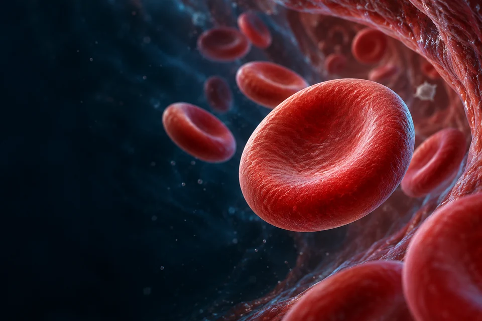
30 SECOND / A LIVING TRANSPORT TISSUE
Read the high-detail tissue first: red cells stay in a lumen; a thin living boundary separates blood from its surroundings. Different mechanisms move gases, water and the blood itself.

Cyan indicates one illustrative along-lumen direction, not measured flow in a named organ. The red cells remain inside the vessel.
Gold selects a teaching region. Direction cues are qualitative explanations on unchanged anatomy, not measured flow, force, pressure or a motion recording.
01 / OVERVIEW
Blood is a living transport medium. Vessels distribute it, exchange material with tissues and return it to the heart. Carrying oxygen is one job; defence, repair, heat and chemical communication are others.
Between organs
Gases, absorbed nutrients, hormones and waste travel by different carriers.
Blood and tissue
Immune cells and soluble proteins have distinct roles.
At a damaged wall
Platelets and coagulation proteins cooperate locally.
Teaching references: NHLBI — How Blood Flows through the Heart · NLM — Physiology, Blood Plasma
02 / PLASMA
Plasma contains water, dissolved ions and small molecules, plus proteins and protein-bound material. The spaces between the selected cells in the illustration represent fluid; they are not empty vacuum.
CONNECT STRUCTURE AND FUNCTION
The detailed formed-element preparation below shows the cellular relationships. It is not a sample count or a fixed fraction diagram.
Liquid: Water, proteins and solutes surround formed elements.
Formed elements: Red cells, white cells and platelets are different categories.
Boundary: Selected pictures do not measure the fraction or abundance.
Serum is obtained after blood clots; some clotting components have been used or removed. It is not simply another name for circulating plasma. Composition and cell fractions vary; a decorative field cannot measure them.
Teaching references: NLM — Physiology, Blood Plasma
03 / PROTEINS
Albumin carries selected substances and contributes strongly to colloid osmotic pressure. Immunoglobulins participate in recognition. Fibrinogen can become fibrin during coagulation. These functions are not interchangeable.
Mainly made in the liver
Carrier and contributor to protein-related fluid exchange.
Made by plasma cells
Antibody functions belong to immune biology.
Soluble plasma protein
Precursor of a local fibrin network.
Teaching references: NLM — Physiology, Blood Plasma · Levick & Michel — Revised Starling principle
04 / FORMED
Selected red cells, one cutaway neutrophil and two platelet fragments reveal different structural jobs. These representative illustrations are not a measured blood smear or true abundance/size comparison; most white-cell types are not shown.

The thin centre and thicker rim belong to one flexible red cell. This organelle-free mature cell is different from its nucleated marrow precursors.
Gold selects a teaching region. Direction cues are qualitative explanations on unchanged anatomy, not measured flow, force, pressure or a motion recording.
Teaching references: OpenStax — Erythrocytes · OpenStax — Leukocytes and Platelets
05 / RBC
A mature human red cell is usually a flexible biconcave disc. Its membrane and internal support permit deformation in small vessels. Mature circulating cells lack a nucleus; this is not a rule for every stage of red-cell development.
Mature circulating cell
The centre is thinner than the rim, not an open hole.
Small vessel passage
Cells can deform and recover; the static model does not simulate this.
Packed with hemoglobin
Loss of the nucleus is part of maturation.
Teaching references: OpenStax — Erythrocytes
06 / HEMOGLOBIN
Adult hemoglobin contains four globin chains, each associated with a heme group. A heme iron can bind an oxygen molecule reversibly. This molecular arrangement is separate from the overall red-cell shape.
CONNECT STRUCTURE AND FUNCTION
One common adult tetramer
4 globin chains · 4 associated heme groups · up to 4 O₂ molecules
One heme-associated iron binds one oxygen molecule reversibly. Occupancy varies.
Locate the red-cell carrier →Globin chains: A common adult hemoglobin tetramer has four chains.
Heme: Each associated heme iron can bind oxygen reversibly.
Boundary: An original relationship model, not atomic geometry or a clinical saturation curve.
The supporting four-site drawing is not an atomic structure, a diagnostic oxygen-saturation chart or a model of every hemoglobin variant.
A red cell contains many hemoglobin molecules. In the common adult tetramer, each of four globin chains has an associated heme group; each heme can bind one oxygen molecule. The molecule can therefore carry up to four oxygen molecules, with occupancy varying rather than every site always being filled.
The cell illustration teaches the carrier compartment. It does not resolve an atomic protein structure or a patient oxygen-saturation reading.
Red-cell and hemoglobin physiologyTeaching references: OpenStax — Erythrocytes
07 / LOADING
Blood reaches lung capillaries where oxygen availability favours loading. At tissues, oxygen can leave hemoglobin and diffuse toward cells. A molecule is not permanently assigned to a single organ.
Locate the red cell and plasma, then compare a using tissue with the lung interface. Oxygen pressure, protein affinity and carbon-dioxide handling answer different questions.
Cyan · O₂ leaves the cellGold · CO₂ enters the cell

Directions identify a selected physiological context, not measured flow. The respiratory or vessel–tissue barrier is outside this cellular window. Original generated anatomy, not microscopy; retained from the carbon-dioxide lesson.
Oxygen use can keep tissue oxygen partial pressure below that in arriving blood. Dissolved oxygen diffuses toward the tissue; reversible release from haemoglobin replenishes dissolved oxygen. The cell does not pump oxygen out.
Mechanism evidence: OpenStax · Gas transport · Tyuma · Bohr and Haldane effects in human haemoglobin · Dash & Bassingthwaighte · Corrected gas-binding model
H⁺ / CO₂ change → oxygen affinity changes.
Oxygenation changes → H⁺ / CO₂ carriage changes.
Vertical direction: greater oxygen-site occupancy. Horizontal direction: greater oxygen partial pressure.
Mechanism evidence: OpenStax · Gas transport · Dash & Bassingthwaighte · Corrected gas-binding model · Cooperative oxygen-binding mechanism · primary study
Oxygen partial pressure describes dissolved oxygen, not the amount bound to haemoglobin. Saturation is the fraction of available haem sites occupied. Blood oxygen content includes haemoglobin-bound and dissolved oxygen.
The same saturation need not mean the same oxygen content if haemoglobin concentration differs. Delivery also depends on blood flow; neither a saturation number nor an affinity change alone establishes tissue supply.
Mechanism evidence: Regulation of Tissue Oxygenation · Oxygen transport · Dash & Bassingthwaighte · Corrected gas-binding model
Adult haemoglobin normally contains two alpha and two beta globin chains, each with a haem. Oxygen binding changes the balance of protein conformations and affects the affinity of other sites: positive cooperativity.
A population of molecules produces the familiar sigmoid oxygen-equilibrium curve. This is not four sites filling in a compulsory spatial order. The four-site summary elsewhere in this chapter counts capacity; it is not an atomic model.
Mechanism evidence: Hemoglobin structure, function and allostery · review · Cooperative oxygen-binding mechanism · primary study
Affinity concerns how readily oxygen binds under stated conditions. Capacity concerns how much can bind when available functional sites are filled. A shift in affinity can change occupancy at the same oxygen pressure without creating extra haem groups.
Lower affinity may support unloading in a tissue yet reduce loading under other conditions. Higher affinity can aid binding but hinder release. Neither direction is a universal improvement.
Mechanism evidence: Regulation of Tissue Oxygenation · Oxygen transport · Dash & Bassingthwaighte · Corrected gas-binding model
Changes in protonation and CO₂ interactions alter haemoglobin’s oxygen equilibrium. Increased H⁺ and CO₂ usually favour lower oxygen affinity; oxygen can unload more readily at a given oxygen partial pressure.
Compare conditions while keeping the other variables stated. A tissue’s lower oxygen pressure and its effect on affinity cooperate physiologically, but they are different explanations.
Mechanism evidence: Tyuma · Bohr and Haldane effects in human haemoglobin · Dash & Bassingthwaighte · Corrected gas-binding model
Deoxygenated haemoglobin can bind more H⁺ and carbamino CO₂ than its oxygenated form. Oxygenation reverses this tendency and helps release carbon dioxide in the lung.
Much carbon travels as bicarbonate rather than directly attached to haemoglobin. Proton buffering therefore matters alongside carbamino binding. The Bohr and Haldane names describe reciprocal aspects of linked binding, not two pumps or two isolated stages.
Mechanism evidence: Tyuma · Bohr and Haldane effects in human haemoglobin · Dash & Bassingthwaighte · Corrected gas-binding model
Higher temperature generally lowers oxygen affinity. The red-cell metabolite 2,3-bisphosphoglycerate, also called 2,3-DPG, preferentially interacts with deoxygenated adult haemoglobin and favours lower-affinity behaviour.
These modifiers do not override oxygen pressure or make every tissue unload a fixed fraction. Isolated haemoglobin experiments establish molecular effects under controlled conditions; they do not give a whole-person oxygen-delivery rate.
Mechanism evidence: Hemoglobin structure, function and allostery · review · Antonini et al. · Isolated human haemoglobin experiment · Dash & Bassingthwaighte · Corrected gas-binding model
Oxygen crosses plasma and the red-cell membrane, and it crosses a separate vessel–tissue or alveolar–capillary interface during exchange. Haemoglobin remains inside the red cell in this normal teaching context.
The existing carbon-dioxide atlas below explains carbonic anhydrase, bicarbonate and chloride exchange in detail. Its chemistry complements oxygen binding; this image cannot resolve an individual haem pocket, transporter or measured gas stream.
Mechanism evidence: OpenStax · Gas transport · Regulation of Tissue Oxygenation · Oxygen transport
Equilibrium studies and mathematical models help separate interacting variables. They do not fully represent a moving capillary, uneven ventilation, changing blood flow or every haemoglobin form.
This comparison supplies qualitative directions, not numerical saturation predictions, clinical thresholds or instructions to change breathing or oxygen treatment. Human medical review has not been performed.
Mechanism evidence: Dash & Bassingthwaighte · Corrected gas-binding model · Antonini et al. · Isolated human haemoglobin experiment · Cooperative oxygen-binding mechanism · primary study
No. Saturation describes site occupancy. Haemoglobin concentration, blood flow and exchange conditions also matter.
No. Carbamino CO₂ forms at globin amino groups. Haemoglobin also buffers H⁺ associated with bicarbonate chemistry.
Bohr asks how H⁺ and CO₂ influence oxygen affinity. Haldane asks how oxygenation influences proton binding and carbon-dioxide carriage. The underlying equilibria are linked.
No. It identifies a net exchange direction in a selected context, not a pump, a flow measurement or a complete respiratory barrier.
Sources checked 21 October 2026. Normal physiological teaching; no clinical thresholds or treatment advice. AI-assisted medical checks are not physician, fine molecular-structure or physical-phone certification.
SUPPORTING PROCESS / ALL STAGES VISIBLE
Lung environment → blood carriage → tissue release
Connect the selected relationship with the anatomical teaching:
Locate the red cell →Separate the molecular binding site →Oxygen availability at lungs favours heme binding.
No permanent molecule-to-organ assignment.
Hemoglobin transports bound oxygen within red cells.
Blood is red, not literally blue.
Local gradients and chemical conditions favour tissue delivery.
No personal saturation/pressure threshold calculated.
Emphasis only: essential facts remain visible. Original supporting explanation, not a measured simulation or patient blood-flow or clotting prediction.
Higher local carbon dioxide, acidity and temperature can favour release in active tissue. These are qualitative relationships; there is no personal oxygen threshold or numerical dissociation curve here. Blood remains red: oxygen-rich blood is brighter red and oxygen-poor blood darker red. Blue in circuit diagrams is a convention.
Teaching references: OpenStax — Erythrocytes · NHLBI — How Blood Flows through the Heart
08 / CARBON-DIOXIDE
Carbon dioxide produced in tissues enters blood. Much is converted into bicarbonate, aided by red-cell carbonic anhydrase. Some remains dissolved or binds to protein sites distinct from the oxygen-binding heme site.
First locate the cell surface, cytosol and plasma. Then compare tissue-side loading with lung-side release, keeping biochemical conversion separate from gas diffusion.
Gold: CO₂ / H⁺ · Cyan: HCO₃⁻ · Pink: Cl⁻. Direction cues are qualitative; names and selected explanations also describe the mechanism.
CO₂ can enter red cells from tissue-side blood. Bicarbonate can leave as chloride enters through anion exchange; haemoglobin buffering is a separate part of the chemistry.
CO₂ + H₂O ⇌ H⁺ + HCO₃⁻
Mechanism sources: OpenStax · normal gas transport · Human erythrocyte band3 / AE1 · primary structure study · NLM · Carbon dioxide transport
No. Dissolved gas and protein-bound forms also contribute. Carbon dioxide binds globin sites rather than the haem iron used by oxygen. The proportions depend on physiological context; this lesson assigns no universal transport fraction.
Normal-physiology sources checked 20 October 2026. Original generated cellular context, not microscopy or physician review. No breathing, drug or infusion instructions.
CONNECT STRUCTURE AND FUNCTION
CO₂ + H₂O ⇌ H⁺ + HCO₃⁻
Tissue-side context: carbon dioxide enters the red cell; carbonic anhydrase accelerates conversion. Bicarbonate can leave as chloride enters.
Lung-side context: the exchange direction reverses and carbon dioxide can leave for exhalation.
Connect the chemical route to lung exchange →Tissue: CO₂ enters RBC; bicarbonate can leave as chloride enters.
Lung: Bicarbonate enters and chloride leaves; CO₂ is regenerated.
Boundary: Dissolved and protein-bound routes coexist; no fixed percentages.
At tissues, bicarbonate can leave a red cell as chloride enters. At lungs this exchange reverses, helping regenerate carbon dioxide for exhalation. Hemoglobin also buffers hydrogen ions. No fixed universal transport percentages are assigned.
Carbon dioxide can enter red cells and be converted to bicarbonate with carbonic-anhydrase assistance. Bicarbonate and chloride exchange across the membrane in opposite directions, reversing as blood passes through lung exchange conditions. Hemoglobin also buffers hydrogen ions.
Oxygen binding to heme is a different interaction from carbon dioxide binding at protein sites or travelling as bicarbonate. These routes coexist; the lesson does not assign fixed percentages to every person or tissue.
Carbon-dioxide transport source · Lung exchangeTeaching references: NLM — Physiology, Carbon Dioxide Transport
09 / FORMATION
Blood-cell lineages arise from hematopoietic progenitors. In adults, red marrow is an important production site. Kidney tissue responds to oxygen conditions with erythropoietin signalling that supports red-cell production.
CONNECT STRUCTURE AND FUNCTION
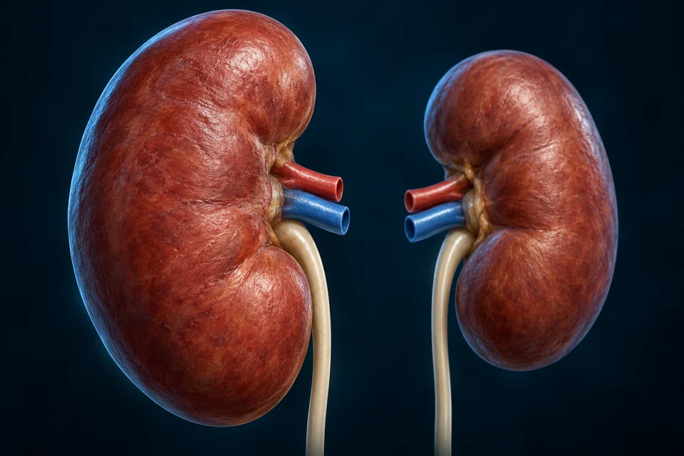
Renal oxygen-sensing context helps regulate erythropoietin. This organ view does not locate or certify individual EPO-producing interstitial cells.
Kidney regulatory roles →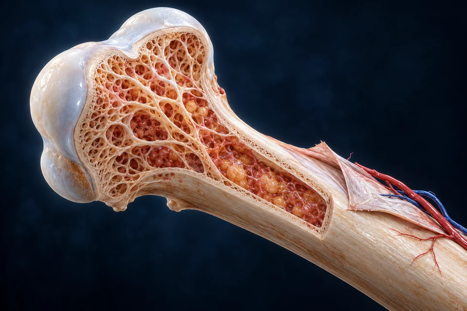
Marrow precursors mature along an erythroid lineage before mature red cells enter circulation. EPO is not the common controller of every blood-cell lineage.
Locate marrow and blood formation →Kidney: Oxygen context affects erythropoietin signalling.
Marrow: Erythroid development increases hemoglobin-bearing output.
Boundary: Blood families have different control paths; not a fixed cell-count prescription.
Compare two normal output routes after locating marrow in the retained anatomical views above. A reticulocyte continues as a whole enucleated cell; a megakaryocyte supplies cytoplasmic elements that become platelets. The independent tissue preparations are not a measured size chart or a complete lineage tree.
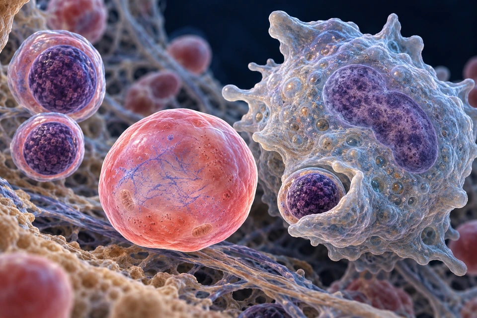
Gold identifies the selected structural context on unchanged tissue. It does not measure flow, secretion rate or molecular signalling. These are original generated teaching preparations, separate from the authentic blood images in this chapter.
A newly enucleated reticulocyte retains a living cytoplasmic compartment. The separate nuclear packet has another fate: the neighbouring macrophage can engulf it. The reticulocyte is not a platelet and is not yet a fully mature red disc.
Normal structure and mechanism: Newton et al. · Erythroblast enucleation (2024) · NLM · Histology, Reticulocytes · NLM · The erythroblastic island
Blood-forming marrow combines developing blood cells with vessels, stromal support and other neighbouring cells. Fat-rich and blood-forming regions differ with age and location; the retained long-bone view locates the compartment without identifying every cell in it.
Stem and progenitor populations replenish specialised output through differentiation and proliferation. The two selected output routes are not a complete lineage tree, and no rule that every stem-cell division makes exactly one stem cell and one committed daughter is required to read them.
Normal structure and mechanism: OpenStax · Production of formed elements · ASH · Megakaryocytes and their marrow niche
During terminal erythroid differentiation, the nucleus becomes compact and is expelled with a small membrane-enclosed rim of material. The remaining enucleated cell is a reticulocyte; the separate nucleus-containing packet is called a pyrenocyte.
Our preparation begins after that separation. A nearby nucleated erythroblast supplies context, not a filmed earlier frame. Nuclear condensation, membrane reorganisation and separation are coordinated cellular events, rather than a nucleus simply dissolving in the circulating cell.
Normal structure and mechanism: Newton et al. · Erythroblast enucleation (2024)
A reticulocyte still contains residual RNA and cellular machinery. Maturation includes removing remaining organellar material and remodelling its membrane and shape before the mature erythrocyte architecture is established.
Fine coloured strands in an atlas are illustrative cytoplasmic context. Reticulocyte identification in a real specimen depends on the acquisition and staining method; this image does not supply a diagnostic percentage, production rate or clinical response.
Normal structure and mechanism: NLM · Histology, Reticulocytes · OpenStax · Erythrocyte structure and production
Macrophages in erythroblastic-island environments interact with developing erythroid cells and can engulf expelled nuclear packets. The large macrophage has its own nucleus: it is not the nucleus that was removed from the reticulocyte.
Supportive niche interactions matter, but mechanisms and their experimental settings must be distinguished. The preparation does not claim that a macrophage must physically perform every enucleation, or that a single idealised island geometry represents all adult human marrow.
Normal structure and mechanism: NLM · The erythroblastic island · Newton et al. · Erythroblast enucleation (2024)
Kidney oxygen-sensing and erythropoietin signalling connect tissue oxygen needs with erythroid development in marrow. The existing organ-feedback summary remains separate from the local cellular preparation.
Producing haemoglobin-bearing cells also requires suitable building materials and cellular conditions, including iron and nutritional support. A signal cannot substitute for all of those requirements. This is normal physiology, not advice to take supplements, alter blood counts or use EPO.
Normal structure and mechanism: OpenStax · Erythrocyte structure and production · OpenStax · Production of formed elements
Megakaryocyte development involves extensive cytoplasmic growth and nuclear changes, often including polyploidisation and a lobulated nuclear outline. A larger membrane and organelle supply supports later cytoplasmic output.
The selected connected nuclear context remains in the producer; it is not divided among the illustrated fragments. A rendered lobe count cannot establish chromosome content, maturation stage or the full range of megakaryocyte development.
Normal structure and mechanism: ASH · Megakaryocytes and their marrow niche · NLM · Platelet production and human marrow observations
In the representative route shown here, a megakaryocyte forms an extension associated with the marrow sinusoidal boundary. A connected intraluminal process supplies smaller elements that undergo further processing.
The nucleus remains outside this cytoplasmic route. The endothelial boundary separates marrow tissue from blood, and the normal process is different from an open wound or a platelet-rich clot. Fine membrane passages and molecular details are not established by this generated tissue.
Normal structure and mechanism: Furniss et al. · Platelet generation in vivo and in vitro (2024) · NLM · Platelet production and human marrow observations
Proplatelet formation is important, but contemporary studies also investigate other protrusions, budding and intravascular processing. Their relative contributions depend on the model, method and biological setting.
Much mechanistic imaging comes from animal or cultured-cell work, alongside human observations. The illustration therefore presents a representative route without claiming that every platelet forms at the same marrow point, that all processing ends there, or that mouse results are automatically measured human physiology.
Normal structure and mechanism: Furniss et al. · Platelet generation in vivo and in vitro (2024) · NLM · Platelet production and human marrow observations
Erythropoietin is associated with erythroid production; thrombopoietin is a major regulator of megakaryocyte development. The liver is a principal source of circulating TPO, while adult kidney EPO signalling belongs to a different physiological axis.
Neither hormone is a tiny cell fragment or a mechanical pair of scissors. Signalling, cell maturation and the final organisation of platelet output are related but distinct; the image does not depict every regulator or give a personal treatment target.
Normal structure and mechanism: NLM · Cytokine control of megakaryopoiesis · NLM · Liver production of TPO compared with EPO · OpenStax · Erythrocyte structure and production
A reticulocyte matures toward oxygen-carrying erythrocyte architecture. A platelet fragment participates in regulated responses such as local vascular repair. Producing either is different from activating a circulating platelet at a damaged surface.
Compare these original preparations with the retained authentic blood observations and the existing gas-carriage and haemostasis lessons. Atlas texture cannot establish a specimen diagnosis, actual cell-output rate, lineage identity by colour or the complete activity of a cell.
Normal structure and mechanism: NLM · Histology, Reticulocytes · Furniss et al. · Platelet generation in vivo and in vitro (2024) · OpenStax · Erythrocyte structure and production
No. The reticulocyte and the membrane-enclosed pyrenocyte are separate after enucleation; the nearby macrophage has a third, independent nuclear compartment.
No. The connected producer retains its nucleus. The extensions and smaller platelet fragments contain cytoplasm, not a share of that nuclear mass.
No. It illustrates a production route at a sinusoidal boundary. Platelet adhesion and fibrin formation at damage belong to the separate haemostasis lesson.
No. Methods, species and alternative routes matter. These preparations teach selected normal relationships without diagnostic counts or a complete lineage map.
Sources checked 21 October 2026. Normal educational relationships; animal, culture and human observations are not interchangeable. Generated fine detail is not acquired microscopy or a diagnostic measurement. AI-assisted checks are not physician review.
Teaching references: OpenStax — Erythrocytes · OpenStax — Leukocytes and Platelets
10 / RECYCLING
Older or damaged red cells can be cleared by macrophages, especially in the spleen, liver and marrow. Protein components and iron can be reused; heme-derived pigment follows a different disposal route.
SUPPORTING PROCESS / ALL STAGES VISIBLE
Selected old red cell → macrophage processing → separate material routes
Connect the selected relationship with the anatomical teaching:
Spleen clearance context →Heme handling and recovery →Phagocytic cells can remove old or damaged red cells.
Sites include spleen, liver and marrow.
Iron and protein components can return to usable pools.
Not a new intact red cell emerging from a macrophage.
Heme pigment handling connects to liver/bile pathways.
Not a bilirubin test or identical cell-lifespan clock.
Emphasis only: essential facts remain visible. Original supporting explanation, not a measured simulation or patient blood-flow or clotting prediction.
Start inside the processing cell, then connect it with the retained spleen and liver preparations. These are different scales and tissues, not three consecutive frames or a single continuous vessel.
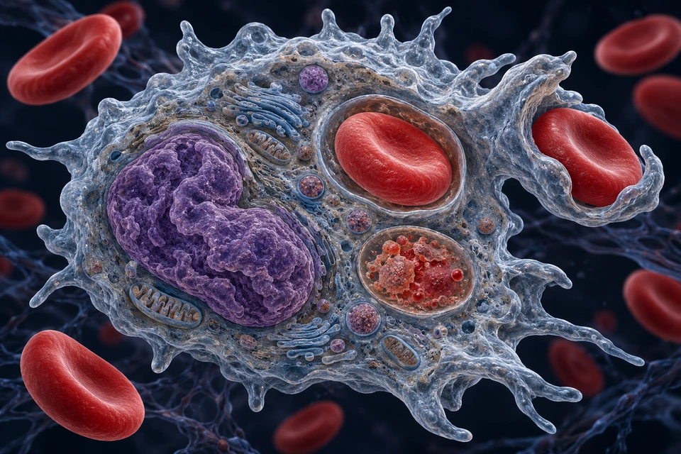
Protein → amino acidsReusable building materials.
Iron → storage or reuseKeep the metal in the available pool.
Heme pigment → liver → bileProcess a different breakdown product.
Gold locates the selected tissue context. The dashed cyan liver cue is a qualitative blood-facing → cellular → bile-facing relationship, not an open duct or a measured molecule track. Colours do not identify molecular cargo. The original atlas preparations remain distinct from the authentic observations elsewhere in this chapter.
The membrane cup engages an outside red cell. Another red cell is enclosed in a separate internal compartment. The large folded purple region is the macrophage’s own nucleus, not a swallowed red-cell nucleus.
Normal mechanism: NLM · Macrophages in erythropoiesis and erythrophagocytosis · NLM · Iron regulation: macrophages in control · NLM · Physiology, bilirubin
A macrophage can surround a selected red cell and enclose it in an internal membrane-bound compartment. Subsequent degradative processing breaks down that cellular cargo. The macrophage retains its own nucleus and metabolism.
The cutaway combines selected processing contexts for comparison, not a timed record. Free haemoglobin handling after intravascular rupture is a different route; this lesson does not present rupture as the required normal first step.
Normal mechanism: NLM · Macrophages in erythropoiesis and erythrophagocytosis
Red cells undergo changes during circulation. Their membrane properties and interactions with clearance tissues contribute to removal. An often-quoted lifespan of about 120 days is a population-level teaching approximation, not an identical expiry time for every cell.
Spleen, liver and marrow contribute in different settings. The gross spleen preparation locates red and white pulp without substituting rodent marginal-zone anatomy or claiming a single universal recognition switch.
Normal mechanism: OpenStax · Erythrocyte lifecycle · NLM · Iron regulation: macrophages in control
Globin proteins can be broken down into amino acids for general reuse. The heme group requires different processing: heme oxygenase releases iron and forms biliverdin, with carbon monoxide also produced; biliverdin reductase forms bilirubin.
Recovering iron does not preserve an intact haemoglobin molecule. Nor does the macrophage manufacture a replacement mature red cell: production and maturation belong to the marrow lesson.
Normal mechanism: OpenStax · Erythrocyte lifecycle · NLM · Physiology, bilirubin
Ferritin safely holds iron within cells. Ferroportin is a membrane exporter; transferrin is the plasma carrier that delivers iron to tissues including developing erythroid cells. These names describe different jobs, not successive kinds of blood cell.
Macrophages can retain recovered iron or release it into the circulating pool. Oxidation and carrier loading help prepare exported iron for transport; the atlas does not identify those molecular reactions by coloured cytoplasmic granules.
Normal mechanism: NLM · Mammalian iron transport · NLM · Iron regulation: macrophages in control
Hepcidin made chiefly by the liver reduces ferroportin-mediated iron export. It can inhibit the exporter and promote its internalisation and degradation, reducing iron entry into plasma from recycling macrophages and other supplying cells.
The regulatory message is separate from an iron atom’s route. Lower or higher hepcidin influence availability within a wider system involving iron supply, erythropoietic demand and other signals; this is not a personal blood-test interpretation or treatment rule.
Normal mechanism: NLM · Hepcidin and iron in health and disease
Unconjugated bilirubin is poorly water soluble and travels mainly bound to albumin. Ordinary binding is noncovalent; the pigment can dissociate for hepatic uptake. This is different from the covalently albumin-bound delta-bilirubin described in some disease settings.
Do not read the image as albumin entering a bile channel with an intact red cell. The tissue view locates interfaces, while the separate text explains chemical transport that is not visibly resolved.
Normal mechanism: PubMed · Human albumin–bilirubin binding study · NLM · Physiology, bilirubin · NLM · Bilirubin metabolism and hepatic transport
Within hepatocytes, UGT1A1 adds glucuronic acid to bilirubin. This produces more water-soluble conjugates. Transport across the canalicular membrane, chiefly involving MRP2, then supplies the bile-facing compartment.
The retained adjacent-cell view shows where that compartment lies, not an atomic model of UGT1A1 or MRP2. Cellular experiments establish particular mechanisms under stated conditions; they do not measure the pathway rate in this generated human teaching preparation.
Normal mechanism: NLM · Bilirubin glucuronidation: experimental mechanisms · NLM · Bilirubin metabolism and hepatic transport
Bile carries pigment to the intestine, where microbes form further products; much pigment-derived material leaves in stool, while some products are reabsorbed and a smaller route contributes to urinary pigments. This is not ordinary filtration of albumin-bound unconjugated bilirubin.
Bile salts instead support fat handling and are largely reclaimed through an enterohepatic loop. Follow the existing Liver and Small Intestine lessons for that loop rather than treating bilirubin itself as the fat-emulsifying agent.
Normal mechanism: NLM · Physiology, bilirubin · NLM · Bilirubin metabolism and hepatic transport
No. It belongs to the macrophage. The red cells have no nuclei; selected ingested material lies in separate membrane compartments.
No. They provide intracellular storage, membrane export and plasma carriage respectively.
No. Iron recovery and the heme-derived pigment pathway separate. Pigment disposal is not the same as loss of the recovered iron.
No. A canalicular bile space and the blood-facing exchange interface are different compartments.
Sources checked 21 October 2026. Normal physiology, without personal diagnostic or treatment thresholds. Experimental molecular evidence and generated tissue are separate. AI-assisted checking is not physician review; fine rendered ultrastructure is not certified.
This is normal recycling context, not a bilirubin test guide or a guarantee that every cell has an identical lifespan.
Teaching references: OpenStax — Erythrocytes
11 / WHITE-CELLS
Neutrophils, eosinophils, basophils, monocytes and lymphocytes differ in shape and activity. The earlier formed-element plate opens one neutrophil; the comparison below adds the other four family contexts. Many leukocytes can enter tissue; detailed immune networks are linked separately.
Start with a nucleus, compare what surrounds it, then ask where the cell works. These normal portraits complement the retained stained blood observations; neither colour nor shape alone establishes a clinical or molecular identity.
Granulocyte · Specialised granule responses

Gold selects the current teaching region on unchanged anatomy. The gallery shows independent enlarged preparations, not relative abundance, calibrated cell size, a smear or stages of one cell changing family.
Two visible lobes are joined by a narrow bridge. This is one bilobed nucleus within one cell; the orange cytoplasmic granules are outside it.
Normal structure and function: University of Leeds · White Blood Cells · Stone et al. · IgE, mast cells, basophils and eosinophils
A mature granulocyte may have a segmented or folded nuclear outline. Narrow connections link lobes within one nuclear system; the cells are not dividing into several small cells.
Read the cell boundary as well as the purple material. The eosinophil bridge is visible here, while basophil granules partly cover its nuclear context.
Normal structure and function: OpenStax · Leukocytes and Platelets · NLM · Histology, White Blood Cell
Conventional staining makes some cytoplasmic granules conspicuous. This underlies the familiar granular/agranular grouping. Monocytes and lymphocytes can still contain less conspicuous granules.
The atlas uses controlled colours and cutaways to separate structures. A stained specimen, a generated granule and a biochemical assay provide different evidence; colour is not a measurement of a protein.
Normal structure and function: OpenStax · Leukocytes and Platelets · University of Leeds · White Blood Cells
A circulating neutrophil first needs the appropriate recruitment context to reach many tissue challenges. Engulfment then encloses material in an intracellular compartment; granule contents can contribute to its handling.
Neither a lobed nucleus nor a granule-rich appearance establishes that these actions are happening. Follow the existing migration lesson for endothelial interactions and tissue guidance.
Normal structure and function: NLM · Histology, White Blood Cell · Immunobiology · Components of the immune system
Its coarse specific granules and commonly bilobed nucleus provide a structural contrast. Granule proteins participate in specialised responses, including contexts involving parasites and allergic inflammation.
Those roles do not mean every eosinophil is damaging tissue or responding to a parasite. The illustration is a normal structural comparison, not a diagnosis from granule colour.
Normal structure and function: Stone et al. · IgE, mast cells, basophils and eosinophils · NLM · Histology, White Blood Cell
Both can participate in mediator-based responses, but a blood basophil is not simply a mast cell waiting to enter tissue. Similar mediators do not make two cell populations interchangeable.
Receptor-bound IgE can contribute to recognition of an appropriate stimulus. Binding or the presence of granules alone does not demonstrate degranulation; this image keeps the contents inside the cell.
Normal structure and function: Stone et al. · IgE, mast cells, basophils and eosinophils · NLM · Histology, White Blood Cell
Some tissue phagocytes arise from circulating monocytes, especially in particular recruitment settings. Other resident populations are established earlier and can maintain themselves locally.
Mouse fate-mapping and parabiosis experiments helped demonstrate this distinction. Their findings correct an overly simple origin story; they do not assign the same turnover or developmental history to every human macrophage.
Normal structure and function: Hashimoto et al. · resident macrophage maintenance (2013)
A small resting lymphocyte can show dense nuclear material and a relatively narrow cytoplasmic rim. That appearance does not identify its antigen receptor, target or developmental programme.
B-cell and T-cell roles need additional functional and molecular information. The existing recognition lesson distinguishes their interactions; the generated portrait is deliberately not assigned one subtype.
Normal structure and function: Molecular Biology of the Cell · Lymphocyte structure and function · Immunobiology · Components of the immune system
Natural killer cells are lymphoid cells with innate effector programmes. Many have a granular lymphocyte appearance; this is not a reason to classify them as neutrophils or basophils.
Nor does a granular lymphocyte picture establish NK identity. Functional context and molecular markers distinguish populations that a generic cellular portrait cannot certify.
Normal structure and function: NLM · Histology, Natural Killer Cells · Immunobiology · Components of the immune system
Adult blood-forming marrow supplies developing blood-cell populations. B-cell development and thymic T-cell development connect those origins with distinct maturation environments.
Later encounters in lymphoid tissues can activate suitable mature lymphocytes. A cell moving through blood is not necessarily developing there, and one simplified lineage does not explain every tissue-resident immune cell.
Normal structure and function: Immunobiology · Components of the immune system · Molecular Biology of the Cell · Lymphocyte structure and function
The retained formed-element preparation includes separate red cells, a neutrophil and platelets. Mature red cells lack a nucleus; a platelet is a membrane-bound fragment derived from a megakaryocyte.
White cells retain nuclei, but nuclear presence alone does not specify their family or role. Use the existing platelet and vessel-repair teaching rather than reading every small purple object as an immune cell.
Normal structure and function: OpenStax · Leukocytes and Platelets
Blood is a transport setting for many leukocytes, while important interactions occur in tissues and lymphoid organs. Endothelial crossing and local guidance require regulated contacts, not an open pipe from blood to a target.
The five-family comparison is a conventional peripheral-blood overview. Dendritic cells, mast cells, macrophages and other tissue populations remain important beyond this selected set.
Normal structure and function: NLM · Histology, White Blood Cell · Immunobiology · Components of the immune system
Smear appearance depends on preparation, stain and viewing plane. Nuclear outlines and granules provide useful clues, but measurements, molecular markers and appropriate clinical context answer other questions.
The existing authentic stained and SEM images remain separate from these generated cutaways. No counts, diagnostic thresholds, receptor assignments or calibrated relative sizes can be extracted from this gallery.
Normal structure and function: University of Leeds · White Blood Cells · Molecular Biology of the Cell · Lymphocyte structure and function · NLM · Histology, Natural Killer Cells
No. This selected eosinophil has one bilobed nucleus within one cell.
No. It refers to the less conspicuous granule appearance in conventional morphology, not the absence of all granules or lysosomes.
No. This generic resting lymphocyte needs additional molecular and functional evidence for subtype assignment.
No. Origins and maintenance differ among tissues and contexts; some populations can self-maintain.
Sources checked 21 October 2026. Original generated cutaways are separate from acquired microscopy and do not certify fine ultrastructure. AI-assisted checking is not physician review. No differential-count thresholds, disease screen or treatment guidance is provided.
Selected cytoplasmic granules
Neutrophils, eosinophils and basophils have different responses.
Adaptive and innate roles
B, T and NK cells are not distinguishable solely by this one illustration.
Blood-to-tissue connections
Some tissue phagocytes derive from monocytes; not all resident macrophages follow one universal origin.
Teaching references: OpenStax — Leukocytes and Platelets
12 / PLATELETS
Platelets are membrane-bound fragments released from marrow megakaryocytes. They have no nucleus, but contain active machinery and granules. Resting discs can change shape after activation; this plate does not show every activation state.
Large marrow megakaryocyte
Fragments enter circulation.
Damaged vessel surface
Adhesion, signalling and aggregation cooperate.
Not a tiny red blood cell
Platelet function differs from gas carriage.
Teaching references: OpenStax — Leukocytes and Platelets · OpenStax — Hemostasis
13 / HEMOSTASIS
Vessel-wall responses, platelet recruitment and coagulation overlap. Platelets form a local plug; thrombin-mediated fibrin formation helps stabilize it. These are coordinated responses, not three isolated events with identical timing.
WALL → ACTIVE FRAGMENTS → PROTEIN NETWORK
Wall response ↔ platelet recruitment ↔ fibrin stabilization
AttachPlatelets bind at an affected surface.
ReinforceFibrin adds an extracellular network.
RegulateIntact lining, inhibitors and lysis constrain the response.

The exposed wall region provides a local context for adhesion and activation. The platelets in this preparation have already changed shape; it is not an earlier time frame.
Gold selects a teaching region. Direction cues are qualitative explanations on unchanged anatomy, not measured flow, force, pressure or a motion recording.
The anatomical preparation locates platelets, extracellular fibres and the adjacent lining. The relationships below explain molecules separately; they are not newly resolved objects within the image.
The missing lining exposes a different surface to flowing blood. Matrix-associated adhesive interactions help capture platelets there; the adjacent intact lining has a different role. A platelet attaches to the wall before a larger aggregate can be supported, but the drawing is not a timed sequence.
Exposed wallPlatelet attachment
Adhesion locates fragments at a reactive surface.
Activated platelet integrinsFibrinogen-mediated bridges
Aggregation links neighbouring platelets.
Local agonistsFurther platelet activation
Signals reinforce the response near the affected region.
Each row is a separate causal relationship. Arrows mean biological action or association, not physical blood flow, a transformation of whole cells into protein, or a universal three-step clock.
Mechanism references: NLM · Platelets in normal haemostasis · NLM · Platelet adhesion under flow · NLM · Integrated understanding of haemostasis
Exposure and signalling support platelet adhesion and aggregation.
Platelets are active fragments, not fibrin strands.
Coagulation generates a fibrin network that supports the local plug.
These events overlap; no complete factor cascade shown.
Intact wall, regulated activity and later removal help localize repair.
Not a medication or bleeding-care recommendation.
Platelets arise from megakaryocytes and lack a nucleus, but retain membranes, granules and machinery for responding to signals. Their change in shape and activity contributes to a local aggregate.
Fibrin is a protein network assembled outside cells. A retained red cell can become caught in that network without producing it. The marrow and platelet sections explain cellular origins; this view follows cooperation at the vessel wall.
Mechanism references: NLM · Platelets in normal haemostasis · OpenStax · Haemostasis overview
When wall-associated substrates become exposed, bound von Willebrand factor can engage the platelet GPIb–IX–V complex. This supports capture in flowing blood; collagen interactions, including those involving GPVI and integrin α₂β₁, contribute to adhesion and activation.
Flow conditions and the exposed substrate affect which interactions matter most. This is not a rule that vWF works only in arteries, or that one receptor alone explains every vascular repair. Individual receptor molecules are not resolved in the tissue image.
Mechanism references: NLM · Platelet adhesion under flow · NLM · Integrated understanding of haemostasis
Adhesion connects a platelet with a surface. Aggregation connects platelets with one another. Activation changes platelet behaviour and receptor function, enabling stronger cooperative interactions.
Activated αIIbβ₃, also called GPIIb/IIIa, binds ligands such as fibrinogen that can bridge adjacent platelets. vWF and other ligands also contribute under relevant conditions. Soluble fibrinogen acting as an adhesive bridge is not yet the same as a polymerised fibrin fibre.
Mechanism references: NLM · Integrated understanding of haemostasis · NLM · Platelet adhesion under flow · NLM · Platelets in normal haemostasis
Platelet agonists include ADP, thromboxane A₂ and thrombin. Their receptors engage intracellular signalling, secretion and integrin activation, reinforcing recruitment around the affected region.
These agonists are not interchangeable and are not all released from one type of granule. Calcium-dependent cellular responses and changes in membrane organisation help couple signalling to platelet function. The lesson describes physiology, not a basis for choosing or stopping medicines.
Mechanism references: NLM · Platelets in normal haemostasis · NLM · Integrated understanding of haemostasis
Exposure of tissue-factor-bearing surfaces supports initiation involving factor VIIa. Subsequent enzyme–cofactor complexes, including factor Xa with Va on suitable membranes, support thrombin generation. Calcium and membrane organisation help localise these interactions.
Only subsets and states of activated platelets provide strongly procoagulant surfaces; aggregation and procoagulant support are not identical functions. Cell-based organisation complements traditional pathway diagrams. The cited surface experiment is a controlled model, not a measurement of this illustrated human vessel.
Mechanism references: NLM · Physiological haemostasis · PubMed · Surface-dependent coagulation experiment · NLM · Platelets in normal haemostasis
Thrombin is an enzyme formed from prothrombin. It removes fibrinopeptides from fibrinogen, allowing fibrin units to associate into a network. The enzyme is not itself the long structural strand.
Thrombin also activates platelets and other coagulation components. Those links explain why the platelet response and coagulation cannot be treated as entirely separate phases, even though they can be described separately for learning.
Mechanism references: NLM · Physiological haemostasis · NLM · Integrated understanding of haemostasis · OpenStax · Haemostasis overview
Fibrin can assemble into fibres before covalent cross-linking is complete. Activated factor XIII, XIIIa, catalyses bonds between fibrin molecules, helping stabilise the network and influence its resistance to breakdown.
Cross-linking does not mean that every fibre becomes impossible to remove. Plasma and cellular factor XIII have additional roles, and some proposed contributions of platelet factor XIII to contraction remain debated. This lesson does not turn those unresolved findings into a universal mechanism.
Mechanism references: NLM · Factor XIII and fibrin cross-links · NLM · Physiological haemostasis
Platelet actomyosin generates contractile forces transmitted through links with fibrin and adhesive receptors. The aggregate can compact as platelets reorganise the surrounding network.
Retraction changes arrangement and mechanical relationships. Fibrinolysis instead involves enzymatic cleavage of fibrin. Neither process is a film recorded by the unchanged anatomical image, and no fixed human timing or force is inferred from it.
Mechanism references: PubMed · Platelet-driven clot contraction experiment · NLM · Factor XIII and fibrin cross-links · NLM · Regulated plasminogen activation
Healthy endothelium separates circulating blood from much of the reactive matrix beneath it. Nitric oxide and prostacyclin restrain platelet activation, while surface nucleotide metabolism reduces the availability of agonists such as ADP.
The balance can change with endothelial activation or injury. An intact-looking edge is a useful location for discussing restraint, but a generated still image cannot measure endothelial mediator release or establish that every local signalling pathway is functioning normally.
Mechanism references: PubMed · Endothelial control of haemostasis · NLM · Platelet adhesion under flow
Antithrombin inhibits thrombin and selected activated clotting factors. The protein C system, supported by endothelial thrombomodulin and protein S, limits the activity of the cofactors Va and VIIIa. Tissue factor pathway inhibitor helps restrain tissue-factor-associated initiation.
These systems are not equivalent to physically covering a breach or cutting existing fibrin into fragments. Thrombin’s interaction with endothelial thrombomodulin links a coagulation enzyme with an inhibitory pathway; its effect depends on molecular and cellular context.
Mechanism references: NLM · Physiological haemostasis · PubMed · Endothelial control of haemostasis
Plasminogen is the inactive precursor of plasmin. Physiological activators, including tissue-type plasminogen activator, support its conversion. Fibrin binding and cell surfaces help organise these interactions; plasmin then cleaves the fibrin network.
PAI-1 inhibits plasminogen activators, while α₂-antiplasmin inhibits plasmin. They act at different levels. Regulated lysis contributes to restoration of patency, but is not synonymous with thrombin inhibition or a prescription for clot-dissolving treatment.
Mechanism references: NLM · Fibrinolysis and coagulation control · NLM · Regulated plasminogen activation
Local haemostasis combines wall responses, platelet activity, fibrin formation, mechanical reorganisation and regulated removal. The supporting repair processes below remain useful, provided that their overlap is retained.
Too little or misplaced activity can have consequences, but this normal lesson does not diagnose bleeding or thrombosis. Clinical thrombus travel and acute treatment are separate topics. Continue to Skin to distinguish a temporary blood-side scaffold from subsequent epithelial covering and connective-tissue rebuilding.
Mechanism references: NLM · Physiological haemostasis · NLM · Fibrinolysis and coagulation control · OpenStax · Haemostasis overview
No. The platelet remains a cell fragment. Coagulation converts soluble fibrinogen into extracellular fibrin; platelet–protein cooperation connects the two materials.
No. Thrombin enables fibrin formation and assembly; XIIIa catalyses covalent cross-links that reinforce the network.
No. Retraction is mechanical compaction associated with platelet force. Plasmin-mediated fibrin cleavage is a distinct process.
No. It supplies a barrier and supports mechanisms that restrain platelets, regulate coagulation and control fibrinolysis.
Sources checked 21 October 2026. Retained original generated anatomy is distinct from microscopy and protein-structure evidence. Molecular relationships are source-supported explanations, not measured geometry or rates. AI-assisted checks are not physician review. Clinical assessment, laboratory interpretation and treatment are outside this normal-mechanism lesson.
Intact endothelium helps restrain unwanted activation. Local repair and fibrin removal also matter after sealing. The supporting lesson omits detailed factor cascades; it does not recommend drugs, supplements or bleeding care.
Adhesion, activation and aggregation recruit platelet fragments at a damaged surface. Coagulation generates fibrin from a soluble precursor, adding an extracellular network that stabilizes the local aggregate. Platelet responses and coagulation reinforce each other rather than following a universal isolated three-frame clock.
Intact endothelium and other control systems help restrict activation, while later fibrin removal contributes to restoration. The illustration locates structures; it does not map every receptor, coagulation factor or repair interval and gives no bleeding-care or medication instructions.
Integrated physiological haemostasis · Normal hemostasis overviewTeaching references: OpenStax — Hemostasis
14 / WALL
The intima faces the blood, the media contains varying muscle and elastic components, and the adventitia supports the vessel externally. This comparison represents a muscular artery and a companion vein, not every artery or vein.

The intimal context includes the blood-facing endothelial interface and supporting material. Endothelium is not the entire thickness of either wall.
Gold selects a teaching region. Direction cues are qualitative explanations on unchanged anatomy, not measured flow, force, pressure or a motion recording.
Teaching references: OpenStax — Structure and Function of Blood Vessels · NLM — Histology, Blood Vascular System
15 / ARTERIES
Arteries carry blood away from the heart. Large elastic arteries accommodate pulsatile ejection and recoil; smaller muscular arteries help distribute blood. Smooth muscle and elastic tissue proportions change along the tree.
Near the pump
Expansion and recoil buffer pulsatile output.
Distribution branches
A relatively developed media participates in diameter control.
Direction of flow
Pulmonary arteries carry oxygen-poor blood away from the heart.
Teaching references: OpenStax — Structure and Function of Blood Vessels · OpenStax — Blood Flow, Blood Pressure and Resistance
16 / VEINS
Veins carry blood toward the heart. Many systemic veins are compliant and contain substantial blood volume at lower pressure than systemic arteries. Wall proportions and shape vary; a round cutaway does not show how a living vein can collapse.
CONNECT STRUCTURE AND FUNCTION
Use the two detailed preparations at their own observation scale. A thick-walled muscular artery is not a magnified capillary or a measured arteriole contraction.
Artery/vein: Three broad tunics, with variable muscle/elastic/support proportions.
Capillary: Thin endothelial interface and site-specific support.
Boundary: Vein direction and artery direction do not specify oxygen content.
Pulmonary veins return oxygen-rich blood. Neither tissue colour nor oxygen content defines whether a vessel is a vein.
Teaching references: OpenStax — Structure and Function of Blood Vessels · OpenStax — Blood Flow, Blood Pressure and Resistance
17 / VALVES
Many limb veins contain valves that help limit reverse flow. Their thin folds move passively with pressure differences. They are not muscle-powered gates, and not all veins contain valves.
CONNECT STRUCTURE AND FUNCTION
Heartward ↑

The two leaflets are apart. The cyan cue shows the indicated heartward route through the local valve.
Gold selects a teaching region. Direction cues are qualitative explanations on unchanged anatomy, not measured flow, force, pressure or a motion recording.
Heartward ↑ · reverse tendency ↓
A potential reverse pressure tendency fills the heartward-facing pockets and brings the cusps together. This compares the same valve context, not two valves in series.
Gold selects a teaching region. Direction cues are qualitative explanations on unchanged anatomy, not measured flow, force, pressure or a motion recording.
Forward pressure: Two thin leaflets separate as blood passes toward the heart.
Reverse pressure: Leaflets meet and limit return in the opposite direction.
Boundary: Selected limb-vein model; not every vein has a valve.
Valve distribution varies along the venous system. Do not extrapolate a paired limb-valve diagram to the vena cava, every venule or every vein. The local muscle pump is taught alongside, not mistaken for contraction of the leaflet itself.
Teaching references: NLM — Normal venous physiology within Mechanisms of Vascular Disease · OpenStax — Structure and Function of Blood Vessels
18 / ARTERIOLES
Arterioles provide adjustable resistance before many exchange beds. Smooth-muscle contraction tends to reduce lumen size; relaxation tends to increase it. Actual flow also depends on pressure gradients and the connected network.
SUPPORTING PROCESS / ALL STAGES VISIBLE
Smooth-muscle state → lumen size → flow distribution
Connect the selected relationship with the anatomical teaching:
Find muscle within a vessel wall →Read the resistance-vessel scope →The wall preparation shows a muscular artery and vein. It is not a contracted-versus-relaxed arteriole simulation.
Arteriolar smooth muscle can narrow the lumen.
A qualitative relation, not a patient flow estimate.
Relaxation can widen the lumen under local conditions.
Pressure and downstream network also matter.
Organ demand, local signals and systemic control interact.
Not all vessels change together.
Emphasis only: essential facts remain visible. Original supporting explanation, not a measured simulation or patient blood-flow or clotting prediction.
An ideal rigid-tube relationship is not an exact calculator for pulsatile living vessels. No blood-pressure diagnosis or numerical flow prediction.
Teaching references: OpenStax — Blood Flow, Blood Pressure and Resistance · OpenStax — Structure and Function of Blood Vessels
19 / CAPILLARIES
The selected continuous microvessel has a thin endothelial lining, basal support and a sparse pericyte context outside its blood-facing surface. Red cells remain within the lumen; surrounding tissue is a different compartment. Wall detail is enlarged for visibility.
Cyan indicates one illustrative along-lumen direction, not measured flow in a named organ. The red cells remain inside the vessel.
Gold selects a teaching region. Direction cues are qualitative explanations on unchanged anatomy, not measured flow, force, pressure or a motion recording.
Teaching references: NLM — Blood Vessels and Endothelial Cells · NLM — Histology, Blood Vascular System
20 / ENDOTHELIUM
Endothelial cells line the vascular lumen and participate in barrier function and local signalling. Continuous, fenestrated and discontinuous microvessels have different exchange arrangements. The same drawing cannot represent every organ.
Start inside the blood. Find the living lining, look outside it for support, then identify the receiving tissue. The three views show distinct arrangements, not stages of one vessel changing shape.
Blood lumen → endothelial interface → tissue environment
Common in muscle and many other tissues; organ-specific specialisations still matter.
Cyan ↔ locates a possible exchange crossing; gold selects the current teaching region. The paired arrowheads do not mean every substance crosses freely or equally in both directions.
The thin endothelial sheet has no fenestral fields. Its cell borders and transport pathways help determine what crosses; continuous does not mean impermeable.
Compare the specialised brain barrier →The route above names consecutive compartments; it is not a flow-rate measurement or a universal path for every molecule. The separate images are not matched magnifications. Cell and pore sizes cannot be compared quantitatively across them.
Structure and function: NLM · Histology, Capillary · Stan et al. · endothelial diaphragms and permeability (2012) · Braet & Wisse · liver endothelial fenestrae (2002)
A fenestra is a transcellular opening: it crosses a thin region of one endothelial cell. An intercellular junction lies where adjacent cells meet. These locations must not be combined into one anonymous gap.
Diffusion through cell membranes, selected carrier-mediated transport, vesicular transport and permitted junctional routes can also contribute. The available routes depend on the molecule and vascular bed; an anatomical pore label alone does not predict a flux.
Structure and function: NLM · Histology, Capillary · Satchell & Braet · glomerular endothelial fenestrations (2009)
Endothelium and its extracellular basal support are distinct structures. In the selected endocrine example, fenestrations interrupt thin endothelial cytoplasm while continuous basal support remains tissue-side.
Basement membranes are organised extracellular material, not another layer of endothelial cells. The liver example differs in its lack of an organised continuous basement membrane; this does not mean that no matrix exists near liver cells.
Structure and function: NLM · Histology, Capillary · Braet & Wisse · liver endothelial fenestrae (2002)
Many fenestrated capillaries have thin diaphragms spanning their fenestrae. PLVAP is important to these structures. Genetic and ultrastructural experiments support a role in maintaining the exchange barrier and plasma-protein balance.
The fine mesh appearance in the new illustration is a qualitative teaching cue, not a reconstruction of PLVAP molecules. Experimental disruption is evidence about function, not an invitation to infer disease or treatment from a picture.
Structure and function: Stan et al. · endothelial diaphragms and permeability (2012)
Mature glomerular endothelium is fenestrated but normally lacks the typical diaphragms found in many other fenestrated beds. The endothelial surface layer, glomerular basement membrane and podocyte interface cooperate in filtration.
A glomerular slit diaphragm is between podocyte foot processes, not a diaphragm across an endothelial fenestra. Passing the endothelial layer does not mean reaching urine: the remaining barrier and subsequent tubular processing matter.
Structure and function: Satchell & Braet · glomerular endothelial fenestrations (2009) · NLM · Histology, Kidney and Glomerulus
Continuous, fenestrated and sinusoidal are useful architectural descriptions. Within each category, surface properties, cell junctions, transport activity and surrounding cells vary with tissue and function.
The brain barrier is a specialised continuous endothelial setting, while endocrine tissue favours exchange with secretory cells. Neither a generic continuous-wall image nor the colour of its lining establishes brain-level barrier properties.
Structure and function: NLM · Histology, Capillary · Satchell & Braet · glomerular endothelial fenestrations (2009)
Between the sinusoidal endothelium and hepatocyte surface lies a specialised perisinusoidal compartment. Plasma constituents exchange across the fenestrated lining toward this interface; hepatocyte microvilli increase the available surface.
Red cells normally remain in the sinusoidal lumen. The retained illustration enlarges the exchange space to show separation. It does not justify moving a red cell into that space or drawing a direct blood-to-bile connection.
Structure and function: Braet & Wisse · liver endothelial fenestrae (2002)
In endocrine tissue, secretory cells lie near a vascular exchange surface. A hormone released on the tissue side reaches the circulation across the intervening environment and vascular interface.
Fenestrae help support exchange without merging the capillary with the secretory cell. The generated surrounding cells are representative endocrine context; their colours and granules do not identify a particular gland or hormone.
Structure and function: NLM · Histology, Capillary · Stan et al. · endothelial diaphragms and permeability (2012)
An anatomical pathway and the force driving movement answer different questions. A solute may diffuse down its own chemical or partial-pressure gradient only where an appropriate route is available.
Bulk water movement depends on pressure-related and protein-related forces as well as barrier properties. A more visibly porous drawing is not a universal numerical ranking of water flux, and arrows are not measured trajectories.
Structure and function: Levick & Michel · microvascular fluid exchange (2010) · NLM · Histology, Capillary
Vascular crossing brings material to a tissue-side extracellular environment. The target cell has its own plasma membrane and transport mechanisms. A molecule need not cross both boundaries by the same route.
Likewise, a secretory product travels outward from a cell before entering blood. Follow the receiving compartment, not only the direction of an arrow; compare membrane transport in Cells rather than treating tissue space as a second cell interior.
Structure and function: NLM · Histology, Capillary · Satchell & Braet · glomerular endothelial fenestrations (2009)
Pericytes associate with selected small vessels and share a close relationship with endothelial basal support. Their arrangement is not the same as a continuous multilayered arterial media.
The retained continuous microvessel locates one sparse supporting-cell context. It does not measure coverage or imply that the endocrine and liver preparations must contain the same supporting-cell pattern.
Structure and function: NLM · Histology, Capillary
Sinusoidal beds occur in several organs, but their specialised architecture and roles differ. The liver preparation teaches exchange toward hepatocytes; it is not a marrow blood-cell release map or a splenic filtration model.
Do not infer that ordinary red cells pass through the visible liver fenestrae. The endothelial windows are much smaller than red cells; their displayed size and the exchange-space width are enlarged for recognition.
Structure and function: Braet & Wisse · liver endothelial fenestrae (2002) · NLM · Histology, Capillary
These illustrations make compartments, layers and structural relationships visible. They are not acquired microscopy, matched magnifications or measurements of pores, diaphragms, concentrations or permeability. Existing authentic blood observations remain separately identified in this chapter.
Ultrastructural studies, molecular experiments and physiological measurements answer different questions. Findings from an experimental animal or isolated preparation are not universal numerical values for every healthy human tissue.
Structure and function: Stan et al. · endothelial diaphragms and permeability (2012) · Satchell & Braet · glomerular endothelial fenestrations (2009) · Braet & Wisse · liver endothelial fenestrae (2002)
No. In the selected endocrine fenestrated example, the basal support remains outside the endothelial sheet.
No. Many endocrine fenestrae have diaphragms; mature glomerular and liver sinusoidal fenestrae normally lack the typical fenestral diaphragm. A podocyte slit diaphragm is a different structure.
No. That is a bile-facing canalicular context. Blood, perisinusoidal exchange space and bile remain distinct.
No. The tissue-cell membrane is another selective interface.
Source check: 21 October 2026. Broad normal relationships checked using the references above; generated fine ultrastructure, human medical review and physical-phone acceptance are not certified. Existing authentic observations retain their original identity and attribution. No patient diagnosis or treatment advice.
Most of the modelled wall
Selective routes and junctions matter.
Specialized exchange sites
Openings differ from a complete absent wall.
Selected sinusoidal beds
Larger gaps and basal support differ by site.
Teaching references: NLM — Blood Vessels and Endothelial Cells · NLM — Histology, Blood Vascular System
21 / EXCHANGE
Oxygen and carbon dioxide move down their own partial-pressure gradients. Other solutes use routes permitted by the wall and their properties. Bulk water movement involves pressure and barrier conditions; it is a different question.
CONNECT STRUCTURE AND FUNCTION
Cyan outward and purple inward cues locate two possible tissue-side gas directions. They are not red cells escaping, universal pore routes or numerical flux.
Gold selects a teaching region. Direction cues are qualitative explanations on unchanged anatomy, not measured flow, force, pressure or a motion recording.
Solute: Diffusion follows a chemical or gas partial-pressure gradient.
Water: Bulk movement depends on pressure and barrier conditions.
Boundary: Neither arrows nor colours are measured transport rates.
Small capillary calibre does not mean zero exchange or zero blood flow. A broad network supplies a large collective exchange area. Flow paths and recruitment vary by tissue; this local segment is not a complete network map.
Teaching references: NLM — Physiology, Carbon Dioxide Transport · NLM — Blood Vessels and Endothelial Cells · Levick & Michel — Revised Starling principle
22 / FLUID-BALANCE
Endothelial surface properties, including the glycocalyx, affect fluid exchange. In many tissues at steady state, sustained net filtration and lymph return are more appropriate than a universal arterial-out, venous-in cartoon.
Find the blood-facing coat, the tissue between the vessels and the closed lymphatic tip. Then distinguish water filtration, solute diffusion and fluid return.
Gold → selected outward-filtration context.Cyan → selected tissue-to-lymph collection context.These separate cues do not describe a direct vessel connection, a flow measurement or the route of an individual molecule.

The endothelial lining and its surface coat contribute to exchange. Pressure-related water movement and molecular diffusion are different questions; the coat is not a solid plug.
The effective protein-related gradient involves the blood-facing barrier and its nearby microenvironment. Bulk interstitial fluid cannot simply substitute for the fluid immediately beneath that surface layer. This is why an older whole-wall cartoon can predict the wrong balance.
Mechanism sources: Levick & Michel · microvascular fluid exchange (2010) · Adamson et al. · rat microvessel oncotic-pressure experiment (2004)
No. Slight net filtration can persist in many tissues at steady state, with lymphatic return helping maintain balance. Absorption may be transient after conditions change, or sustained in specialised settings. These statements are qualified physiological relationships, not a fluid-prescribing rule.
Mechanism sources: Levick & Michel · microvascular fluid exchange (2010)
A study of perfused rat mesenteric microvessels combined albumin measurements with structural reconstruction and modelling. It supports a local surface-barrier explanation. Its vessel conditions and numerical results must not be presented as measurements of every human tissue.
Mechanism sources: Adamson et al. · rat microvessel oncotic-pressure experiment (2004)
Specialised initial-lymphatic cell borders include short junctional regions beside overlapping wall margins. Those entry arrangements differ from the larger internal valves of collectors. Their detailed organisation is known from experimental tissue studies; individual junction proteins are not resolved in this generated preparation.
Mechanism sources: Baluk et al. · specialised lymphatic junctions (2007) · Lymphatic endothelial junction regulation · review (2020)
Sources checked 20 October 2026. AI-assisted source and illustration checks do not constitute physician review. This normal-physiology lesson gives no infusion, diagnosis or treatment instructions.
SUPPORTING PROCESS / ALL STAGES VISIBLE
Microvessel barrier → tissue fluid → lymph return
Connect the selected relationship with the anatomical teaching:
Locate the microvessel interface →Continue with fluid return →Pressure, protein-related forces and barrier properties affect water movement.
Diffusion of a solute is a separate process.
Many tissues show steady-state net filtration; absorption has context-specific exceptions.
Not a fixed arterial-out / venous-in universal model.
Lymphatics return tissue fluid and proteins to venous circulation.
No universal daily litre or 90% recovery quota.
Emphasis only: essential facts remain visible. Original supporting explanation, not a measured simulation or patient blood-flow or clotting prediction.
Absorption can be transient or sustained in specialized settings. Protein-related pressure is not interchangeable with total solute osmolarity. No universal daily litre quota or fixed 90% reabsorption rule is used.
Bulk water movement depends on pressure differences, protein-related forces and the properties of the endothelial barrier. Solute diffusion is a separate process. In many tissues, steady-state exchange includes net filtration with fluid and proteins returning through lymphatics; specialized beds and transient conditions have important exceptions.
The illustration does not resolve the endothelial glycocalyx or measure its permeability. A thin painted lining is not evidence for a universal daily volume, a fixed recovery percentage or a clinical fluid prescription.
Revised Starling principle · Lymphatic returnTeaching references: Levick & Michel — Revised Starling principle · NLM — Physiology, Blood Plasma
23 / VENOUS-RETURN
Blood returns toward lower downstream pressure. Skeletal-muscle compression and breathing-related pressure changes can assist venous return; regional valves can limit reversal. Neither help replaces the underlying pressure relationship.
SUPPORTING PROCESS / ALL STAGES VISIBLE
Regional filling → muscle compression → pressure-directed return
Filling follows pressure differences. The visible open valve is one selected configuration, not a rule that every valve stays open throughout a whole filling phase.
Gold selects a teaching region. Direction cues are qualitative explanations on unchanged anatomy, not measured flow, force, pressure or a motion recording.
Lower-pressure venous segments receive blood.
Regional valves respond passively to pressure.
Surrounding skeletal muscle raises local venous pressure.
Valve leaflets are not muscles.
Open downstream routes and closed reverse-facing valves favour return.
Not every vein contains valves; breathing also contributes.
Emphasis only: essential facts remain visible. Original supporting explanation, not a measured simulation or patient blood-flow or clotting prediction.
Muscle outside a vein can compress a regional venous segment and raise its pressure. A favourable downstream gradient allows ejection; appropriately oriented valves help limit reverse passage. In a system with valves on both sides of a compressed segment, their configurations need not be the same.
The paired illustrations compare one local valve open and closed, with heartward upward in both. They are not two valves in series or recorded muscle-contraction phases. Breathing can also alter regional pressure relationships. Not all veins contain valves, and this preparation does not certify a named deep-vein dissection or an individual flow rate.
Muscle and respiratory-pump context · Normal venous physiologyTeaching references: NLM — Normal venous physiology within Mechanisms of Vascular Disease · OpenStax — Blood Flow, Blood Pressure and Resistance
24 / REGULATION
Vessel tone changes with tissue activity, endothelial signals and autonomic/hormonal input. Control differs by organ and vessel class. Local dilation does not simply mean every vessel in the body becomes wider.
Tissue metabolism and endothelial response
Flow distribution can adapt to a regional demand.
Autonomic connections
Many resistance and capacity vessels receive regulatory input.
Circulating signals
Kidney/endocrine volume and tone control operate on connected systems.
Teaching references: OpenStax — Blood Flow, Blood Pressure and Resistance · NLM — Blood Vessels and Endothelial Cells
25 / CONNECTIONS
The pump, airway, marrow, liver, kidney, immune tissue and microvessel each solve a different part of the transport problem. This chapter supplies the missing blood/wall connections while preserving their existing lessons.
Teaching references: NHLBI — How Blood Flows through the Heart · NLM — Physiology, Blood Plasma
26 / EVIDENCE
The original models and illustrations show selected relationships. Surface texture, colours, dimensions, granules and precise cellular abundance are illustrative. Fine structures cannot be diagnosed or measured from this artwork.
Begin with three visible contrasts: pale-centred red discs, one darker lobed nucleus, and a much smaller platelet profile. Then compare a surface view. These are observed preparations, separate from the generated anatomical illustrations elsewhere in this chapter.
LIGHT MICROSCOPY · STAINED PREPARATION

Wright-stained peripheral blood field · Ajay Kumar Chaurasiya · CC BY-SA4.0. Original source colours/grayscale retained; lossless full-size encoding and proportional small-screen derivative only. New labels are separate and editable.
SCANNING ELECTRON MICROSCOPY · SURFACE PREPARATION

SEM blood cells · Bruce Wetzel and Harry Schaefer / National Cancer Institute · Public domain · source record. Original source colours/grayscale retained; lossless full-size encoding and proportional small-screen derivative only. New labels are separate and editable.
| View | What it helps reveal | What it cannot establish here |
|---|---|---|
| Stained blood film | Cell outlines, stain response and a nuclear pattern in a flattened field. | Live motion, three-dimensional depth or a clinical count from this selected uncalibrated field. |
| Grayscale SEM | Prepared outer surfaces, relief and overlap. | A stained nuclear interior, exact immune subtype or real-time cellular activity. |
| Generated atlas | Selected structure–function relationships with controlled cutaways and editable explanation. | An observed specimen, measured geometry or independent proof of ultrastructure. |
A smear spreads a sample onto a slide and staining changes optical contrast. SEM uses a prepared specimen and electron-derived surface information. Neither is a natural-colour live view of all circulating blood. Field choice, overlap, focus and preparation affect appearance.
These supplied photographs have no verified specimen scale bar for this lesson. Screen width, pixel size and an objective quoted for a different slide cannot create one. No new physical size, cell count or diagnostic comparison is calculated.
The photographs show what their methods recorded. The atlas can open a selected structure or isolate a mechanism that these views do not reveal. Compare the same broad concepts, without treating the different images as one specimen, a time sequence or evidence that every model detail has been observed.
Independent normal-morphology sources, checked 20 October 2026: Histology Guide · human blood-cell morphology · OpenStax · red-cell form and function · OpenStax · leukocytes and platelets.
Original photographs retain their credited rights. New accompanying observed-image labels and explanations are offered under CC BY-SA4.0; no photographer endorsement is implied. Educational comparison, not a laboratory report, disease screen or physician review.
Observed preparation; scale must be verified
Two credited authentic preparations in this lesson complement the generated anatomy; no calibrated cell count or diagnostic interpretation is supplied.
Source-bounded teaching relationship
Editable callouts identify broad locations, not measured boundaries.
Patient-specific context
Blood counts, clotting studies and imaging are outside this normal teaching scope.
Human medical review: not performed. No clinical diagnosis, transfusion decision, treatment or medication advice.
Teaching references: NLM — Histology, Blood Vascular System · OpenStax — Erythrocytes
EDITORIAL RECORD
Blood-cell roles, muscular artery/vein wall contexts, gas transport and bounded normal microvascular exchange. Supporting models are not clinical measurements.
Four qualified original text-free illustrations and their 17 existing targets are retained. Two additional original preparations show local platelet–fibrin repair and paired venous leaflet configurations. Independent names, leaders, qualitative direction cues and selected explanations share stable anatomical targets. Chemical equations and counts remain compact supporting summaries, not substitute anatomy. Main visible cell/wall relationships are source-verified at stated educational scope; fine generated cutaway detail remains illustrative / not independently verified. These are not genuine microscopy or measured histology. Historical native guides/source files remain preserved; no source artwork or old assets deleted. AI-assisted content and visual production. Human medical review: not performed. Educational only.
Revision: blood-vessels-normal-2026-10-21-hemostasis-coupling-v1. Stable local identity: ahb_ch_blood_vessels_english_v1; not a new CMS entity. Historical master locations remain in the original manifest; the two new masters are versioned with their prompts and derivative hashes. Labels and paths remain editable. Migration physical-phone acceptance: Pending, non-blocking. No patient diagnosis, transfusion decision, medication advice or physician approval claim. Observed microscopy and clinical disease remain separate work.