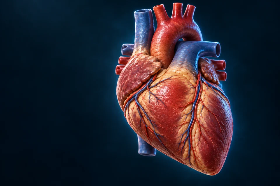
Start at the heart, then trace selected major vessels in body context. Compare regional supply, venous convergence and the portal route through the liver.
▷
Watch chapter overviewFilm planned · not yet available
Current new-family Heart master reused · fine detail illustrative
02 / SYSTEMIC
Distribution and return are different networks
Compare arterial distribution with venous convergence in two detailed regional teaching preparations. The separate expandable native overview retains selected systemic arteries on the left and veins on the right from one unchanged source frame. Neither display is a complete vascular tree.
Original generated regional preparations, not CT, a patient dissection or the same individual as the source-native reference. Separate arterial and venous studies omit the opposite network for clarity. Front views: subject right is image left in each window. Branching variants exist. Fine wall layers, small branches and hidden cardiac insertions are not independently certified. Blue is a display convention, not blue blood. Human medical review: not performed.Structure notes · further context
Ascending aorta
The proximal systemic outlet rises before curving into the arch.
Brachiocephalic trunk
One right-sided trunk precedes the split toward the right head/neck and upper limb.
Left common carotid
A separate direct origin from the arch distinguishes it from the right route.
Left subclavian
The third usual origin turns laterally toward the left upper limb.
Away from the heart
A usual proximal branching pattern supplies different destinations. The local arrows are not a complete circulation.
Venous convergence
Original generated regional preparations, not CT, a patient dissection or the same individual as the source-native reference. Separate arterial and venous studies omit the opposite network for clarity. Front views: subject right is image left in each window. Branching variants exist. Fine wall layers, small branches and hidden cardiac insertions are not independently certified. Blue is a display convention, not blue blood. Human medical review: not performed.Structure notes · further context
Right brachiocephalic vein
This relatively short route contributes to superior caval return.
Left brachiocephalic vein
The longer crossing route joins its partner on the subject’s right.
Superior vena cava
The converged route continues toward the right atrium; the hidden atrial junction is not mapped precisely.
Toward the heart
Separate upper-body inputs combine into a superior caval approach. Blue display colour does not mean blue blood.
Source-native reference · selected adult male network
Compare source identity and regional orientation. This unchanged surface model is a location reference alongside the detailed teaching preparation, not fine tissue histology or a claim that both images depict the same individual.
Selected adult male reference, not every human vessel or branching pattern. Front view: the model’s right is on your left. Native source positions and laterality retained; red/blue is teaching convention, not the literal colour of blood. Source mesh seams and simplified fine surfaces remain visible. New source-native Golden-family rendering: studio key/rim lights and proportional supersampling, no geometry deformation or invented microscopic tissue. Fine model surface detail is coarser than generated Hero artwork.Structure notes · further context
Left common carotid artery
Existing FMA4058 / FJ3483 source identity. Anchor raycasts this visible source surface. Native adult male reference; selected branches, simplified surface and source mesh seams are not a patient angiogram, surgical map or independently verified microscopic anatomy. Human medical review not performed. New framing reprojection and first-visible-surface raycast passed; source-native seams remain disclosed.
Abdominal aorta
Existing FMA3789 / FJ1932 source identity. Anchor raycasts this visible source surface. Native adult male reference; selected branches, simplified surface and source mesh seams are not a patient angiogram, surgical map or independently verified microscopic anatomy. Human medical review not performed. New framing reprojection and first-visible-surface raycast passed; source-native seams remain disclosed.
Right femoral artery
Existing FMA70249 / FJ2143 source identity. Anchor raycasts this visible source surface. Native adult male reference; selected branches, simplified surface and source mesh seams are not a patient angiogram, surgical map or independently verified microscopic anatomy. Human medical review not performed. New framing reprojection and first-visible-surface raycast passed; source-native seams remain disclosed.
Superior vena cava
Existing FMA4720 / FJ3645 source identity. Anchor raycasts this visible source surface. Native adult male reference; selected branches, simplified surface and source mesh seams are not a patient angiogram, surgical map or independently verified microscopic anatomy. Human medical review not performed. New framing reprojection and first-visible-surface raycast passed; source-native seams remain disclosed.
Inferior vena cava
Existing FMA10951 / FJ3441 source identity. Anchor raycasts this visible source surface. Native adult male reference; selected branches, simplified surface and source mesh seams are not a patient angiogram, surgical map or independently verified microscopic anatomy. Human medical review not performed. New framing reprojection and first-visible-surface raycast passed; source-native seams remain disclosed.
Right great saphenous vein
Existing FMA21379 / FJ2145 source identity. Anchor raycasts this visible source surface. Native adult male reference; selected branches, simplified surface and source mesh seams are not a patient angiogram, surgical map or independently verified microscopic anatomy. Human medical review not performed. New framing reprojection and first-visible-surface raycast passed; source-native seams remain disclosed.
The optional native adult male source frame is viewed from the front. Its right appears on your left. Colours separate routes; blood in veins is not literally blue. Selected native branches stop where this teaching selection stops, not where blood normally ends.
Anatomical position
Side convention
Left/right belongs to the body, not the screen.
Selected anatomy
Not a complete vascular tree
Missing tiny branches and capillary networks are not absent from living tissue.
Context
Ghosted organs or bones
Transparent surrounding anatomy locates vessels; it is not another blood channel.
Branching distributes supply across regions. A named artery is a route to a territory, not a promise that every cell depends on one unchanging vessel. Microscopic exchange belongs to smaller vessel networks.
Head and neck
Carotid and vertebral routes
The brain also has vertebral supply; the neck drawing does not display every cerebral branch.
Limbs
Upper and lower networks
Vessel names change along the path and near anatomical boundaries.
Organs
Abdominal branches
Renal and digestive routes have different destinations.
The usual arch gives a right-sided brachiocephalic trunk, a left common carotid and a left subclavian artery. The brachiocephalic trunk then divides on the right. Common variants exist; this model is one reference pattern.
Original generated regional preparations, not CT, a patient dissection or the same individual as the source-native reference. Separate arterial and venous studies omit the opposite network for clarity. Front views: subject right is image left in each window. Branching variants exist. Fine wall layers, small branches and hidden cardiac insertions are not independently certified. Blue is a display convention, not blue blood. Human medical review: not performed.Structure notes · further context
Ascending aorta
The proximal systemic outlet rises before curving into the arch.
Aortic arch
The curve turns toward the subject’s left and continues posteriorly downward.
Brachiocephalic trunk
One right-sided trunk precedes the split toward the right head/neck and upper limb.
Right common carotid
The upward continuation comes from the brachiocephalic trunk in this usual pattern.
Right subclavian
The lateral branch also comes from the right-sided trunk.
Left common carotid
A separate direct origin from the arch distinguishes it from the right route.
Left subclavian
The third usual origin turns laterally toward the left upper limb.
Descending aortic context
The distal arch continues downward; this does not map all thoracic branches.
One usual pattern has three distinct origins
Find the right-sided brachiocephalic trunk, then the separate left carotid and left subclavian origins. The illustration does not imply that this is the only normal arrangement.
Source-native reference · selected adult male network
Compare source identity and regional orientation. This unchanged surface model is a location reference alongside the detailed teaching preparation, not fine tissue histology or a claim that both images depict the same individual.
Selected adult male reference, not every human vessel or branching pattern. Front view: the model’s right is on your left. Native source positions and laterality retained; red/blue is teaching convention, not the literal colour of blood. Source mesh seams and simplified fine surfaces remain visible. New source-native Golden-family rendering: studio key/rim lights and proportional supersampling, no geometry deformation or invented microscopic tissue. Fine model surface detail is coarser than generated Hero artwork.Structure notes · further context
Arch of aorta
Existing FMA3768 / FJ3411 source identity. Anchor raycasts this visible source surface. Native adult male reference; selected branches, simplified surface and source mesh seams are not a patient angiogram, surgical map or independently verified microscopic anatomy. Human medical review not performed. New framing reprojection and first-visible-surface raycast passed; source-native seams remain disclosed.
Brachiocephalic artery
Existing FMA3932 / FJ3417 source identity. Anchor raycasts this visible source surface. Native adult male reference; selected branches, simplified surface and source mesh seams are not a patient angiogram, surgical map or independently verified microscopic anatomy. Human medical review not performed. New framing reprojection and first-visible-surface raycast passed; source-native seams remain disclosed.
Right common carotid artery
Existing FMA3941 / FJ3564 source identity. Anchor raycasts this visible source surface. Native adult male reference; selected branches, simplified surface and source mesh seams are not a patient angiogram, surgical map or independently verified microscopic anatomy. Human medical review not performed. New framing reprojection and first-visible-surface raycast passed; source-native seams remain disclosed.
Left common carotid artery
Existing FMA4058 / FJ3483 source identity. Anchor raycasts this visible source surface. Native adult male reference; selected branches, simplified surface and source mesh seams are not a patient angiogram, surgical map or independently verified microscopic anatomy. Human medical review not performed. New framing reprojection and first-visible-surface raycast passed; source-native seams remain disclosed.
Right subclavian artery
Existing FMA3953 / FJ3579 source identity. Anchor raycasts this visible source surface. Native adult male reference; selected branches, simplified surface and source mesh seams are not a patient angiogram, surgical map or independently verified microscopic anatomy. Human medical review not performed. New framing reprojection and first-visible-surface raycast passed; source-native seams remain disclosed.
Left subclavian artery
Existing FMA4694 / FJ3479 source identity. Anchor raycasts this visible source surface. Native adult male reference; selected branches, simplified surface and source mesh seams are not a patient angiogram, surgical map or independently verified microscopic anatomy. Human medical review not performed. New framing reprojection and first-visible-surface raycast passed; source-native seams remain disclosed.
Why is the usual arch not a mirror image?
The right head/neck and upper-limb routes usually share a proximal brachiocephalic trunk. The left common carotid and left subclavian usually start separately. A normal variation can change the number or arrangement of visible origins, so learning this representative pattern does not establish a person’s branching anatomy.
The two studies separate arteries from veins to make different relationships visible. Matching screen positions do not mean that the two networks have identical branches.
Common carotid arteries divide into external routes for much of the face and neck and internal routes toward cerebral and ocular supply. Vertebral arteries provide another important route to the brain. The circle of Willis does not guarantee adequate collateral flow in every person.
Do not treat a surface region of the brain as a rigid one-vessel territory. Detailed cerebral circulation is linked in the Brain chapter; this overview does not establish stroke diagnosis or a surgical route.
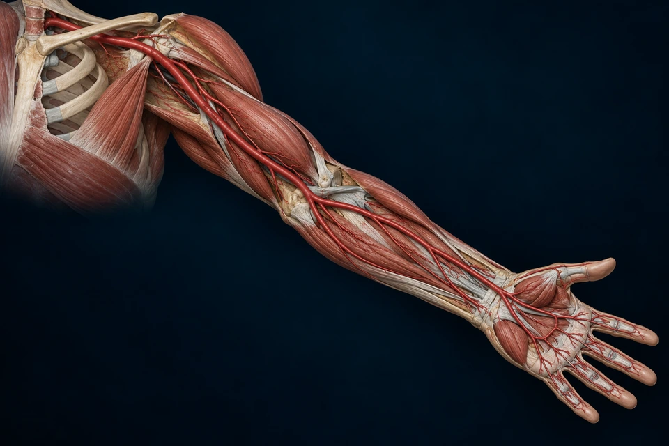
The subclavian artery continues as the axillary artery beyond the lateral border of the first rib. Beyond the lower border of teres major it continues as the brachial artery. These names describe regional anatomy along a connected route.
CONNECT REGIONAL STRUCTURE AND FUNCTION
A continuous route has regional names
Original generated left upper-limb context, separate from the adult male native reference. Selected tissue is opened to reveal an anterior route. Gross landmarks and named continuations are teaching context; fine branch counts, palmar arches, vessel calibre and variants are not a complete certified map. The main artery is not a superficial vein. Human medical review: not performed.Structure notes · further context
Subclavian context
The proximal route passes beneath the clavicular region toward the first-rib boundary.
Axillary artery
Beyond the lateral first-rib boundary, the connected route is named axillary.
Brachial artery
Beyond the lower border of teres major, the route continues through the arm under this name.
Forearm division
Near the elbow the main route divides toward the radial and ulnar territories.
Radial artery
The selected forearm route is on the thumb side.
Ulnar artery
The other main route continues on the little-finger side.
Connected hand circulation
Branches contribute to palmar connections; this is not every digital branch or a guarantee of collateral capacity.
Follow the continuous main route
The regional name changes before the forearm division. The arterial route is distinct from a superficial venous pattern.
Shoulder and arm
Original generated left upper-limb context, separate from the adult male native reference. Selected tissue is opened to reveal an anterior route. Gross landmarks and named continuations are teaching context; fine branch counts, palmar arches, vessel calibre and variants are not a complete certified map. The main artery is not a superficial vein. Human medical review: not performed.Structure notes · further context
Subclavian context
The proximal route passes beneath the clavicular region toward the first-rib boundary.
Axillary artery
Beyond the lateral first-rib boundary, the connected route is named axillary.
Brachial artery
Beyond the lower border of teres major, the route continues through the arm under this name.
Forearm division
Near the elbow the main route divides toward the radial and ulnar territories.
Forearm and hand
Original generated left upper-limb context, separate from the adult male native reference. Selected tissue is opened to reveal an anterior route. Gross landmarks and named continuations are teaching context; fine branch counts, palmar arches, vessel calibre and variants are not a complete certified map. The main artery is not a superficial vein. Human medical review: not performed.Structure notes · further context
Forearm division
Near the elbow the main route divides toward the radial and ulnar territories.
Radial artery
The selected forearm route is on the thumb side.
Ulnar artery
The other main route continues on the little-finger side.
Connected hand circulation
Branches contribute to palmar connections; this is not every digital branch or a guarantee of collateral capacity.
Start: Subclavian continues beyond the first rib as axillary.
Arm: Axillary continues beyond teres major as brachial.
Forearm: Radial and ulnar routes contribute to connected hand circulation.
Why can one continuous artery have several names?
A regional boundary can change the name without creating a break, a valve or a new pump. Subclavian becomes axillary at the lateral first-rib boundary; axillary becomes brachial beyond the lower border of teres major. The whole connected route stays inside anatomical tissue, not between disconnected boxes.
The new gross preparation and the optional native reference have different purposes and are not registered to one patient. Exact muscle borders, small branches and palmar connections need their own detailed anatomical assessment.
Radial lies on the thumb side and ulnar on the little-finger side in anatomical position. Their branches participate in the hand’s arterial arches. Neither route alone should be drawn as the complete hand circulation.
Radial
Thumb-side route
The source selection includes a main forearm segment.
Ulnar
Little-finger-side route
A companion route reaches the hand.
Arches
Connected branches
Fine palmar/digital vessels and anatomical variation are outside this plate.
The descending aorta passes through the thorax and continues below the diaphragm as the abdominal aorta. Different branches serve the chest wall, abdominal organs and lower body. Exact levels and distances vary; no universal numerical map is implied.
Thorax
Wall and organ branches
Intercostal and other branches distribute supply in the chest.
Abdomen
Visceral and paired branches
Celiac, mesenteric and renal routes have distinct territories.
Lower body
Iliac division
The main trunk divides toward the two common iliac arteries.
Abdominal branches are named for useful destinations
The celiac trunk gives branches toward foregut organs. Superior and inferior mesenteric arteries serve different intestinal territories. Paired renal arteries supply the kidneys. This is a regional guide, not a complete branch catalogue.
Celiac
Foregut context
Common hepatic, splenic and left gastric branches have different destinations.
Mesenteric
Intestinal context
Superior and inferior mesenteric territories differ and have connections.
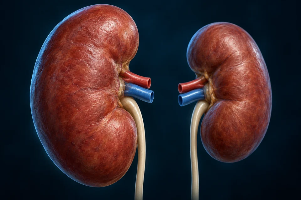
Renal
Kidney context
Arterial blood arrives before the kidney’s smaller filtration circulation.
Each common iliac artery divides toward an internal iliac route serving pelvic structures and an external iliac route continuing toward the lower limb. A simplified branch guide accompanies the native anatomy; it is not a precise pelvic surgical map.
The detailed preparations compare a proximal-thigh route with a separate posterior-knee window. An expandable right-sided native reference preserves selected artery, deep vein and great saphenous vein together. Small tributaries, perforators and complete foot branches are not invented to fill gaps.
Pelvis → proximal thigh
Original generated regional windows: anterior-oblique pelvic/proximal-thigh context and a separate posterior knee context. They are not two time frames or an aligned patient scan. Tissue openings expose representative main routes; fine fascial, nerve, venous tributary and hiatus-edge relationships are not certified surgical anatomy. Human medical review: not performed.Structure notes · further context
Common iliac context
The proximal route precedes the division toward pelvic and lower-limb destinations.
Pelvic arterial branch
The internal iliac route serves pelvic structures; its full branching tree is omitted.
External iliac artery
The main limb-directed continuation approaches the inguinal boundary.
Inguinal ligament context
The arterial route passes deep to this fibrous boundary rather than through its surface.
Femoral artery
Below the inguinal boundary, the limb-directed artery has the femoral name.
Deep femoral venous context
A companion deep vein remains separate from the arterial lumen.
A named route crosses an anatomical boundary
The limb-directed route passes deep to the inguinal boundary before the femoral name is used. Pelvic branches follow a separate destination.
Behind the knee
Original generated regional windows: anterior-oblique pelvic/proximal-thigh context and a separate posterior knee context. They are not two time frames or an aligned patient scan. Tissue openings expose representative main routes; fine fascial, nerve, venous tributary and hiatus-edge relationships are not certified surgical anatomy. Human medical review: not performed.Structure notes · further context
Proximal arterial continuation
After passing the adductor hiatus, the femoral route becomes popliteal; the exact aperture edge is not resolved here.
Popliteal arterial context
The named route lies behind the knee, not across the patella at the front.
Companion venous context
Deep return accompanies this region; it is not the superficial great saphenous route.
Distal femoral context
The bone helps distinguish a posterior regional view from the anterior knee.
Calf-muscle context
The selected window stops before a complete distal leg and foot vascular tree.
The popliteal context is posterior
The view changes to a separate posterior knee window. The artery is not crossing the patella or continuing along the anterior joint surface.
Source-native reference · selected adult male network
Compare source identity and regional orientation. This unchanged surface model is a location reference alongside the detailed teaching preparation, not fine tissue histology or a claim that both images depict the same individual.
Selected adult male reference, not every human vessel or branching pattern. Front view: the model’s right is on your left. Native source positions and laterality retained; red/blue is teaching convention, not the literal colour of blood. Source mesh seams and simplified fine surfaces remain visible. New source-native Golden-family rendering: studio key/rim lights and proportional supersampling, no geometry deformation or invented microscopic tissue. Fine model surface detail is coarser than generated Hero artwork.Structure notes · further context
Right femoral artery
Existing FMA70249 / FJ2143 source identity. Anchor raycasts this visible source surface. Native adult male reference; selected branches, simplified surface and source mesh seams are not a patient angiogram, surgical map or independently verified microscopic anatomy. Human medical review not performed. New framing reprojection and first-visible-surface raycast passed; source-native seams remain disclosed.
Right femoral vein
Existing FMA21188 / FJ2144 source identity. Anchor raycasts this visible source surface. Native adult male reference; selected branches, simplified surface and source mesh seams are not a patient angiogram, surgical map or independently verified microscopic anatomy. Human medical review not performed. New framing reprojection and first-visible-surface raycast passed; source-native seams remain disclosed.
Right great saphenous vein
Existing FMA21379 / FJ2145 source identity. Anchor raycasts this visible source surface. Native adult male reference; selected branches, simplified surface and source mesh seams are not a patient angiogram, surgical map or independently verified microscopic anatomy. Human medical review not performed. New framing reprojection and first-visible-surface raycast passed; source-native seams remain disclosed.
Right popliteal artery
Existing FMA77380 / FJ2170 source identity. Anchor raycasts this visible source surface. Native adult male reference; selected branches, simplified surface and source mesh seams are not a patient angiogram, surgical map or independently verified microscopic anatomy. Human medical review not performed. New framing reprojection and first-visible-surface raycast passed; source-native seams remain disclosed.
Right popliteal vein
Existing FMA44328 / FJ2171 source identity. Anchor raycasts this visible source surface. Native adult male reference; selected branches, simplified surface and source mesh seams are not a patient angiogram, surgical map or independently verified microscopic anatomy. Human medical review not performed. New framing reprojection and first-visible-surface raycast passed; source-native seams remain disclosed.
Right posterior tibial artery
Existing FMA43898 / FJ2172 source identity. Anchor raycasts this visible source surface. Native adult male reference; selected branches, simplified surface and source mesh seams are not a patient angiogram, surgical map or independently verified microscopic anatomy. Human medical review not performed. New framing reprojection and first-visible-surface raycast passed; source-native seams remain disclosed.
After the femoral artery passes through the adductor hiatus, it continues as the popliteal artery behind the knee. Leg routes then continue toward anterior and posterior territories. The optional native view is frontal/oblique; its vessel may lie behind ghosted bone. The separate new tissue window shows posterior knee context.
Deep venous return near the knee continues toward the femoral vein. This is not a guide to finding, compressing or operating on a vessel.
Most systemic organ beds are parallel destinations
Blood leaving the aorta can reach different organ networks without first passing through every other organ. Distribution changes with local and whole-body conditions. A selected connection guide expresses this relationship without simulating pressure or flow.
ANATOMY AND RELATIONSHIP / ALL BASIC FACTS VISIBLE
Several organs receive parallel supply
Brain territory
Carotid and vertebral supply reaches cerebral networks.
A systemic arterial route divides toward several organ networks.
Exchange
Each selected bed has smaller vessels; one organ is not simply upstream of every other organ.
Return
Venous paths converge toward the heart. Branches here are relationship teaching, not measured flow.
All basic facts stay visible. Image windows and selected explanations locate anatomy; they are not a timed recording, measured flow or collateral-perfusion prediction. Human medical review: not performed.
What does parallel supply mean for a living circulation?
Branches distribute systemic supply to different tissue networks, with venous drainage converging later. Regional resistance and demand help determine distribution; the pictures do not give each organ an equal share. The lung and body pumps form a connected circulation, while portal routes add an important serial exchange relationship within it.
The organ cards identify different destinations. Their size and highlights are not a percentage of cardiac output, a pressure reading or a simulation of a person’s response.
Internal jugular and subclavian veins join into a brachiocephalic vein on each side. The two brachiocephalic veins then form the superior vena cava. Pulmonary veins do not join this upper-body pathway.
Original generated regional preparations, not CT, a patient dissection or the same individual as the source-native reference. Separate arterial and venous studies omit the opposite network for clarity. Front views: subject right is image left in each window. Branching variants exist. Fine wall layers, small branches and hidden cardiac insertions are not independently certified. Blue is a display convention, not blue blood. Human medical review: not performed.Structure notes · further context
Right internal jugular
An upper-body venous input approaches the right confluence.
Right subclavian vein
Upper-limb return joins the jugular route near the thoracic inlet.
Right brachiocephalic vein
This relatively short route contributes to superior caval return.
Left brachiocephalic vein
The longer crossing route joins its partner on the subject’s right.
Left internal jugular
A separate left-sided input approaches the left confluence.
Left subclavian vein
Return from the other upper limb contributes on this side.
Superior vena cava
The converged route continues toward the right atrium; the hidden atrial junction is not mapped precisely.
Jugular and limb return approach each side
The internal jugular and subclavian inputs contribute to the two brachiocephalic routes. Smaller tributaries and superficial drainage are not a complete inventory here.
Source-native reference · selected adult male network
Compare source identity and regional orientation. This unchanged surface model is a location reference alongside the detailed teaching preparation, not fine tissue histology or a claim that both images depict the same individual.
Selected adult male reference, not every human vessel or branching pattern. Front view: the model’s right is on your left. Native source positions and laterality retained; red/blue is teaching convention, not the literal colour of blood. Source mesh seams and simplified fine surfaces remain visible. New source-native Golden-family rendering: studio key/rim lights and proportional supersampling, no geometry deformation or invented microscopic tissue. Fine model surface detail is coarser than generated Hero artwork.Structure notes · further context
Left internal jugular vein
Existing FMA4762 / FJ3485 source identity. Anchor raycasts this visible source surface. Native adult male reference; selected branches, simplified surface and source mesh seams are not a patient angiogram, surgical map or independently verified microscopic anatomy. Human medical review not performed. New framing reprojection and first-visible-surface raycast passed; source-native seams remain disclosed.
Right internal jugular vein
Existing FMA4754 / FJ3585 source identity. Anchor raycasts this visible source surface. Native adult male reference; selected branches, simplified surface and source mesh seams are not a patient angiogram, surgical map or independently verified microscopic anatomy. Human medical review not performed. New framing reprojection and first-visible-surface raycast passed; source-native seams remain disclosed.
Left subclavian vein
Existing FMA4763 / FJ3486 source identity. Anchor raycasts this visible source surface. Native adult male reference; selected branches, simplified surface and source mesh seams are not a patient angiogram, surgical map or independently verified microscopic anatomy. Human medical review not performed. New framing reprojection and first-visible-surface raycast passed; source-native seams remain disclosed.
Right subclavian vein
Existing FMA4755 / FJ3587 source identity. Anchor raycasts this visible source surface. Native adult male reference; selected branches, simplified surface and source mesh seams are not a patient angiogram, surgical map or independently verified microscopic anatomy. Human medical review not performed. New framing reprojection and first-visible-surface raycast passed; source-native seams remain disclosed.
Left brachiocephalic vein
Existing FMA4761 / FJ3482 source identity. Anchor raycasts this visible source surface. Native adult male reference; selected branches, simplified surface and source mesh seams are not a patient angiogram, surgical map or independently verified microscopic anatomy. Human medical review not performed. New framing reprojection and first-visible-surface raycast passed; source-native seams remain disclosed.
Right brachiocephalic vein
Existing FMA4751 / FJ3583 source identity. Anchor raycasts this visible source surface. Native adult male reference; selected branches, simplified surface and source mesh seams are not a patient angiogram, surgical map or independently verified microscopic anatomy. Human medical review not performed. New framing reprojection and first-visible-surface raycast passed; source-native seams remain disclosed.
Superior vena cava
Existing FMA4720 / FJ3645 source identity. Anchor raycasts this visible source surface. Native adult male reference; selected branches, simplified surface and source mesh seams are not a patient angiogram, surgical map or independently verified microscopic anatomy. Human medical review not performed. New framing reprojection and first-visible-surface raycast passed; source-native seams remain disclosed.
External and internal iliac veins contribute to common iliac veins, which converge toward the inferior vena cava. Renal veins also enter the inferior systemic route. The left/right renal arrangements are not exact mirror images.
The portal vein is different: it enters the liver before hepatic venous return reaches systemic circulation. Do not route intestinal portal blood directly from the gut into the renal vein.
The great saphenous vein is a superficial route that joins the femoral system. Deep veins and communicating connections also matter. The femoral vein is a deep vein; calling it a superficial femoral vein would be misleading.
Superficial
Great saphenous context
A long route lies superficial to the deep fascial compartment.
Deep
Femoral and popliteal context
Return also follows deep routes near muscles and arteries.
Connections
Not all shown
Perforating veins and numerous tributaries are missing from this selection.
Why does “superficial” not describe every long visible vein?
Superficial and deep refer to relationships with fascial compartments. The great saphenous is a superficial route, while the femoral vein belongs to deep return. A rendered vessel visible through transparent tissue does not become anatomically superficial because it is easy to see.
The source selection leaves out many tributaries and perforators. It cannot establish valve function, an individual drainage pattern or a procedure location.
In the detailed upper-abdominal preparation, distinguish portal inflow, a separate hepatic arterial input and hepatic venous outflow. The expandable native abdominal reference retains aortic, portal and systemic routes with ghosted liver/spleen/kidney context from the same source model. Some small arterial branches and organ microcirculation are not visible.
Original generated anatomy, not angiography or a patient scan. The pancreas is selectively opened/transparent to reveal a posterior vein, not to move it onto the anterior surface. Portal branches and hepatic veins are distinct networks within an opened liver. Fine sinusoidal channels, biliary anatomy, fixed flow fractions and inferior mesenteric variants are not mapped. Human medical review: not performed.Structure notes · further context
Splenic vein
The route is seen through the opened pancreatic context; its normal course is posterior to the pancreatic body.
Opened pancreatic context
Residual translucent tissue provides the front–back reference around the posterior vessel.
Portal confluence context
Superior mesenteric and splenic return join behind the pancreatic neck. Inferior mesenteric joining patterns vary.
Superior mesenteric vein
Selected intestinal drainage approaches the confluence from below.
Portal vein
The combined venous inflow reaches the liver before systemic venous return.
Separate hepatic arterial context
An arterial input also reaches liver tissue; the portal route is not the only inflow.
Hepatic venous outflow
Separate venous branches carry liver outflow toward the systemic caval route.
Posterior caval context
The inferior vena cava receives hepatic outflow; portal inflow is not a direct shortcut into it.
Drainage reaches a confluence behind the pancreas
The opened pancreatic tissue is a depth window. The splenic route has not been moved onto the anterior pancreatic surface.
Source-native reference · selected adult male network
Compare source identity and regional orientation. This unchanged surface model is a location reference alongside the detailed teaching preparation, not fine tissue histology or a claim that both images depict the same individual.
Selected adult male reference, not every human vessel or branching pattern. Front view: the model’s right is on your left. Native source positions and laterality retained; red/blue is teaching convention, not the literal colour of blood. Source mesh seams and simplified fine surfaces remain visible. New source-native Golden-family rendering: studio key/rim lights and proportional supersampling, no geometry deformation or invented microscopic tissue. Fine model surface detail is coarser than generated Hero artwork.Structure notes · further context
Celiac trunk
Existing FMA50737 / FJ1846 source identity. Anchor raycasts this visible source surface. Native adult male reference; selected branches, simplified surface and source mesh seams are not a patient angiogram, surgical map or independently verified microscopic anatomy. Human medical review not performed. New framing reprojection and first-visible-surface raycast passed; source-native seams remain disclosed.
Superior mesenteric artery
Existing FMA14749 / FJ2011 source identity. Anchor raycasts this visible source surface. Native adult male reference; selected branches, simplified surface and source mesh seams are not a patient angiogram, surgical map or independently verified microscopic anatomy. Human medical review not performed. New framing reprojection and first-visible-surface raycast passed; source-native seams remain disclosed.
Hepatic portal vein
Existing FMA71904 / FJ3082 source identity. Anchor raycasts this visible source surface. Native adult male reference; selected branches, simplified surface and source mesh seams are not a patient angiogram, surgical map or independently verified microscopic anatomy. Human medical review not performed. New framing reprojection and first-visible-surface raycast passed; source-native seams remain disclosed.
Superior mesenteric vein
Existing FMA14332 / FJ3647 source identity. Anchor raycasts this visible source surface. Native adult male reference; selected branches, simplified surface and source mesh seams are not a patient angiogram, surgical map or independently verified microscopic anatomy. Human medical review not performed. New framing reprojection and first-visible-surface raycast passed; source-native seams remain disclosed.
Splenic vein
Existing FMA14331 / FJ3641 source identity. Anchor raycasts this visible source surface. Native adult male reference; selected branches, simplified surface and source mesh seams are not a patient angiogram, surgical map or independently verified microscopic anatomy. Human medical review not performed. New framing reprojection and first-visible-surface raycast passed; source-native seams remain disclosed.
Inferior vena cava
Existing FMA10951 / FJ3441 source identity. Anchor raycasts this visible source surface. Native adult male reference; selected branches, simplified surface and source mesh seams are not a patient angiogram, surgical map or independently verified microscopic anatomy. Human medical review not performed. New framing reprojection and first-visible-surface raycast passed; source-native seams remain disclosed.
The superior mesenteric and splenic veins usually meet to form the portal vein. The inferior mesenteric connection varies and is not forced into one universal joining point. Portal blood reaches liver sinusoids before leaving through hepatic veins toward the inferior vena cava.
ANATOMY AND RELATIONSHIP / ALL BASIC FACTS VISIBLE
Portal drainage reaches another exchange network
Original generated anatomy, not angiography or a patient scan. The pancreas is selectively opened/transparent to reveal a posterior vein, not to move it onto the anterior surface. Portal branches and hepatic veins are distinct networks within an opened liver. Fine sinusoidal channels, biliary anatomy, fixed flow fractions and inferior mesenteric variants are not mapped. Human medical review: not performed.Structure notes · further context
Splenic vein
The route is seen through the opened pancreatic context; its normal course is posterior to the pancreatic body.
Opened pancreatic context
Residual translucent tissue provides the front–back reference around the posterior vessel.
Portal confluence context
Superior mesenteric and splenic return join behind the pancreatic neck. Inferior mesenteric joining patterns vary.
Superior mesenteric vein
Selected intestinal drainage approaches the confluence from below.
Collect behind the pancreas
A different window into the same unchanged anatomical preparation. This is an observation sequence, not blood particles jumping between disconnected tissues or a timed recording.
Drainage
Selected digestive and splenic drainage reaches portal inflow.
Liver exchange
Portal blood enters liver sinusoids; hepatic arterial supply also contributes.
Outflow
Hepatic veins carry blood toward the inferior vena cava. This is not a direct gut-to-kidney path.
All basic facts stay visible. Image windows and selected explanations locate anatomy; they are not a timed recording, measured flow or collateral-perfusion prediction. Human medical review: not performed.
Why is this not the same pattern as most parallel organ beds?
Portal drainage from selected abdominal territories reaches another exchange network in the liver before hepatic venous return reaches the caval system. That is different from requiring blood to pass through every systemic organ one after another. The liver also receives a separate hepatic arterial input.
The splenic vein’s normal pancreatic relationship is posterior. Transparency exposes that route in the new illustration; it does not turn it into an anterior vessel. The confluence behind the pancreatic neck and the separate hepatic outflow should remain distinct when following the route.
The liver receives portal venous inflow and hepatic arterial inflow. Portal blood carries material from drained abdominal organs and still contains oxygen; it is not oxygen-free blood. No fixed universal flow proportion is assigned.
Portal
Venous inflow
Material arrives after drainage from digestive and splenic networks.
Hepatic artery
Systemic arterial inflow
Arterial oxygen delivery also reaches liver tissue.
Hepatic veins
Outflow
Blood leaves the liver toward the inferior vena cava.
Vessel direction and oxygen status are different facts
Pulmonary arteries carry blood away from the right heart toward lungs. Pulmonary veins return blood to the left heart. The lungs also have a smaller systemic bronchial supply for supporting tissue, distinct from the main gas-exchange circuit.
ANATOMY AND RELATIONSHIP / ALL BASIC FACTS VISIBLE
Direction and oxygen status are separate properties
Generated organ context, not CT or a complete vessel-root reconstruction. Red/blue convention distinguishes relative oxygen content; hidden pulmonary veins and fine lung vessel trees are not individually mapped.
Right pump → lungs
The retained qualified cardiopulmonary image locates the organ and proximal vessel context. Fine pulmonary exchange is taught in the Lung chapter, not measured here.
Right heart
Pulmonary arteries leave the right heart toward the lungs.
Lungs
Gas exchange changes oxygen/carbon-dioxide carriage; blood is still red.
Left heart
Pulmonary veins return to the left heart, which sends blood into systemic arteries.
All basic facts stay visible. Image windows and selected explanations locate anatomy; they are not a timed recording, measured flow or collateral-perfusion prediction. Human medical review: not performed.
Anastomoses connect selected routes, but their size, arrangement and physiological contribution vary. The source model cannot establish whether a particular person has adequate collateral supply. Anatomical variation is normal and is not automatically disease.
CONNECT REGIONAL STRUCTURE AND FUNCTION
Connections exist; capacity is not guaranteed
A bounded window from the same original gross upper-limb illustration. It locates a connected hand network, not a certified complete palmar arch, collateral capacity or fine digital anatomy.Structure notes · further context
Connected hand circulation
Branches contribute to palmar connections; this is not every digital branch or a guarantee of collateral capacity.
Compare connected branches with the full upper-limb preparation. A visible connection alone cannot establish adequate perfusion if another route is impaired.
The broad routes lead toward smaller arteries, arterioles and exchange beds, then venous return. Material crosses selected microvascular interfaces; fluid recovery also connects with lymph. A large-vessel render cannot show every microscopic exchange process.
Local arteriolar tone and nervous/hormonal controls affect distribution. The route map is not a resting-flow percentage chart or a blood-pressure calculator. Learn regulation at the scales where it occurs.
The optional native reference plates preserve selected source shapes, positions and laterality. They support broad regional orientation. Source selection, raycasts and rendering are not human anatomical or medical review, and the processed meshes are not a patient scan.
The existing display-system grouping places cerebral ventricles under cardiac and some liver parenchymal segments under venous. This production selection does not trust those grouping names as factual anatomy: exact structures are selected, unrelated brain meshes are excluded and liver context is ghosted as tissue. The original atlas manifest is unchanged.
Source mesh seams, simplified surfaces, missing branches and unverified fine detail remain explicit limitations. A raycast confirms a label lands on a named source mesh; independent factual sources support what the label teaches. Neither alone establishes surgical accuracy.
Selected systemic arterial and venous routes, typical arch origins, major regional destinations and portal versus systemic return. Supporting models are not flow measurements.
Revision: vascular-routes-normal-2026-10-19-eyes-v2. Stable local identity: ahb_ch_vascular_routes_english_v1; not a new CMS entity. Historical native masters remain at their recorded locations; four new generated masters, prompts and derivative hashes are separately versioned. Labels and paths remain editable. Migration physical-phone acceptance: Pending, non-blocking. No patient diagnosis, surgical instruction, medication advice or physician approval claim. Observed microscopy and clinical disease remain separate work.