Not a lymph node in miniature
The normal thymus does not have a lymph node’s incoming lymph route, follicles or sinus map. Its compartments support developing T cells.
Compare the node’s separate organisation →THYMUS / THE 30-SECOND PICTURE
Behind the sternum, living epithelial tissue supports developing T cells. Tissue organisation, receptor interactions and selection connect a small chest organ with immunity throughout the body.
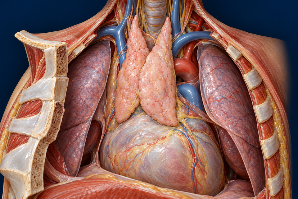
Locate the paired thymic lobes between the lung margins, anterior to the upper-heart region. Then identify the retracted sternum that normally covers this view.
The outline locates the related teaching structure. It is not a measured selection signal, cell trajectory or activity simulation.
Structure and mechanism references: Kenhub · Normal thymic location and vessels · OpenStax · Primary and secondary lymphoid organs · Baldwin & Robey · Thymic tuning and regulatory development (2024)
01 / OUTER CORTEX · INNER MEDULLARY CORE · LIVING SUPPORT
Find dense cortex, paler medullary tissue and partial connective septa. The retained high-detail view is a local preparation; the real medullary core continues in three dimensions rather than forming isolated sealed pockets.

Look for the dark outer field and its supporting epithelial environment. Conventional receptor development and positive selection have an important cortical context, but colour alone does not identify a molecular stage.
The outline locates the related teaching structure. It is not a measured selection signal, cell trajectory or activity simulation.
The normal thymus does not have a lymph node’s incoming lymph route, follicles or sinus map. Its compartments support developing T cells.
Compare the node’s separate organisation →Branching epithelial cells meet thymocytes within a living network. A darker nucleus cannot tell you its receptor, co-receptor or developmental history.
Look closer at cell contacts →Structure and mechanism references: Columbia University · Thymus · Duke University · Normal lymphatic histology · Histology Guide · MH079 thymic tissue
02 / CELL SURFACES MEET · DIFFERENT OUTCOMES REMAIN DISTINCT
Locate the epithelial–thymocyte interface, then the separate presenting-cell contact and clearance example. The cells are not arranged as a compulsory assembly line, and a living contact is not cell fusion.

In the cortical positive-selection context, developing cells need suitable self-peptide–MHC engagement and survival signals. This contact picture introduces that relationship; it does not measure binding or identify each molecule.
The outline locates the related teaching structure. It is not a measured selection signal, cell trajectory or activity simulation.
Conventional alpha-beta T cells recognise peptide with MHC. Positive selection supports useful engagement and survival; “self” is part of the display context, not something every surviving cell must ignore completely.
Follow receptor development and selection →Deletion can occur in more than one compartment. Regulatory development differs from deletion, and peripheral tolerance remains important after export.
Continue with immune memory and restraint →Structure and mechanism references: Baldwin & Robey · Thymic tuning and regulatory development (2024) · Takaba & Takayanagi · Mechanisms of T-cell selection (2017) · Histology Guide · EM379 human thymic medulla
GO DEEPER / STRUCTURE · SIGNALS · DEVELOPMENT · CONNECTED IMMUNITY
The thymus occupies the anterior and superior mediastinal region, behind the sternum and in front of the upper pericardial and great-vessel region. The lungs frame this central space rather than containing the gland. Its paired soft lobes can extend upward toward the lower neck.
The new organ view exposes a selected youthful preparation. The retracted cut bone is anterior; the heart continues deeper and inferiorly. This is orientation, not a surgical approach or a fixed map of every adult gland.
Structure and mechanism references: Kenhub · Normal thymic location and vessels
Blood-forming tissue supplies lymphoid precursors. In the thymus, developing T cells interact with a specialised environment before joining the circulating repertoire. The thymus is therefore a primary lymphoid organ, alongside bone marrow, rather than another node filtering incoming tissue lymph.
After export, naïve T cells can encounter relevant antigen in secondary lymphoid organs. Activation, expansion, effector activity and memory are a later context. A cell completing thymic development has not already fought every infection it may meet.
Structure and mechanism references: OpenStax · Primary and secondary lymphoid organs · Immunobiology · Developmental compartments
Connective septa enter from the capsule but do not create a set of fully sealed miniature organs. Dense cortical tissue surrounds paler medullary regions; within a lobe the medulla forms a connected three-dimensional core.
A thin section can cut that core into apparently separate pale islands. The retained tissue picture explains local contrast; separate pale profiles do not establish separate enclosed chambers. The normal thymus has no lymph-node-style follicles, germinal centres or lymph sinuses.
Structure and mechanism references: Columbia University · Thymus · Duke University · Normal lymphatic histology
Thymic epithelial cells extend processes among developing lymphocytes. This cellular scaffold is different from imagining thymocytes suspended in an empty collagen sieve. Dendritic cells and macrophages contribute other interactions; each cell shape in the artwork remains an explanatory selection.
In the medulla, layered epithelial whorls form Hassall corpuscles. They are tissue structures, not shells around every thymocyte or receptacles into which all rejected cells are poured. The external human TEM is useful for observing epithelial processes at a different scale.
Structure and mechanism references: Histology Guide · EM379 human thymic medulla · Histology Guide · MH079 thymic tissue
Developing conventional alpha-beta T cells rearrange receptor genes while progressing through several stages. Early cells do not yet express both CD4 and CD8; a later cortical double-positive population expresses both co-receptors before conventional single-positive lineages emerge.
The large number of small dark nuclei in tissue is not a readout of those molecular stages. Receptor specificity, CD4/CD8 phenotype and developmental age require evidence beyond the colour of a generated cell.
Structure and mechanism references: Immunobiology · Developmental compartments · Kenhub · Thymic histology and normal connections
Cortical epithelial interactions help select thymocytes capable of responding to self-peptide displayed by MHC. Cells without adequate survival signals can be lost. The selected repertoire must work with the body’s presentation system; selection is not a search for cells that never interact with self.
Signal strength and cellular tuning matter. The control highlights a contact site rather than assigning a universal numerical affinity threshold or making every cell pass an identical timed examination.
Structure and mechanism references: Baldwin & Robey · Thymic tuning and regulatory development (2024) · Takaba & Takayanagi · Mechanisms of T-cell selection (2017)
Strongly self-reactive thymocytes can undergo deletion. The medulla provides additional encounters with diverse self material, but deletion is not restricted to one medullary checkpoint: cortical selection can also remove cells.
Appropriate signals can support regulatory T-cell development. Survival, deletion and regulation are not interchangeable names, nor does thymic selection guarantee zero autoimmune risk. Peripheral control remains important after cells leave.
Structure and mechanism references: Baldwin & Robey · Thymic tuning and regulatory development (2024)
Medullary epithelial programmes help expose developing cells to a wider range of self antigens. AIRE contributes to this expression, but is not the only regulator; FEZF2-dependent programmes also contribute.
The point is controlled cellular presentation, not whole liver, skin or pancreas tissue physically entering the thymus. Antigen-presenting interactions connect intracellular processing with what a thymocyte encounters at a cell surface.
Structure and mechanism references: Takaba & Takayanagi · Mechanisms of T-cell selection (2017)
Tight endothelial junctions and surrounding cellular and basement-membrane layers contribute to the cortical blood–thymus barrier. This limits exposure to circulating material in the developing-cell environment.
The barrier is regionally specialised. It does not mean that the thymus has no blood supply, that nutrients cannot arrive, or that all blood vessels and medullary regions are sealed in the same way.
Structure and mechanism references: Columbia University · Thymus · Histology Guide · EM379 human thymic medulla
Small arterial branches reach the gland, commonly from internal thoracic and thyroid-region vessels. Venous routes return toward nearby thoracic and neck veins. Vascular access supports the tissue and cellular traffic; the gland is not an inline lymph filter.
The thymus lacks afferent lymphatic vessels of the lymph-node type. Its lymph drains outward toward regional thoracic nodes. Perivascular autonomic connections belong to the wider organ context; this chapter does not claim to map every fibre or a universal direct human parasympathetic circuit.
Structure and mechanism references: Kenhub · Normal thymic location and vessels · Kenhub · Thymic histology and normal connections
With age, adipose tissue becomes more prominent and active thymic epithelial and lymphoid regions occupy less space. A young densely cellular preparation should not be mistaken for the expected appearance of every adult chest.
The Duke adult human specimen illustrates one involutional pattern. Residual thymic tissue and function can remain. Normal ageing is not an abrupt off-switch at puberty or a reason to infer an individual’s immune capacity from one illustration.
Structure and mechanism references: Duke University · Normal lymphatic histology · Columbia University · Thymus
A routine section can reveal dense cortex, paler medulla, septa, fat and epithelial whorls. It does not establish peptide specificity or the signalling history of each lymphocyte. Human acquired material and generated teaching preparations must remain clearly distinguished.
The external EM379 record identifies adult human tissue. MH079 provides neonatal/adult comparisons but its page does not confirm species, so it is not silently labelled human here. No institutional slide pixels or textbook figures are copied into this lesson.
Structure and mechanism references: Histology Guide · EM379 human thymic medulla · Histology Guide · MH079 thymic tissue · Duke University · Normal lymphatic histology
ACQUIRED TISSUE / A COMPLEMENTARY FORM OF EVIDENCE
Duke’s 0076_E record identifies an adult human H&E specimen with involutional change. Histology Guide’s EM379 identifies thymic medulla from a 63-year-old human and describes epithelial processes, lymphocytes and an epithelial whorl.
External specimen references remain on their institutional sites; their pixels are not redistributed. MH079 species is not specified on its page, so it is not silently presented as confirmed human tissue. The original illustration and preserved atlas preparation are generated teaching images.
Structure and mechanism references: Duke University · Normal lymphatic histology · Histology Guide · EM379 human thymic medulla · Histology Guide · MH079 thymic tissue
No. Developing cells arrive in a specialised primary lymphoid environment; the normal thymus does not have a node’s afferent lymph route and sinus plan.
No. Useful engagement is needed for conventional development. Excessive self-reactivity raises different selection and regulatory outcomes.
No. Context and signalling matter. Deletion and regulatory development differ, and peripheral tolerance remains necessary.
No. Involution changes tissue proportions; remaining tissue and function must not be erased by an all-or-none diagram.
Targeted normal anatomy and cell-development checks: 20 October 2026. Existing FB102 whole-book research guided coverage. Independent anatomy, university histology and peer-reviewed selection sources were compared; source simplifications and retrieval limits are stated separately.
Premium medical bases, semantic names, anatomical targets and teaching states remain separate layers for future translation. English is the current chapter language. This normal chapter provides no personal diagnosis or treatment. Physician review, precise generated cell/protein geometry, physical-phone acceptance and independent post-publication review are not certified.