Surface anatomy is not the whole map
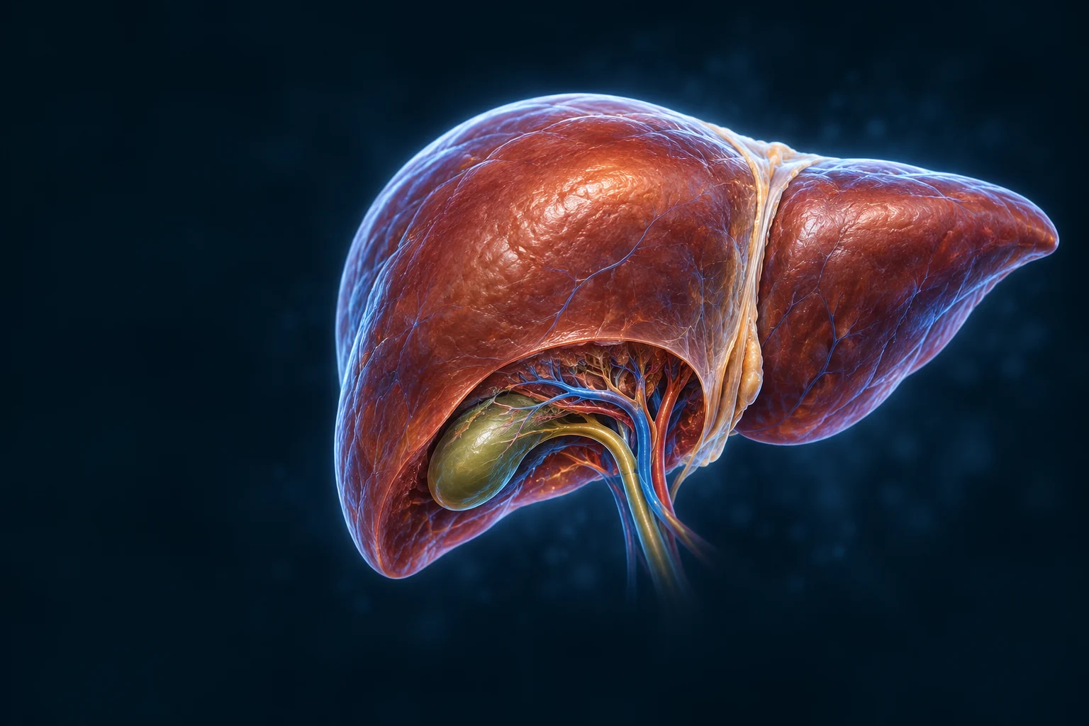
The anterior right and left lobes are easy to locate. The smaller caudate and quadrate lobes lie on surfaces hidden here. Functional segments follow vascular territories rather than simply the falciform ligament.
CHAPTER / DIGESTIVE SYSTEM
A crossing point. A working tissue.
Follow two blood supplies into a repeating tissue network, then move to the cells that handle fuel, make proteins and send bile toward the intestine.
Follow blood, cells and bile →Chapter overview film planned. No playable video is currently available.
02 / ORGAN IN CONTEXT
The liver lies mainly in the right upper abdomen beneath the diaphragm. A fibrous capsule covers it; peritoneal folds help hold it in place. The gallbladder is a separate organ against its underside.

The anterior right and left lobes are easy to locate. The smaller caudate and quadrate lobes lie on surfaces hidden here. Functional segments follow vascular territories rather than simply the falciform ligament.
At the porta hepatis on the inferior surface, portal-vein and hepatic-artery branches enter while hepatic ducts leave. Hepatic veins use a different exit toward the vena cava behind the liver.
03 / TWO BLOOD SUPPLIES
Many absorbed nutrients reach the liver before joining the wider circulation. The portal vein brings blood from abdominal organs; the hepatic artery adds arterial blood. Their small branches feed the same sinusoidal network.
SAME ANATOMY · CHANGING DEMAND
The portal vein is the return from abdominal organs, not a food-only pipe. Both inlets keep supplying the liver between meals; hepatic veins carry blood onward.
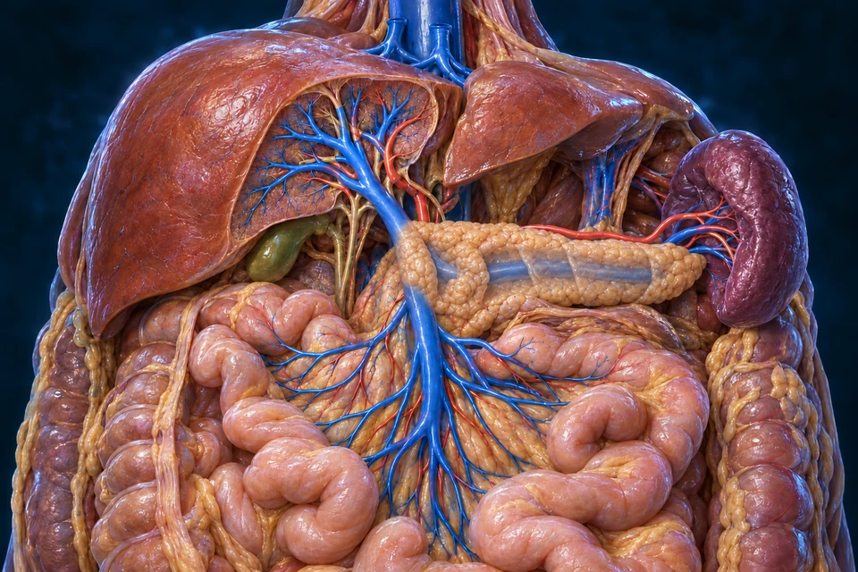
Rings locate structures to compare; they do not encode flow rate, pressure, vessel dilation or moving particles. The anatomy and all six targets stay fixed in every state and enlarged view.
Still delivers blood from the gut and spleen, even when no new meal is being absorbed.
Adds arterial blood to the same sinusoidal network. The two inlets are not separate destinations.
Fuel handling shifts toward supporting the wider body. Compare glycogen and glucose production below.
Keep the distinction. A change of function does not switch off the circulation.
Blood flow is volume per unit time. Delivery of a substance also depends on its concentration. Net release depends on the difference between what arrives and what leaves, as well as flow. A higher glucose output can therefore coexist with lower regional blood flow.
The meal MRI study measured portal velocity, hepatic arterial flow and liver tissue perfusion separately. This lesson uses its healthy-volunteer findings, including an arterial decrease after food. The study also included patients; their results are not used as a normal response here. No universal healthy-person percentage or diagnosis can be read from this illustration.
Human intraoperative measurements also found an arterial increase when portal inflow was temporarily interrupted. That supports the buffer relationship; observations under anesthesia in surgical patients are not normal meal or exercise measurements. This lesson does not assign their numerical response to a healthy reader.
The exercise study estimated flow across the hepatosplanchnic region and measured arteriohepatic venous substrate exchange. It did not measure each blue tributary separately. Another study measured the superior mesenteric artery during brief exercise after a large meal and found its flow maintained. Different feeding states, protocols and measurement sites matter.
Meal MRI methods and results ↗ · Exercise flow and substrate exchange ↗ · Brief pre-/post-meal exercise comparison ↗ · Human arterial/portal interaction ↗
Follow the two inlets into sinusoids → · Compare the separate fuel-storage mechanism → · Connect pancreatic glucose feedback →
Directed normal-physiology source check: 16 October 2026. Qualitative teaching contexts, not measured patient flow or exercise advice. Source checking and AI-assisted illustration review are not physician review.
Water-soluble nutrients such as glucose and amino acids enter intestinal blood. Gut-derived substances and splenic venous blood also reach the liver. Most long-chain dietary fat takes an initial lymphatic route, so “all nutrients go straight to the liver” would be misleading.
Hepatocytes can store, transform or release arriving molecules. Some orally absorbed compounds are substantially altered before reaching systemic blood. This is a physiological concept, not dosing or treatment advice.
Both bring blood into liver sinusoids.
Blood exchanges materials beside hepatocytes.
Blood returns to the systemic circulation.
Cells secrete into small spaces between neighbors.
Bile leaves through ducts, not the blood channels.
The cystic duct connects storage to the main intestinal route.
Supporting route map: both basic routes remain visible. Highlighting changes emphasis, not the anatomy or access to information.
Inflow, drainage and posterior venous relations: Merck Manual · Liver Structure and Function · Manipal Anatomy Museum · Portal vein and pancreatic relationsTargeted source check: 15 October 2026. This is not human medical review.
04 / TISSUE ORGANIZATION
The classical lobule groups cell plates around a central venule. At its edge, a portal tract brings blood branches and a bile duct close together. Proximity does not connect their contents.
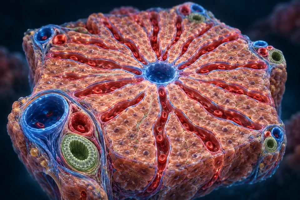
Enter at the peripheral portal regions.
Carry mixed blood alongside hepatocytes.
Collects blood for hepatic venous drainage.
Bile enters canaliculi between adjacent cells.
Route bile separately from sinusoidal blood.
Collect it into progressively larger ducts.
These opposed tissue-level routes are supporting direction maps, not a claim that every canaliculus is a straight radial pipe. Oxygen and metabolic activity vary across the lobule; this view does not map all functional zones.
Blood and bile use different tissue routes: Merck Manual · Liver Structure and Function · PubMed · Boyer, Bile formation and secretionTargeted source check: 15 October 2026. This is not human medical review.
05 / CELLULAR EXCHANGE
A hepatocyte handles molecules from blood while secreting bile into a different space. This polarity matters: bile canaliculi are formed between neighboring cell membranes, not by another blood vessel.
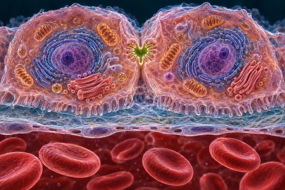
Controlled supporting inset, not to scale. Downward bile arrow follows the local channel only; larger-scale drainage runs toward portal ducts. Individual organelles and microscopic dimensions are not depicted.
Process nutrients, make plasma proteins and secrete bile; different membrane faces keep blood exchange separate from bile output.
A thin, porous lining separates flowing blood from the exchange space beside hepatocytes.
Resident macrophages associated with sinusoids clear particles and participate in immune surveillance.
Cells in the perisinusoidal space store vitamin A and help maintain the supporting matrix.
Bile-duct epithelial cells modify bile with water and electrolytes as it passes through the duct system.
Making a plasma protein and making bile are different outputs. Newly made secreted proteins use the cell’s protein-processing and vesicle machinery before release toward blood. Bile components are secreted across the canalicular membrane into a separate space between neighboring hepatocytes.
This is why the two working faces matter. A canaliculus does not carry newly made albumin back into a sinusoid, and the blood-facing exchange surface is not the bile outlet. The atlas locates the compartments; it does not identify individual transport proteins from generated texture.
Follow the maintained cell protein-secretion lesson → · See where the separate bile route leads →
Five complementary cell roles, not five cells identified in the generated lobule texture. Immune, stromal and duct cells support the hepatocytes; the liver is more than a collection of one cell type.
Hepatocyte polarity and ductal modification: PubMed · Boyer, Bile formation and secretion · University of Leeds · Hepatocyte function and cell interfacesTargeted source check: 15 October 2026. This is not human medical review.
06 / WORKING FUNCTIONS
The liver helps distribute fuel, builds important proteins and handles molecules destined for elimination. It works with muscle, fat, gut and kidneys; it is neither the body’s only store nor a mechanical blood filter.
Bile salts help fats become accessible to digestion and absorption; they are not digestive enzymes. Albumin and many clotting proteins leave in blood, while bile follows the separate duct route.
07 / A CHANGING FUEL BALANCE
Pancreatic insulin and glucagon help coordinate liver metabolism. Their balance favors storage after a meal and glucose supply between meals. Other hormones, substrate availability and time also influence the response.
With nutrients available, insulin favors glycogen synthesis and suppresses glucose production. The liver also processes amino acids and lipids; this simplified view isolates carbohydrate handling.
As the insulin-to-glucagon balance changes, glycogen breakdown and gluconeogenesis support blood glucose. Their relative contributions change with time; this is not fasting advice or a glucose measurement.
Glycogen breakdown supplies stored glucose. Gluconeogenesis makes glucose from substrates such as lactate, glycerol and selected amino acids. These are different mechanisms; the kidney also contributes to glucose production.
Liver cells can release free glucose into blood. Muscle glycogen mainly fuels the muscle itself. A shared fuel name does not mean the two tissues have identical roles.
Follow insulin and glucagon feedback in the pancreas →
Hepatic storage and glucose supply: OpenStax A&P 2e · Insulin, glucagon and hepatic glucose balanceTargeted source check: 15 October 2026. This is not human medical review.
08 / METABOLISM MEETS EXCRETION
Amino-acid metabolism releases nitrogen. Hepatic urea synthesis packages much of it into a form carried by blood to the kidneys. Urea is not bile, and the liver does not make urine.
The liver’s urea pathway connects metabolism with excretion. Start with the cell and its blood-facing surface, then change scale to a mitochondrial compartment and back to the tissue circulation.
Amino-group transfer ≠ ammonia release
Amino acids support new proteins and other molecules. When further metabolism is needed, their carbon skeletons and amino groups have different fates. Aminotransferases move amino groups between molecules; that transfer alone is not the same reaction as releasing free ammonia.
Read the image: Locate the hepatocyte as a working cell. Its visible organelles provide context, not identified aminotransferase molecules.
Mechanism evidence: OpenStax · Protein and carbon handling · IUBMB · Alanine aminotransferase reaction · NCBI Bookshelf · Ammonia biochemistry
This supporting reaction guide explains chemistry, not an anatomical pipe or a molecular image. The selected context emphasises related reactions; it does not assign a fixed elapsed time.
Ammonia + bicarbonate → carbamoyl phosphate, with two ATP used and N-acetylglutamate required.
Mechanism evidence: Reactome · Mitochondrial CPS1 reaction
Carbamoyl phosphate + ornithine → citrulline. Citrulline proceeds to the cytosol.
Mechanism evidence: Reactome · Matrix OTC reaction · Reactome · Ornithine/citrulline exchange
Citrulline + aspartate → argininosuccinate. ATP is converted to AMP and pyrophosphate.
Mechanism evidence: Reactome · Cytosolic ASS1 reaction
Argininosuccinate → arginine + fumarate. Fumarate links nitrogen handling to other metabolism.
Mechanism evidence: Reactome · Argininosuccinate cleavage
Arginine + water → urea + ornithine. Ornithine returns for reuse, rather than leaving with the urea.
Mechanism evidence: Reactome · Cytosolic ARG1 reaction · Reactome · Ornithine/citrulline exchange
Protein turnover supplies amino acids as well as food digestion. Some are reused; others supply intermediates for further metabolism. Nitrogen disposal does not mean every amino acid is first burned for energy or that all carbon skeletons become glucose.
An amino-group transfer commonly gathers nitrogen onto glutamate while changing the partner carbon compounds. Separate reactions can release ammonia. Keeping transfer and release distinct prevents a misleading picture of every aminotransferase directly pouring ammonia into blood.
Mechanism evidence: OpenStax · Protein and carbon handling · IUBMB · Alanine aminotransferase reaction · NCBI Bookshelf · Ammonia biochemistry
Glutamine can carry nitrogen that has been incorporated into its amide group. Glutaminase can release that amide nitrogen as ammonia while forming glutamate. Alanine carries an amino group together with a carbon skeleton that can be transferred back to pyruvate in the liver.
These exchanges connect different tissues and metabolic needs. They are not two sealed transport pipes, and the routes overlap with synthesis, intestinal metabolism and renal functions. The illustration locates the blood–cell interface without claiming to track individual nitrogen atoms.
Mechanism evidence: NCBI Bookshelf · Ammonia biochemistry
CPS1 prepares carbamoyl phosphate from an ammonia-derived nitrogen and bicarbonate-derived carbon. This entry reaction uses two ATP. OTC then combines the carbamoyl group with ornithine to form citrulline. The location is the mitochondrial matrix, not the space between the membranes.
The inner membrane separates compartments. Citrulline export and ornithine return involve a carrier-mediated exchange. That transport is part of connecting the cycle across the membrane; it is not the same process as making ATP at the inner membrane.
Mechanism evidence: Reactome · Mitochondrial CPS1 reaction · Reactome · Ornithine/citrulline exchange
ASS1 uses citrulline, aspartate and ATP to form argininosuccinate. ASL produces arginine and fumarate, and ARG1 produces urea and ornithine. The named compounds are chemical intermediates, not compartments that blood flows through.
Ornithine is regenerated and can re-enter the mitochondrial part. Urea leaves the cycle as a product. A circular chemical scheme therefore expresses repeated reactions and carrier regeneration; it should not be mistaken for an anatomical tube bent into a ring.
Mechanism evidence: Reactome · Cytosolic ASS1 reaction · Reactome · Argininosuccinate cleavage · Reactome · Cytosolic ARG1 reaction · Reactome · Ornithine/citrulline exchange
Urea contains two nitrogen atoms. In the standard hepatic pathway, one enters through ammonia and the other through aspartate; bicarbonate supplies its carbon. “Ammonia becomes urea” is a useful summary, but it leaves out the second nitrogen source.
Amino groups can be exchanged before these entry points. An atlas mechanism does not imply that both nitrogens came directly from one newly eaten amino acid, or that an individual molecule’s prior route can be read from a generated tissue image.
Mechanism evidence: NCBI Bookshelf · Urea-cycle physiology · Reactome · Mitochondrial CPS1 reaction · Reactome · Cytosolic ASS1 reaction
CPS1 consumes two ATP, while the ASS1 reaction converts another ATP to AMP and pyrophosphate. Counting the subsequent pyrophosphate hydrolysis gives the familiar four high-energy phosphate equivalents for the combined sequence. This accounting describes coupled reactions, not energy released merely by breaking an isolated bond.
NAGS produces N-acetylglutamate, the required CPS1 activator. Arginine can stimulate NAGS and connect amino-acid availability with entry control. Substrate supply and longer-term enzyme regulation remain relevant; the activator does not make every hepatocyte behave identically.
Mechanism evidence: Reactome · Mitochondrial CPS1 reaction · Reactome · Cytosolic ASS1 reaction · Reactome · N-acetylglutamate formation · OpenStax · ATP and coupled reactions
Urea synthesis is associated predominantly with periportal hepatocyte functions. A small pericentral population rich in glutamine synthetase can capture residual ammonia into glutamine. These complementary roles help explain why one “average liver cell” cannot represent the entire tissue.
The cited perfusion study investigated rat liver. It supports a mechanistic discussion, not exact human cell rows or numerical zone boundaries. No enzyme-expression map has been painted onto the retained lobule image; the existing labels locate vessels and tissue only.
Mechanism evidence: Experimental hepatic nitrogen zonation · Liver zonation, revisited · Review
A substance can be filtered, reabsorbed or secreted as it passes through the nephron. Urea transport and recycling also contribute to the renal concentrating system, so filtered urea is not all immediately discarded. Urea production by the liver and its eventual excretion are different parts of nitrogen balance.
The kidneys also generate ammonia from glutamine and can excrete ammonia nitrogen in urine. This participates in acid-base regulation and should not be described as a miniature complete hepatic urea cycle. Neither route means that the liver makes urine or that bile carries all unwanted nitrogen.
Mechanism evidence: Weiner, Mitch & Sands · Urea and renal ammonia · NIDDK · Filtration and tubular handling
Reaction identities come from biochemical evidence; a tissue render cannot identify CPS1, NAGS or a particular carrier from a coloured dot. The retained mitochondrial preparation is a compartment reference, not a molecular reconstruction of a specific hepatocyte.
This course concerns normal physiology and learning links. It does not interpret blood tests, diagnose inherited enzyme disorders or recommend protein intake, ammonia-lowering treatment or personal liver and kidney care. Source checks are not physician review.
Mechanism evidence: Reactome · Curated human urea pathway · Weiner, Mitch & Sands · Urea and renal ammonia
No. It transfers an amino group between molecules. Ammonia release is a separate reaction and should not be drawn as the inevitable direct output of every transfer.
No. The entry reactions are in the hepatocyte mitochondrial matrix; the cycle continues in the cytosol and requires transport between these compartments.
No. Ornithine is regenerated. Urea is a product released while the regenerated carrier can support another turn.
No. Urea circulates, and renal filtration and tubular handling contribute to its urinary fate. Renal ammonia handling is related to acid-base balance but is a different pathway.
Normal physiology · source checks 21 October 2026. Qualified original wordless atlas images and semantic geometry retained. Not acquired microscopy, individual enzyme localisation, physician review, personal diet guidance or physical-phone acceptance.
Nitrogen is collected during amino-acid processing.
Reactions spanning mitochondria and cytoplasm form urea.
Renal handling ultimately excretes most urea.
Supporting mechanism overview, not the complete enzyme cycle. Explore renal filtration, recovery and urine concentration →
Hepatic mitochondrial/cytoplasmic urea synthesis and renal excretion: NCBI Bookshelf · Physiology, Urea CycleTargeted source check: 15 October 2026. This is not human medical review.
09 / THE DIGESTIVE OUTPUT
Bile contains water, electrolytes, bile salts and other substances, including conjugated bilirubin. Hepatocytes start its formation; duct cells modify it. The gallbladder stores and concentrates a portion before release into the small intestine.
Into storage through the cystic ductCystic duct → gallbladder
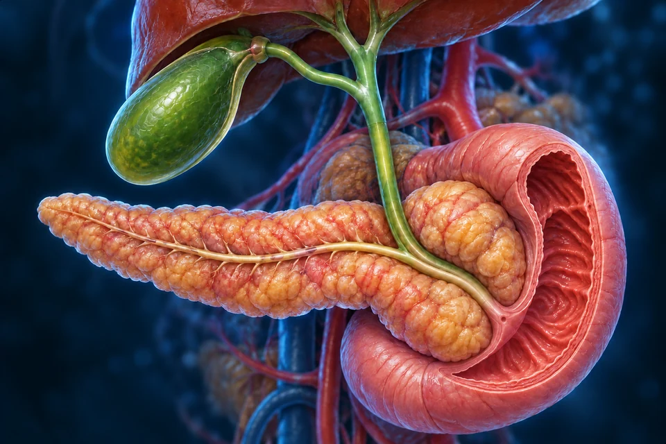
Gold indicates local bile direction on visible duct segments. It is not a full flow trace, a measured volume or a map of a hidden sphincter. The liver continues forming bile in both states; not all bile must pass through the gallbladder.
Greater resistance at the intestinal outlet favors diversion through the cystic duct into the gallbladder, where bile is concentrated. The liver continues making bile.
CCK from the small intestine helps contract the gallbladder and relax the outlet. Bile returns through the cystic duct and continues down the common bile duct into the duodenum.
Supporting direction diagram, not an anatomical shape. Secretin, released when acid enters the duodenum, promotes ductal water and bicarbonate secretion. CCK and secretin are intestinal signals, not hormones made by the gallbladder.
Bile production, storage and release: NIDDK / NIH · OpenStax. Local direction and anatomy checked 17 October 2026; prior qualified gross anatomy retained.
Bile arrives from liver and gallbladder. Pancreatic fluid supplies digestive enzymes and bicarbonate. Their ducts commonly share a short terminal channel, but their origins and contents remain different.
CCK links food in the small intestine with gallbladder emptying. Secretin helps ductal cells add bicarbonate-rich fluid in response to acid. These signals coordinate delivery; the simplified control map does not show every neural and hormonal input.
See how bile salts work with intestinal fat digestion → · Connect CCK and secretin with pancreatic delivery →
Bile formation, storage and meal-related delivery: PubMed · Boyer, Bile formation and secretion · OpenStax A&P 2e · Liver, pancreas and gallbladderTargeted source check: 15 October 2026. This is not human medical review.
10 / REUSE AND DISPOSAL
Bile salts aid fat handling and mostly return for reuse. Bilirubin arises from heme breakdown and follows an elimination pathway. They share a vehicle, not an identical purpose.
Bile salts help disperse lipids and support their absorption.
Most are reabsorbed near the end of the small intestine.
Hepatic uptake and resecretion conserve the pool. A small loss is replaced by new synthesis.
Unconjugated bilirubin travels bound to albumin after heme is broken down, much of it from aging red cells.
The liver changes bilirubin into a more water-soluble form and secretes it into bile.
Gut microbes transform pigments; much leaves in stool. Some products are reabsorbed and a small amount reaches urine.
Supporting pathways. The bile-salt cycle is not a diagram of every bile constituent, and red-cell clearance occurs in the spleen and other tissues as well as the liver.
Follow bile salts from the ileum back to the liver →
Ileal bile-salt return and the separate bilirubin pathway: OpenStax A&P 2e · Liver, pancreas and gallbladder · Merck Manual · Liver Structure and FunctionTargeted source check: 15 October 2026. This is not human medical review.
11 / READING REAL TISSUE
In a real section, the plane of the cut changes what can be recognized. Central venules and portal tracts need surrounding tissue context; a perfectly outlined hexagon is not a requirement.
The institutional H&E slide shows cell plates, sinusoids and vascular/duct regions in real tissue. Follow the small blood channels rather than expecting the generated block’s exact cell pattern.
Open the source’s human liver slide ↗External microscopy reference. No image pixels are copied or embedded: redistribution rights were not established. Its stain and specimen are identified by the source, not inferred from our artwork.
Electron microscopy resolves membrane faces, pores and organelles that gross renderings cannot prove. The separate controlled cell inset teaches the relationships; the University of Leeds reference provides tissue context.
Explore the institutional cell reference ↗No generated texture is presented as micrograph, patient imaging or measured histology. Clinical imaging, disorders and diagnostic interpretation remain a later phase.
Explore source geometry · connect the scale
The existing source-labelled model establishes gross form. It does not show lobules, sinusoidal flow, hormone levels or cellular membranes. An isolated model cannot establish body position by itself.
THE CONNECTED ORGAN
Follow blood to the lobule, exchange to the hepatocyte, bile to the intestine and urea toward the kidneys. Anatomy explains why those routes remain distinct.
Return to AllHumanBodyEDITORIAL RECORD
Source scope. Source-verified educational relationships: gross location; portal/arterial supply and hepatic outflow; classical lobule organization; separate blood and bile routes; cell interfaces; core metabolic, hormonal, urea and biliary processes. The sources below support teaching claims, not every generated pixel.
Review state. English public educational chapter. AI-assisted visual/content production. Fine generated branching, cellular texture, proportions and hidden anatomy remain illustrative / not independently verified. Human medical review: not performed. Educational purpose only; not an exhaustive hepatology encyclopedia or clinical guide.
English revision: liver-english-2026-10-17-connected-quality. Existing Liver knowledge identity is unchanged. Current migration physical-phone acceptance Pending, non-blocking. Existing source-labelled liver geometry is linked below; it cannot resolve microscopic tissue, diagnosis or treatment. No clinical imaging or treatment guidance is claimed. Labels and supporting arrows remain editable English layers, separate from the text-free anatomy masters.