Digestive / VISUAL EXPLAINER
How Liver Fibrosis Develops Over Time
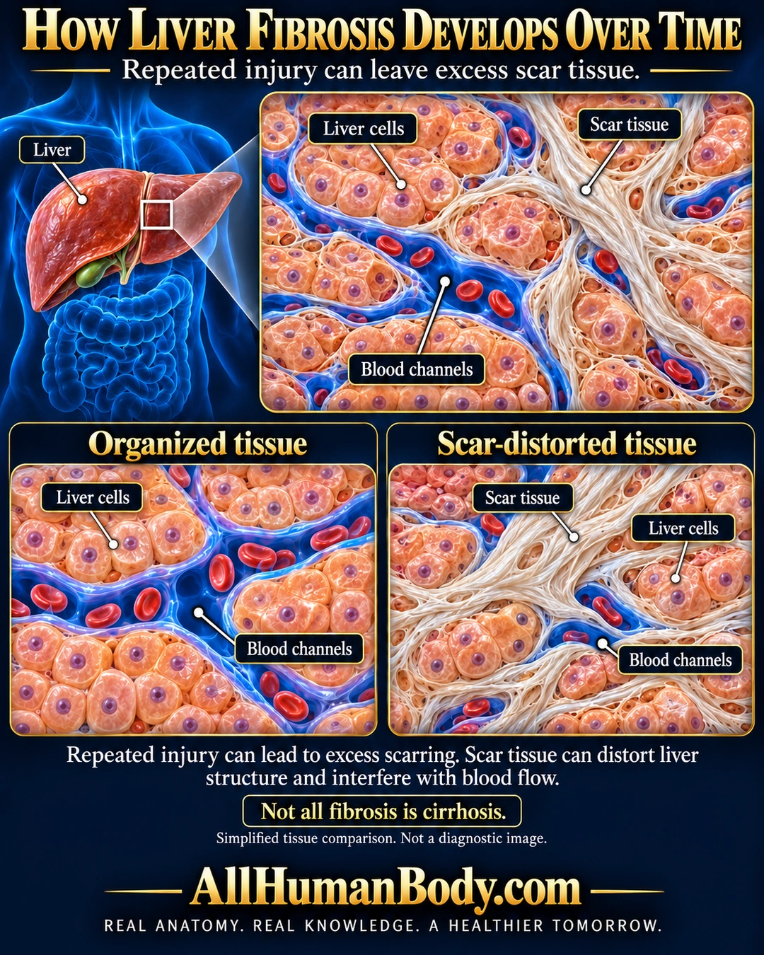
Repeated or continuing liver injury can leave excess scar tissue. This can distort tissue organization and interfere with blood flow. Fibrosis is not always cirrhosis and does not follow one fixed timetable.
READ THE IMAGE
What the illustration shows
Navy and gold poster with a liver locator in a translucent blue torso. An enlarged tissue region shows peach liver cells, blue blood channels containing red cells, and pale fibrous bands between cell groups. Lower panels compare organized tissue with scar-distorted tissue, including locally narrowed channels. Leaders point to liver cells, scar tissue and blue channel spaces. Text explains that repeated injury can lead to scarring, altered structure and impaired blood flow, and says not all fibrosis is cirrhosis. Gold AllHumanBody.com and the full slogan appear on a plain navy footer.
How to interpret this poster
- Not patient biopsy, complete lobule map, fixed progression, cirrhosis stage or claim about cause/reversibility. No treatment or personal alcohol inference.
- AI-assisted educational illustration; not physician certification, a diagnosis or individualized medical advice.
- Fine annotations require enlargement; preserve the full uncropped illustration and its qualifiers.
AI-assisted educational illustration, adapted into web-sized images without changing its composition. This is general education, not an individual diagnosis or treatment recommendation.
EVIDENCE / CONTEXT
Sources and what they support
- MSD: Fibrosis of the liver ↗
Repeated injury, scarring, structural distortion and flow effects.