Follow growing bone, tooth succession and changing signals. Then explore maintenance, lens mechanics, tissue variation and cellular states across a lifetime.
▷
Watch chapter overviewFilm planned · not yet available
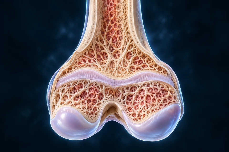
Growing bone context · cartilage activity and replacement support lengthening
Original gross developing-bone illustration, not X-ray or exact age. Fine trabeculae and microscopic zones are illustrative; the independently explained cell-zone inset is separate. New Golden-family text-free preparation; broad visible relationships source-supported, fine generated dimensions/textures illustrative. Human medical review: Not performed.Structure notes · further context
Scope of these landmarks
Names locate broad visible structures in an original generated teaching preparation. Fine tissue, cell counts and microscopic attachments are not measurements or physician-certified anatomy.
A growth plate is separate from the joint-facing cap.
Locate the cartilage between the epiphysis and metaphysis. Tissue activity and replacement support lengthening; the articular surface has another job.
Growth changes size; development changes organization and capacity. Maintenance continues when rapid growth ends, and age-related changes vary between tissues and people.
Growth
Tissue size and form
Regions do not grow at identical rates.
Development
Maturation and experience
Coordination and function emerge through several interacting systems.
Aging
Change over time
Chronological age does not describe every organ’s condition.
In the immature long bone, cartilage at the physis separates epiphysis and metaphysis. The articular cartilage at the joint surface has a different location and purpose.
Original gross developing-bone illustration, not X-ray or exact age. Fine trabeculae and microscopic zones are illustrative; the independently explained cell-zone inset is separate. New Golden-family text-free preparation; broad visible relationships source-supported, fine generated dimensions/textures illustrative. Human medical review: Not performed.Structure notes · further context
Scope of these landmarks
Names locate broad visible structures in an original generated teaching preparation. Fine tissue, cell counts and microscopic attachments are not measurements or physician-certified anatomy.
Why can lengthening stop while living bone keeps changing?
The growth plate is cartilage between epiphyseal and metaphyseal regions. Cartilage-cell proliferation, enlargement and matrix changes contribute to lengthening while replacement and bone formation occur toward the metaphysis. The smooth cartilage facing the joint is a different structure with a different job.
Maturation changes the plate and eventual fusion ends this route of lengthening. It does not end bone life: surfaces can still be modelled and existing material remodelled. The adult bone illustration therefore belongs to a different teaching context from an open growth plate.
The gross bone view and enlarged tissue view may use different orientations. Read their epiphyseal and metaphyseal names before following a direction arrow. Neither image provides a fusion birthday, a height forecast or a measured map of every cell fate.
Resting, proliferating and enlarging chondrocytes occupy different parts of the plate. The epiphyseal-to-metaphyseal sequence explains growth without claiming every generated pixel is a cellular zone.
AUXILIARY RELATIONSHIP / READ WITH THE ANATOMICAL CONTEXT
Plate regions have a teaching direction
Epiphyseal side: Resting/reserve context lies toward the epiphysis.
Within cartilage: Proliferating columns and enlarging chondrocytes occupy different regions.
Metaphyseal side: Matrix changes, vascular invasion and bone deposition occur toward replacement; no thickness measured.
Optional symbolic relationship
Independent supporting orientation; not measured geometry, histology, optical ray tracing or a diagnostic timetable. Human medical review not performed.
The inset has its own orientation: epiphyseal side at top, metaphyseal replacement below. The main distal-femur plate has the epiphysis below; these are separately labeled views, not aligned photographs.
Cartilage cells make matrix and change size; matrix calcification, vascular invasion and bone deposition occur toward the metaphyseal side. Subsequent remodeling produces working architecture.
MECHANISM / ALL ESSENTIAL STAGES VISIBLE
Cartilage production and replacement work together
Cartilage activity → matrix change → bone deposition and remodeling
Original generated teaching views of a growing bone, not a mature adult bone or a measured tissue section. The larger panel explains broad regional order; cell fate, exact zone thickness and fine cartilage–bone boundaries are not certified by generated texture.Structure notes · further context
Growth is not adult turnover
Growth plates contribute to lengthening before maturation. Adult bone continues surface modelling and remodelling after plates have closed.
Not one universal fate
Lineage evidence is more complex than a claim that every hypertrophic chondrocyte always dies or always becomes a bone cell; much experimental evidence comes from animal models.
First locate the growth plate in the bone.
Original generated teaching views of a growing bone, not a mature adult bone or a measured tissue section. The larger panel explains broad regional order; cell fate, exact zone thickness and fine cartilage–bone boundaries are not certified by generated texture.Structure notes · further context
Growth is not adult turnover
Growth plates contribute to lengthening before maturation. Adult bone continues surface modelling and remodelling after plates have closed.
Not one universal fate
Lineage evidence is more complex than a claim that every hypertrophic chondrocyte always dies or always becomes a bone cell; much experimental evidence comes from animal models.
Then read the tissue from epiphyseal to metaphyseal side.
Original generated teaching views of a growing bone, not a mature adult bone or a measured tissue section. The larger panel explains broad regional order; cell fate, exact zone thickness and fine cartilage–bone boundaries are not certified by generated texture.Structure notes · further context
Growth is not adult turnover
Growth plates contribute to lengthening before maturation. Adult bone continues surface modelling and remodelling after plates have closed.
Not one universal fate
Lineage evidence is more complex than a claim that every hypertrophic chondrocyte always dies or always becomes a bone cell; much experimental evidence comes from animal models.
Find the growth region
The small organ view locates an open growth plate below the epiphysis. In the larger tissue view, epiphyseal-side cartilage lies above the metaphyseal replacement region.
Gold outlines identify selected teaching locations in the full view. They do not measure activity, speed or a universal sequence; the narrow windows retain the same anatomical targets.
The enlarged tissue window uses its own epiphyseal-to-metaphyseal orientation. It is not a magnification measured from the separate gross femur illustration.
Chondrocytes divide, change size and produce matrix within organized plate regions.
Produce cartilage
Chondrocytes divide, change size and produce matrix within organized plate regions.
This is a qualitative sequence, not a microscopic image.
Change the scaffold
Matrix mineralization and vascular invasion accompany replacement toward the metaphysis.
Some chondrocyte-derived cells also contribute to bone lineages; no universal fate is asserted.
Remodel bone
Bone deposition and later remodeling shape the working trabecular framework.
Bone is not simply inert calcified cartilage.
Optional symbolic guide · compare the relationship
Original editable relationship, separate from the anatomical preparations. Dimensions and timing are qualitative, not microscopy, a measured simulation or a personal aging forecast.
Cell-lineage studies also show that some chondrocyte-derived cells contribute to bone/stromal lineages. Neither every cell turns into bone nor every chondrocyte must die is a valid universal rule; no human fraction is asserted.
Plate activity supports elongation. Periosteal bone formation and internal remodeling alter thickness and shape; articular cartilage is not a second growth plate.
Length
Epiphyseal–metaphyseal interface
Growth-plate activity contributes to long-bone elongation.
Width and form
Outer and internal surfaces
Formation and resorption cooperate to shape living bone.
Growth hormone and IGF signaling cooperate with thyroid and sex-steroid context. Local cell-to-cell factors help regulate plate activity; one hormone alone does not determine a person’s final height.
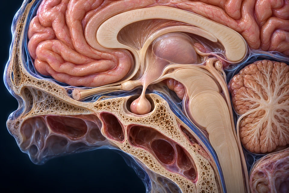
New original Golden-family text-free teaching preparation. Expanded near-midline regional preparation; anterior is image left. The exposed brain and skull contours, hidden optic relations and exact dimensions remain illustrative. This is not a surgical map or patient scan. Human medical review: Not performed.Structure notes · further context
Hypothalamic region
The region belongs to the base of the diencephalon. Individual nuclei and the generated ventricle/optic contours are not identified or claimed verified here.
Pituitary stalk
The infundibulum carries neural processes and vessels. It is not a duct carrying a brain fluid into the pituitary.
Pituitary gland
The anterior endocrine and posterior neural regions are enlarged in the next distinct view. This regional view establishes location, not all vascular junctions.
Bony seat
The pituitary lies in this bony depression below the brain. Bone and adjacent cavity contours are illustrative, not operative boundaries or a measured cross-section.
This qualified brain–pituitary context locates the start of distant signalling. It does not depict the gonads, every feedback route or an individual’s pubertal state; follow the adjacent stages and endocrine lesson for the distinct jobs.
GH and IGF pathways cooperate with thyroid and sex-steroid context.
Distant signals
GH and IGF pathways cooperate with thyroid and sex-steroid context.
No one hormone determines final height.
Local activity
Cells respond through proliferation, maturation and matrix behavior.
Local regulators do not act independently of the tissue environment.
Maturation
Plate activity changes and eventual fusion limits further elongation.
No fixed closure age or intervention recommendation.
Optional symbolic guide · compare the relationship
Original editable relationship, separate from the anatomical preparations. Dimensions and timing are qualitative, not microscopy, a measured simulation or a personal aging forecast.
Growth-plate activity slows and cartilage is eventually replaced as the plate fuses. Estrogen signaling contributes in both sexes; timing differs between bones and individuals.
AUXILIARY RELATIONSHIP / READ WITH THE ANATOMICAL CONTEXT
Open cartilage and fused context differ
Open plate: Cartilage remains between bony regions during elongation.
Maturation: Plate activity changes; estrogen contributes in both sexes.
Fused context: Cartilage continuity is eventually replaced; bone maintenance continues.
Optional symbolic relationship
Independent supporting orientation; not measured geometry, histology, optical ray tracing or a diagnostic timetable. Human medical review not performed.
No fixed birthday, guaranteed remaining growth, drug recommendation or personal height prediction. Bone remains metabolically active after elongation ends.
Head and trunk–limb proportions change as regions grow differently. Skull sutures and the developing brain are related but do not share one universal completion date.
Regional rates
Head, trunk and limbs
Changing proportions reflect different growth patterns.
Growing vault
Sutures and membranous junctions
The newborn chapter provides the age-specific vault context.
No adult geometry was stretched into child anatomy. A rights-cleared measured age-series remains separate visual debt; these cards do not pretend to be one.
Primary teeth are followed by permanent teeth, and a mixed-dentition period includes both. The local cutaway teaches one replacement relationship rather than certifying a complete jaw or child age.
Original localized succession context, not a complete dentition or a measured child age. Fine cusps, root shapes, follicular/periodontal detail and neighboring tooth identity are illustrative. New Golden-family text-free preparation; broad visible relationships source-supported, fine generated dimensions/textures illustrative. Human medical review: Not performed.Structure notes · further context
Scope of these landmarks
Names locate broad visible structures in an original generated teaching preparation. Fine tissue, cell counts and microscopic attachments are not measurements or physician-certified anatomy.
A permanent premolar succeeds a primary molar. The local illustration places the developing successor beneath the primary tooth, within the surrounding bone. Its relationship to the primary roots helps explain why tooth development involves changes in the tooth and its supporting tissues together.
Permanent molars are a different case: they are added farther back rather than replacing primary molars. Counting a visible tooth as a molar is therefore not enough to decide which replacement relationship it belongs to.
This is one selected local relationship, not a complete dentition timetable or a prediction of an individual child’s eruption. Continue to the dental chapter for tooth tissues and attachment; fine cusp and root details in the generated view are not clinical identification evidence.
Permanent incisors and canines replace their primary predecessors; premolars replace primary molars. Permanent molars develop behind the primary dentition rather than replacing a primary molar.
AUXILIARY RELATIONSHIP / READ WITH THE ANATOMICAL CONTEXT
Addition: Permanent molars form posteriorly, not underneath a primary molar to replace it.
Scope: One local illustration is not a complete dental formula or child age estimate.
Optional symbolic relationship
Independent supporting orientation; not measured geometry, histology, optical ray tracing or a diagnostic timetable. Human medical review not performed.
A developing crown is not a second erupted row. Exact tooth counts and individual eruption schedules are not read from this localized picture.
Eruption involves the tooth follicle, surrounding bone and attachment tissues, not only roots pushing a crown upward. Primary-root resorption and successor eruption are related but variable.
Develop
Within the jaw
A crown develops before appearing in the mouth.
Move and remodel
Bone and attachment context
Surrounding tissues change along the eruption path.
Function
At the gum line
The erupted tooth becomes part of the chewing apparatus.
No clinical eruption threshold or treatment plan; the illustration is not a timed sequence.
Muscles, joints, sensory input and neural circuits cooperate in posture and coordinated action. Practice and environment matter alongside tissue maturation.
MECHANISM / ALL ESSENTIAL STAGES VISIBLE
A movement links sensing, planning and action
Sensory input ↔ neural integration ↔ muscle action and feedback
Body position and environmental information reach the nervous system.
Sense
Body position and environmental information reach the nervous system.
Not every developmental skill is one reflex.
Integrate
Neural activity and experience coordinate a response.
This map is not an exact developmental wiring diagram.
Act and learn
Muscles move the body; new sensory information contributes to future action.
Practice and maturation interact without one rigid milestone clock.
Optional symbolic guide · compare the relationship
Original editable relationship, separate from the anatomical preparations. Dimensions and timing are qualitative, not microscopy, a measured simulation or a personal aging forecast.
Motor activity, communication and social interaction develop through different experiences and capacities. One observed skill cannot stand in for the whole child or a complete neurological examination.
AUXILIARY RELATIONSHIP / READ WITH THE ANATOMICAL CONTEXT
Development does not use one clock
Motor: Posture, movement and sensory coordination.
Communication: Language and other communication capacities.
Social learning: Interaction and experience; domains overlap without a single ranking scale.
Optional symbolic relationship
Independent supporting orientation; not measured geometry, histology, optical ray tracing or a diagnostic timetable. Human medical review not performed.
This is a qualitative learning map, not a milestone screening tool, age deadline or causal judgement about family income, structure or language.
Increasing pulsatile GnRH signaling influences pituitary LH and FSH, which act on gonadal tissues. The same axis has earlier-life activity; puberty is not its first existence.
New original Golden-family text-free teaching preparation. Expanded near-midline regional preparation; anterior is image left. The exposed brain and skull contours, hidden optic relations and exact dimensions remain illustrative. This is not a surgical map or patient scan. Human medical review: Not performed.Structure notes · further context
Hypothalamic region
The region belongs to the base of the diencephalon. Individual nuclei and the generated ventricle/optic contours are not identified or claimed verified here.
Pituitary stalk
The infundibulum carries neural processes and vessels. It is not a duct carrying a brain fluid into the pituitary.
Pituitary gland
The anterior endocrine and posterior neural regions are enlarged in the next distinct view. This regional view establishes location, not all vascular junctions.
Bony seat
The pituitary lies in this bony depression below the brain. Bone and adjacent cavity contours are illustrative, not operative boundaries or a measured cross-section.
This qualified brain–pituitary context locates the start of distant signalling. It does not depict the gonads, every feedback route or an individual’s pubertal state; follow the adjacent stages and endocrine lesson for the distinct jobs.
Pulsatile GnRH contributes to changing pituitary output.
Hypothalamus
Pulsatile GnRH contributes to changing pituitary output.
The axis has activity earlier in life; puberty is not its first existence.
Pituitary
LH and FSH act on different gonadal cell populations.
This is not a single chemical acting identically on every cell.
Gonads and feedback
Steroid and inhibin signals contribute to regulation.
Adrenal maturation is a distinct process; no clinical hormone threshold.
Optional symbolic guide · compare the relationship
Original editable relationship, separate from the anatomical preparations. Dimensions and timing are qualitative, not microscopy, a measured simulation or a personal aging forecast.
Why distinguish gonadal signalling from adrenal maturation?
The hypothalamic–pituitary–gonadal route involves pulsatile GnRH, pituitary LH and FSH, and responses in different gonadal cell populations. Steroid and inhibin signals contribute to feedback. A three-step diagram is a guide to communication, not three structures acting without returning information.
Adrenarche refers to a distinct adrenal maturation process. It can be dissociated from activation of the gonadal route, so the two processes should not be collapsed into one switch or one visible sign.
Growth, tissue responses and developmental domains do not share one identical clock. These normal relationships do not provide hormone thresholds, a diagnosis, an intervention recommendation or a definition of personal identity.
Gonadal steroids and inhibin participate in feedback that helps regulate the axis. Existing reproductive lessons explain cell targets and cycle phases in greater detail.
The adrenal cortical zona reticularis increases production of androgen precursors during adrenarche. This differs from gonadal-axis activation; hair development alone does not establish the whole gonadal maturation state.
Sex-steroid, GH and IGF signaling interact with growth-plate biology during pubertal growth. Increased activity and later plate maturation are related processes, not a permanent acceleration switch.
Activity
Growth-related signals
Influence cartilage and other tissues.
Maturation
Plate biology changes
Later fusion limits further longitudinal growth.
No intervention to increase height or delay fusion; variation remains explicit.
Voice, hair, muscle and reproductive tissues respond to changing signals in different sequences and extents. These normal changes are descriptions of physiology, not a measure of identity, worth or fixed social behavior.
Voice
Laryngeal/vocal apparatus
Growth can change pitch.
Hair and skin
Androgen-responsive sites
Responses vary by site and person.
Muscle/body composition
Multiple tissue responses
Hormone context interacts with activity and other influences.
No sexualized imagery, body-rating chart or universal trait checklist.
Menstrual phases connect follicular activity, endometrial tissue and hormonal feedback. Early-cycle patterns and individual durations vary; one 28-day drawing cannot be a universal timetable.
Living bone continues remodeling; skin and many other tissues renew and repair. Different cell types have different turnover and proliferative capacity.
MECHANISM / ALL ESSENTIAL STAGES VISIBLE
Tissues continue working after growth slows
Use / local conditions ↔ maintenance ↔ remodeling and repair
Original generated 3D teaching preparation, not microscopy. Selected resorption and formation contexts are brought together for comparison, not a universal spatial conveyor or a measured time sequence. Fine processes, boundaries and proportions remain illustrative.Structure notes · further context
Two different lineages
Osteoclasts arise from haematopoietic monocyte/macrophage-related precursors. Osteoblast-lineage cells form bone; an osteoclast does not transform into an osteoblast.
Coupling is communication
Signals coordinate removal and formation across local contexts. The illustration does not show all signals or prove a fixed interval between the two jobs.
Coordinate a surface transition
Removal and formation are different jobs linked by communication. The two contexts do not depict a cell changing from osteoclast into osteoblast.
Gold outlines identify selected teaching locations in the full view. They do not measure activity, speed or a universal sequence; the narrow windows retain the same anatomical targets.
Bone-surface renewal is one tissue example. The selected emphasis is a location cue, not a universal repair sequence, rate or guarantee.
Different cell populations support matrix and tissue turnover.
Not all cells divide at the same rate or have the same capacity.
Adapt
Remodeling and repair change structures over time.
No universal rate or automatic recovery guarantee.
Optional symbolic guide · compare the relationship
Original editable relationship, separate from the anatomical preparations. Dimensions and timing are qualitative, not microscopy, a measured simulation or a personal aging forecast.
Compare a separate mature-bone contextRetained original adult-bone illustration, not a child specimen, scan or longitudinal age series. Growth cartilage is shown separately in this chapter.
The outer epithelium lies above a connective dermis and subcutaneous context. A qualitative comparison makes these relationships visible; it cannot measure a person’s age or predict every skin site.
Original qualitative comparison, not a universal younger/older phenotype or scale measurement. Fine cells, vessel branches, fiber identities and thickness illustrative. Intrinsic aging and sun-related change are distinguished in text. New Golden-family text-free preparation; broad visible relationships source-supported, fine generated dimensions/textures illustrative. Human medical review: Not performed.Structure notes · further context
Scope of these landmarks
Names locate broad visible structures in an original generated teaching preparation. Fine tissue, cell counts and microscopic attachments are not measurements or physician-certified anatomy.
Dermal collagen organization and cell–matrix interactions can change with age. Fibroblast activity and matrix condition influence one another; decorative fiber texture is not histology.
AUXILIARY RELATIONSHIP / READ WITH THE ANATOMICAL CONTEXT
Cell response: Fibroblast behavior and matrix condition interact.
Variation: Intrinsic and photoaging patterns differ; fine generated fibers are not histology.
Optional symbolic relationship
Independent supporting orientation; not measured geometry, histology, optical ray tracing or a diagnostic timetable. Human medical review not performed.
Fine generated cells/fibers are illustrative. This is not an observed biopsy or quantified collagen-loss percentage.
Chronological tissue changes interact with lifetime exposure. Photoaging can alter elastic tissue differently from intrinsic aging; one simple rule that all fibers decrease is misleading.
Intrinsic context
Time and biology
Age-related changes occur within tissues.
Exposure context
UV and other influences
Can produce different tissue patterns.
Individual variation
Site and history
The two original blocks are examples, not deterministic outcomes.
No treatment, cosmetic intervention or race-based prediction.
The lens sits behind the iris, held by zonular fibers and surrounded by ciliary tissue. The cornea bends light first; the lens then contributes adjustable optical power.
Original gross anterior-segment context, not complete globe. Fine insertions, fiber counts, lens texture and rear cutaway boundaries illustrative. No rays or measured elasticity baked into this image. New Golden-family text-free preparation; broad visible relationships source-supported, fine generated dimensions/textures illustrative. Human medical review: Not performed.Structure notes · further context
Scope of these landmarks
Names locate broad visible structures in an original generated teaching preparation. Fine tissue, cell counts and microscopic attachments are not measurements or physician-certified anatomy.
What changes when near focusing becomes more difficult?
In the conventional accommodation relationship, ciliary-muscle contraction reduces zonular tension and permits a responsive lens to become more rounded. The change increases optical power for nearer viewing. The ciliary tissue, suspension fibres and lens are different parts of that relationship.
Age-related reduction in lens reshaping can make near focus more difficult. This is not the same statement as saying that the ciliary muscle disappears, that every difficulty is myopia, or that a lens has become cloudy with cataract.
The retained anterior-eye image locates the structures. Its detailed surface does not measure elasticity, fibre insertion or an individual’s focusing ability. The Eyes & Vision lesson supplies the fuller near/far optical comparison.
In the conventional accommodation model, ciliary contraction reduces zonular tension and allows the lens to become more rounded for near focus. Age-related lens biomechanics reduce this response.
AUXILIARY RELATIONSHIP / READ WITH THE ANATOMICAL CONTEXT
Tension and lens response are different steps
Near effort: Ciliary contraction reduces zonular tension in the conventional model.
Lens response: A responsive lens becomes more rounded and increases optical power.
Age-related limitation: Reduced lens reshaping can limit near focus; not equivalent to cataract or myopia.
Optional symbolic relationship
Independent supporting orientation; not measured geometry, histology, optical ray tracing or a diagnostic timetable. Human medical review not performed.
The drawing is qualitative, not ray tracing or a refractive prescription. Presbyopia is not equivalent to myopia, cataract or disappearance of the ciliary muscle.
Inner-ear changes, neural pathways and lifetime exposures can affect hearing. Age-related loss is not inevitable complete deafness or proof that every cochlear hair cell is absent.
Sensory tissue
Cochlear context
Sound transduction depends on living sensory structures.
Some people take longer to learn or recall information as they age. Dementia is not a normal, inevitable stage of aging; this normal-function lesson does not supply a diagnostic symptom checklist.
The menopausal transition can involve variable cycles and hormone fluctuations; later ovarian estrogen/progesterone production is reduced. It is a normal reproductive transition, not total failure of all endocrine function.
Transition
Variable ovarian activity
Patterns can fluctuate rather than falling smoothly.
Later state
Reduced ovarian hormone output
Feedback and other tissues adjust.
Variation
Different experiences
No fixed personal date or universal symptom bundle.
Resting function and reserve are different questions
A system may meet ordinary demands while its response to greater demands changes. For example, heart-rate response during activity can change with age even when resting rate changes little.
Ordinary demand
Resting function
Does not measure the maximum response.
Greater demand
Reserve capacity
A different question from the resting observation.
Why separate resting performance from physiological reserve?
An organ’s ordinary resting work and its capacity to respond to an increased demand are different questions. Maintenance, adaptation and reserve should therefore be considered together rather than assuming that a functioning organ has unlimited additional capacity.
Changes occur across tissues and vary between people. Chronological age is not a hidden measurement that can be read from the skin, eye or bone illustrations, and a qualitative teaching comparison cannot predict a person’s response to stress.
Senescence describes a cell state, not simply cell death. Such cells can have useful roles in repair and tumor suppression, and persistent states can also contribute to age-related problems.
MECHANISM / ALL ESSENTIAL STAGES VISIBLE
Division arrest differs from cell death
Cell state change → altered active behavior → context-dependent effects
Cellular activity and the ability to divide are different questions. No cell in these gross tissue illustrations is identified as senescent by appearance. Continue cellular structure and work →
Selected focus: Stop dividing
A senescent cell enters a state of sustained division arrest.
Stop dividing
A senescent cell enters a state of sustained division arrest.
Not every nondividing cell is senescent.
Remain active
Metabolism and signaling can continue in these cells.
Senescence is not simply an empty dead cell.
Affect tissue
Repair/tumor suppression and adverse persistent effects depend on context.
No universal marker, aging score or therapeutic recommendation.
Optional symbolic guide · compare the relationship
Original editable relationship, separate from the anatomical preparations. Dimensions and timing are qualitative, not microscopy, a measured simulation or a personal aging forecast.
No universal marker, personal biological-age calculation, antiaging treatment or claim that all aged cells are senescent.
How can a cell stop dividing without being a dead cell?
Senescence describes a state of sustained division arrest in which metabolism and signalling can continue. Division arrest and loss of cellular activity are therefore different claims.
Not every cell that is currently not dividing is senescent. Likewise, one cell state cannot explain every change in an aging organ. Effects of senescent cells can be beneficial or adverse depending on the biological context and persistence.
The chapter’s tissue images do not identify individual senescent cells. No universal visible marker, personal aging score or antiaging treatment recommendation is inferred from their appearance.
Age is not a measurement hidden in an illustration
Main plates are original gross teaching views. Imaging, microscopy and longitudinal observations answer different questions; none is fabricated as a patient-derived measurement here.
Gross teaching
Original anatomy
Fine generated detail remains illustrative at stated scope.
Mechanism
Editable supporting map
Relationships are qualitative, not exact simulation.
Observation
Separate resource
Rights-cleared Xray, microscopy and photographic age series remain debt.
Inheritance & Cell Division covers how information is copied and combined. This chapter does not claim complete life-course coverage, clinical diagnosis or human medical review.
Source-supported gross growth-cartilage, tooth-successor and anterior-eye relationships; normal maturation, endocrine feedback, qualitative skin/lens/cell change and reserve. No clinical prediction, individual age measurement or treatment.
Methods and limits
Retained detailed text-free preparations show growing bone, tooth succession, the anterior eye and qualitative skin organisation. Independent names and explanations identify visible structures. Enlarged growth tissue, adult renewal and brain–pituitary contexts connect anatomy with normal mechanisms. The separate mature-bone reference is not an immature specimen or an age series. Fine trabeculae, tooth roots, zonular insertions, cell counts, skin vessels and textures remain illustrative / not independently verified. No observed patient imaging or histology. Human medical review: not performed. Educational only.
Revision: growth-lifespan-eyes-context-2026-10-19-v2. Stable content identity: ahb_ch_growth_lifespan_english_v1. Names, explanations and paths are independent editable layers. New physical-phone acceptance: not performed. No image-based age prediction, no clinical protocols and no claim of physician approval. Rights-cleared age-series imaging are separate work.