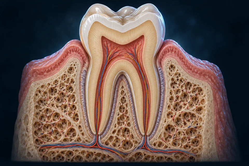
New original Golden-family text-free teaching preparation. Educational molar section, not universal root anatomy or a dental scan. Enamel covers the crown; the thin root coat represents cementum. Fine tubules, nerves, vessels, fibers and tissue thicknesses remain illustrative. Human medical review: Not performed.Structure notes · further context
Enamel
Mature enamel is acellular. It is not bone, and it is distinct from root cementum.
Dentin
Odontoblast cell bodies lie near the pulp-dentin boundary; their processes extend into tubules. The generated fine radial texture is not a verified tubule specimen.
Pulp
The pulp chamber continues into root canals. Exact neural branches and capillary patterns are illustrative.
Cementum
It provides attachment for periodontal-ligament fibers. The pale root coat should not be read as enamel extending to the apex.
Periodontal ligament
It distributes load and contributes sensory information. The tooth is not directly fused to its alveolus.
Alveolar bone
Bone and ligament remodel. Exact trabecular geometry in this generated preparation is not patient evidence.
Gingiva
It belongs to the supporting oral tissues, rather than being a hard layer within the crown.
Enamel outside; dentin beneath
A hard outer crown and underlying dentin form a layered structure. Root cementum is another outer tissue, not enamel extending down the root.
Gold outlines locate teaching regions on one unchanged preparation. They do not show a measured force, secretion rate or moving tissue.
01 / LOWER-ARCH
A tooth row is a sequence of different working surfaces
Look down toward this selected lower permanent row. Narrow anterior crowns and broader posterior crowns occupy a continuous curved arrangement. Fourteen teeth are present in this source row; third molars are absent from the downloaded selection, not from the definition of permanent dentition.
Open the source-native tooth-row reference
The named meshes preserve this adult male source selection and its native positions. Their coarse outer surfaces locate teeth; they do not resolve enamel, dentin, pulp or ligament microstructure. Third molars are absent from the two fourteen-tooth rows.
A curved row presents different crown shapes
1
Left lower central permanent incisor
A narrow anterior crown presents a cutting edge.
More about left lower central permanent incisor
Fresh native projection and first named target surface raycast, transparent named target retained; not microscopic tissue, canal topology or clinical imaging. Broad selected roles independently source-supported; model detail is not human medical review.
3
Left lower permanent canine
A pointed crown assists contact and division.
More about left lower permanent canine
Fresh native projection and first named target surface raycast, transparent named target retained; not microscopic tissue, canal topology or clinical imaging. Broad selected roles independently source-supported; model detail is not human medical review.
2
Left lower lateral permanent incisor
A narrow anterior crown presents a cutting edge.
More about left lower lateral permanent incisor
Fresh native projection and first named target surface raycast, transparent named target retained; not microscopic tissue, canal topology or clinical imaging. Broad selected roles independently source-supported; model detail is not human medical review.
123456
6
Left lower second permanent molar
A broader posterior crown participates in crushing and grinding.
More about left lower second permanent molar
Fresh native projection and first named target surface raycast, transparent named target retained; not microscopic tissue, canal topology or clinical imaging. Broad selected roles independently source-supported; model detail is not human medical review.
5
Left lower first permanent molar
A broader posterior crown participates in crushing and grinding.
More about left lower first permanent molar
Fresh native projection and first named target surface raycast, transparent named target retained; not microscopic tissue, canal topology or clinical imaging. Broad selected roles independently source-supported; model detail is not human medical review.
4
Left lower first permanent premolar
A broader posterior crown participates in crushing and grinding.
More about left lower first permanent premolar
Fresh native projection and first named target surface raycast, transparent named target retained; not microscopic tissue, canal topology or clinical imaging. Broad selected roles independently source-supported; model detail is not human medical review.
A curved row presents different crown shapes · names and basic functions are visible
The oblique upper row exposes whole tooth components, including portions normally hidden by supporting tissue. Names retain side and arch: a left upper tooth is not interchangeable with a left lower one. Coarse source cusps are not a clinical occlusal-contact model.
Open the source-native tooth-row reference
The named meshes preserve this adult male source selection and its native positions. Their coarse outer surfaces locate teeth; they do not resolve enamel, dentin, pulp or ligament microstructure. Third molars are absent from the two fourteen-tooth rows.
Upper tooth classes occupy a different arch
3
Left upper permanent canine
A pointed crown assists contact and division.
More about left upper permanent canine
Fresh native projection and first named target surface raycast, transparent named target retained; not microscopic tissue, canal topology or clinical imaging. Broad selected roles independently source-supported; model detail is not human medical review.
2
Left upper lateral permanent incisor
A narrow anterior crown presents a cutting edge.
More about left upper lateral permanent incisor
Fresh native projection and first named target surface raycast, transparent named target retained; not microscopic tissue, canal topology or clinical imaging. Broad selected roles independently source-supported; model detail is not human medical review.
1
Left upper central permanent incisor
A narrow anterior crown presents a cutting edge.
More about left upper central permanent incisor
Fresh native projection and first named target surface raycast, transparent named target retained; not microscopic tissue, canal topology or clinical imaging. Broad selected roles independently source-supported; model detail is not human medical review.
123456
6
Left upper second permanent molar
A broader posterior crown participates in crushing and grinding.
More about left upper second permanent molar
Fresh native projection and first named target surface raycast, transparent named target retained; not microscopic tissue, canal topology or clinical imaging. Broad selected roles independently source-supported; model detail is not human medical review.
5
Left upper first permanent molar
A broader posterior crown participates in crushing and grinding.
More about left upper first permanent molar
Fresh native projection and first named target surface raycast, transparent named target retained; not microscopic tissue, canal topology or clinical imaging. Broad selected roles independently source-supported; model detail is not human medical review.
4
Left upper first permanent premolar
A broader posterior crown participates in crushing and grinding.
More about left upper first permanent premolar
Fresh native projection and first named target surface raycast, transparent named target retained; not microscopic tissue, canal topology or clinical imaging. Broad selected roles independently source-supported; model detail is not human medical review.
Upper tooth classes occupy a different arch · names and basic functions are visible
Incisors bring a cutting edge, canines a pointed crown, and premolars and molars broader contacts. Jaw movement, tongue and cheeks combine these surfaces during chewing; no tooth class performs every task in isolation.
Crown contacts and coordinated movement work together
Selected relationship: Divide. Anterior edges and pointed crowns help divide food.
Original generated crown-form comparison, not extracted specimens or an individual dental chart. These examples teach contact surfaces, not a measured scale, exact cusp count for every tooth, or a universal root-number rule. Root anatomy and tooth presence vary. Human medical review: Not performed.Structure notes · further context
Shape works with position and movement
The tongue and cheeks reposition food between the teeth while the jaw brings opposing surfaces into contact. A tooth is not an independent cutting machine.
Crown form is not a full tooth atlas
These selected examples do not identify side, tooth number, occlusal relationships, eruption stage or a treatment need. Use the following tooth/socket preparation for enamel, dentin, pulp and supporting-tissue relationships.
Divide
Anterior edges and pointed crowns help divide food.
Process
Broader posterior contacts assist crushing and grinding.
Reposition
Tongue and cheeks help guide material during jaw movement.
Selection locates a teaching context. It is not an individual bite simulation, a developmental film or a measured renewal rate.
Central and lateral incisors differ by position within each front pair. First and second premolars sit before the molars in the permanent row. These spatial names are easier to learn before introducing an individual dental numbering chart.
Side
Your left or right
Body laterality is retained regardless of camera direction.
The usual primary set has twenty teeth and no premolars. The complete permanent pattern includes thirty-two with third molars. Permanent premolars succeed primary molars; permanent molars are added posteriorly. Presence and eruption vary.
Why are twenty primary teeth not simply replaced by thirty-two successors?
A primary quadrant normally has two incisors, one canine and two molars. Permanent premolars succeed the primary molars, while permanent molars are posterior additions. The complete permanent pattern therefore differs in both number and classes. Third-molar presence and eruption vary.
The two source-native rows on this page each contain fourteen selected permanent teeth. Their absent third molars describe this dataset, not a new definition of normal dentition.
This local posterior enlargement removes surrounding jaw context so the two source teeth can be inspected. Root divisions differ among teeth and people. A visible source root is not a validated root-canal count, apical opening or microscopic layer.
Open the source-native root-form reference
The named meshes preserve this adult male source selection and its native positions. Their coarse outer surfaces locate teeth; they do not resolve enamel, dentin, pulp or ligament microstructure. Third molars are absent from the two fourteen-tooth rows.
Two posterior teeth expose their root form
1
Left lower first permanent molar
A broader posterior crown participates in crushing and grinding.
More about left lower first permanent molar
Fresh native projection and first named target surface raycast, transparent named target retained; not microscopic tissue, canal topology or clinical imaging. Broad selected roles independently source-supported; model detail is not human medical review.
12
2
Left lower second permanent molar
A broader posterior crown participates in crushing and grinding.
More about left lower second permanent molar
Fresh native projection and first named target surface raycast, transparent named target retained; not microscopic tissue, canal topology or clinical imaging. Broad selected roles independently source-supported; model detail is not human medical review.
Two posterior teeth expose their root form · names and basic functions are visible
Enamel covers the anatomical crown; cementum covers the root. Dentin forms much of the structure around a living pulp space. The existing molar cutaway provides the large-scale relationship; it is linked rather than repeated as another full-page picture.
Enamel is built from oriented mineral crystallites arranged in rod and interrod regions. Rod paths and crystal direction vary through the tissue; some surface regions lack a clear rod pattern. The enlarged supporting guide explains organization rather than reproducing a microscopic specimen.
Rod and interrod regions organize mineral direction
Original generated developmental tissue illustration, not observed histology. The local secretory front is not an erupted tooth retaining an ameloblast layer. Mineral bundles and organelles are enlarged teaching representations, not a universal crystal pattern, cell count or measured scale. Human medical review: Not performed.Structure notes · further context
Local detail from the same preparation
This view shows the lower mineral region of the following developmental front. It is not an extra microscope image, a measured crystal angle or a universal enamel-rod cross section.
Rods
Mineral crystallites are organized in rod regions.
Interrod
Neighbouring regions have related material with different orientation.
Boundary
This pattern is a qualitative local enlargement, not a universal keyhole shape or measured section.
Illustrative anatomy and editable explanation are distinct from acquired microscopy. The gross preparation and local windows answer different questions; detailed sensory transduction remains uncertain.
Living builders create an acellular mature surface
Developing ameloblasts secrete a matrix and support its maturation as mineral grows and much matrix material is removed. Mature erupted enamel does not retain an enamel-building cell layer to remodel it like bone. This is a formation relationship, not a whole-tooth regeneration claim.
Matrix production and mineral maturation are different jobs
Selected relationship: Secrete. Developing ameloblasts provide an enamel matrix.
Original generated developmental tissue illustration, not observed histology. The local secretory front is not an erupted tooth retaining an ameloblast layer. Mineral bundles and organelles are enlarged teaching representations, not a universal crystal pattern, cell count or measured scale. Human medical review: Not performed.Structure notes · further context
Secretory ameloblast
A polarized epithelial cell participates in forming enamel before eruption.
Basal nuclear region
The cell body and nucleus remain on the cellular side of the forming material.
Apical secretion front
The Tomes-process region helps organize secretion at the enamel-forming interface.
Rod-region mineral
Many fine crystallites share a related orientation within a rod region.
Interrod organization
Neighbouring enamel has a different crystallite orientation; it is not an empty gap.
Secrete
Developing ameloblasts provide an enamel matrix.
Mature
Mineral growth and matrix processing change the developing material.
Erupted enamel
The mature surface has no enamel-building cell layer to remodel it like bone.
Selection locates a teaching context. It is not an individual bite simulation, a developmental film or a measured renewal rate.
Why is forming enamel different from maintaining erupted enamel?
Secretory ameloblasts provide an extracellular matrix and help organize the forming mineral. During maturation, matrix processing, ion handling and crystal growth change the material. These activities belong to tooth development; mature erupted enamel does not retain an enamel-building layer that remodels it like living bone.
The illustrated local front explains cell polarity and mineral organization. It is not an exact timetable, a reconstruction of every enamel-organ layer or evidence for whole-tooth regeneration.
Dentinal tubules extend outward from the pulp boundary. Matrix around a tubule differs from matrix between tubules. Odontoblast bodies stay at the pulpal boundary and extend processes into tubules; their full extent is not fixed in this guide.
Cells remain by the pulp; processes extend into tubules
Original generated tissue illustration, not microscopy. This is a local window, not the full thickness of dentin or a measured tubule/process length. The mineralization front and predentin are discussed in the text; this image does not certify their exact boundary or thickness. Human medical review: Not performed.Structure notes · further context
Pulp connective tissue
A living vascular compartment supplies the cells at the dentin boundary.
Odontoblast body
The nucleated cell body stays on the pulp side, rather than being embedded throughout dentin.
Odontoblast process
A slender extension enters a tubule; the picture does not show its full extent through the tooth.
Dentinal tubule
A narrow fluid-containing pathway lies within a continuous mineralized matrix.
Dentin matrix
Matrix between tubules differs from the more mineralized material around their walls.
Pulp blood vessels
The vascular profiles are in pulp. Dentin is not shown as a blood-filled lattice.
Pulp sensory fibres
Neural fibres are distinct from odontoblast processes; this is not an axon in every tubule.
The cell body remains by the pulp
Follow the nucleated odontoblast row, then its finer extensions entering the dentin. The whole cell is not inside a tubule.
Gold outlines locate teaching regions on one unchanged preparation. They do not show a measured force, secretion rate or moving tissue.
Cell bodies
Odontoblast bodies occupy the pulpal boundary.
Tubules
Fluid-filled pathways extend through dentin; process extent varies.
Matrix
Peritubular matrix surrounds the lumen; intertubular matrix lies between neighbouring tubules.
Illustrative anatomy and editable explanation are distinct from acquired microscopy. The gross preparation and local windows answer different questions; detailed sensory transduction remains uncertain.
How can a mineralized tissue have a living inner boundary?
Odontoblast cell bodies remain along the pulp side and extend processes into tubules. They produce an unmineralized predentin matrix before it becomes mineralized dentin. Peritubular material around a lumen differs from intertubular material between neighbouring tubules.
Dentin has no ordinary internal blood-vessel network. Vessels in the pulp support living tissue at its boundary; this differs from a vessel directly opening into every tubule. The length of a cellular process and the reach of a sensory fibre cannot be read from a generated local window.
A hard exterior surrounds living connective tissue
Pulp contains cells, vessels and nerves. Its chamber continues into root canals, with supply approaching through root-end and sometimes accessory openings. Pulp is not just a red filling, and mature enamel is not a blood-fed tissue.
Vessels
Living centre
Supply supports pulpal cells and nearby dentin maintenance.
Odontoblasts
Boundary with dentin
Cell bodies line the pulpal edge rather than being trapped throughout dentin.
Sensory nerves
Pulp and inner dentin
Trigeminal pathways carry information toward the nervous system.
A fibrous periodontal ligament connects root cementum with alveolar bone. Gingiva adds the surrounding soft-tissue interface. These are related tissues, not a tooth fused directly to the socket or a rigid cable hanging from a gum alone.
A fibrous interface connects two mineralized tissues
Original generated normal root-flank illustration, not microscopy or a clinical socket section. Fibre directions, dimensions and cell counts are representative, not measured. The isolated window does not include the whole gingival attachment, root apex or a complete sensory map. Human medical review: Not performed.Structure notes · further context
Root dentin
The root body is a different tissue from its thin external attachment coat.
Cementum
A mineralized root coat receives the tooth-side attachment of ligament fibres.
Ligament collagen
Bundles span from cementum toward socket bone and distribute changing loads.
Ligament cell context
Spindle-shaped connective-tissue cells lie among the bundles; appearance alone does not certify subtype.
Ligament blood supply
Small vascular profiles occupy the connective tissue between bundles.
Socket-facing bone
The bone-side wall receives ligament attachment. The root is not fused directly to bone.
Deeper alveolar bone
Trabeculae support the surrounding jaw region beyond the socket wall.
Cementum is the root coating that receives fibres
Root dentin supplies much of the tooth body. Cementum, on its outside, provides the tooth-side attachment for ligament fibres.
Gold outlines locate teaching regions on one unchanged preparation. They do not show a measured force, secretion rate or moving tissue.
Cementum
Root coating provides attachment for ligament fibres.
Ligament
Collagen-rich connective tissue transmits and distributes load.
Bone
Alveolar bone provides the socket-side attachment. Gingiva is another supporting component.
Illustrative anatomy and editable explanation are distinct from acquired microscopy. The gross preparation and local windows answer different questions; detailed sensory transduction remains uncertain.
Why does a thin living ligament matter during contact?
Fibres attach on the cementum and alveolar-bone sides, while cells, vessels and other connective-tissue components occupy the space between them. Changes in loading are accommodated by a living, hydrated attachment complex rather than a rigid tooth-to-bone union.
Periodontal mechanoreceptors contribute feedback to chewing control, but the new root-flank image does not map their precise endings. Distinguish that load feedback from sensory events within pulp and inner dentin. Bone turnover, cementum maintenance and ligament collagen turnover are different tissue jobs.
Ligament fibroblasts maintain connective tissue; cementoblasts contribute cementum, and bone-forming and bone-resorbing cells participate in bone turnover. Dentin can be added at its inner boundary. These activities do not mean enamel renews like living bone.
Original generated normal root-flank illustration, not microscopy or a clinical socket section. Fibre directions, dimensions and cell counts are representative, not measured. The isolated window does not include the whole gingival attachment, root apex or a complete sensory map. Human medical review: Not performed.Structure notes · further context
Root dentin
The root body is a different tissue from its thin external attachment coat.
Cementum
A mineralized root coat receives the tooth-side attachment of ligament fibres.
Ligament collagen
Bundles span from cementum toward socket bone and distribute changing loads.
Ligament cell context
Spindle-shaped connective-tissue cells lie among the bundles; appearance alone does not certify subtype.
Ligament blood supply
Small vascular profiles occupy the connective tissue between bundles.
Socket-facing bone
The bone-side wall receives ligament attachment. The root is not fused directly to bone.
Deeper alveolar bone
Trabeculae support the surrounding jaw region beyond the socket wall.
Periodontal sensory endings respond to loading. Their feedback interacts with jaw-muscle control, while intradental sensory pathways arise within the tooth. Neither system is represented by a single nerve painted through every dentinal tubule.
Tooth sensation and contact feedback are related but distinct
Trigeminal sensory endings contribute to tooth sensation; not every tubule contains a full-length axon.
Ligament
Mechanoreceptor feedback contributes information about loading.
Uncertainty
Detailed transduction mechanisms remain debated; an odontoblast is not simply a neuron joined to every ending.
Illustrative anatomy and editable explanation are distinct from acquired microscopy. The gross preparation and local windows answer different questions; detailed sensory transduction remains uncertain.
Mineralized tissues, collagen-rich attachment and living pulp do not have the same cell populations or renewal ability. Colour on a source mesh is only a display material; it is not evidence of enamel thickness, dentinal fibres or sensory endings.
Enamel
Mature acellular tissue
Mineral organization withstands repeated contact.
Dentin–pulp
Connected living boundary
Odontoblasts and supply maintain inner tissue relationships.
Periodontium
Support complex
Ligament, cementum, bone and gingiva work together.
A reference plate and microscopy answer different questions
The native tooth surfaces show location and external form. Observed microscopy can resolve rods, tubules and cells at other scales; these editable guides are not microscopy. Decalcified sections may lose enamel, so absence in one preparation cannot be treated as absence in the tooth.
No source micrograph, X-ray, CT, clinical scan or diagnostic comparison is presented here. Licensed measured imaging remains separate observation work. The existing source does not contain distinct enamel, dentin, pulp, ligament or cell meshes.
This selection omits third molars. Fine cusp patterns, root and canal shapes, missing teeth and eruption state vary. These native adult male positions do not define all adults, children or clinical bite relationships.
Each view preserves tooth positions within the source reference. Exposed roots would normally sit within supporting tissue; their display here explains external form, not an individual bite or a canal map.
Chewing prepares food for swallowing, while epithelial digestion and nutrient uptake occur farther along the tract. Cranial nerves and jaw muscles connect this small region with larger control networks. Follow the existing detailed lessons instead of repeating their plates here.
Revision: dental-support-english-2026-10-02-deep-v1. Stable local identity: ahb_ch_dental_support_english_v1; not a new CMS entity. Native-render masters remain archived; new tissue masters and editable labels have separate versioned records. Migration physical-phone acceptance: Pending, non-blocking. No patient diagnosis, surgical instruction, medication advice or physician approval claim. Observed microscopy and clinical disease remain separate work.