L5 → disc → sacral base
The upper joint receives spinal loading. The front promontory is an edge of the sacral body, not a separate support under L5.
SACRUM / A SPINAL BASE WITHIN A PELVIC RING
A broad base. Two lateral joints. Protected passages.
The sacrum forms the posterior central part of the pelvic ring and transfers load between the spine and the two hip bones.
Spine → sacral base → sacroiliac connections → hip bones.
Start with the connected anatomy →30-second orientation: find the spine, central sacrum and closed pelvic ring.
Closer bone anatomy: compare the front and back before identifying an opening.
Deeper learning: distinguish the bony canal from roots, branches and the spinal cord.
Return to 3D anatomy30-SECOND ORIENTATION / STRUCTURE AND FUNCTION
Follow the broad base and lateral connections. A selected focus adds a qualitative cue; all structure names and short explanations remain visible.
Selected focus: From the spine

The upper joint receives spinal loading. The front promontory is an edge of the sacral body, not a separate support under L5.
The lateral joints and their supporting ligaments share load with the hip bones. The arrows show a relationship, not an isolated force calculation.
The pubic symphysis joins the hip bones in front. The whole ring participates; the sacrum is neither an immobile block nor a freely swinging hinge.
These are teaching emphases on one unchanged bone illustration. No stress map, movement amplitude, ligament attachment or patient-specific loading is simulated.
Teaching references: OpenStax · Vertebral column · OpenStax · Pelvic girdle and pelvis · Cho and Kwak · Sacroiliac-joint movement review
3-MINUTE UNDERSTANDING / POSITION AND LOAD
A bone can be fused internally while the joints around it retain limited movement.
The adult sacrum usually combines five vertebral segments. Its superior base is broad; the inferior apex narrows toward the coccyx. Broad upper lateral regions, the alae, spread away from the first sacral body.
Above it, L5 articulates with the sacrum across the lumbosacral disc and paired posterior joints. At either side, an ear-shaped auricular surface meets the ilium at a sacroiliac joint. These are different connections with different orientations.
Load from the trunk reaches the sacrum and is shared with the hip bones through the sacroiliac region. Strong ligaments help restrain separation and shear. The anterior pelvic ring also contributes to a connected structure; a simple downward arrow is only a qualitative orientation cue.
Normal sacroiliac movement is limited rather than absent. Reported values depend on the study method, position and structures measured. No fixed angle, ideal posture or individual movement diagnosis can be read from this illustration.
Teaching references: OpenStax · Vertebral column · OpenStax · Pelvic girdle and pelvis · Cho and Kwak · Sacroiliac-joint movement review
GROSS ANATOMY / ANTERIOR OR PELVIC VIEW
The openings and fusion ridges tell different parts of the story.

Four transverse ridges mark junctions between the five typical fused bodies. They are bony landmarks, not four remaining movable intervertebral discs.
Four pairs of anterior sacral foramina usually flank the midline. Ventral primary rami and accompanying vessels pass through them. A visible hole is an opening through bone; it is not itself a nerve, ganglion or sensory receptor.
The promontory is the projecting anterior margin of the superior first sacral body. The broad base includes the upper body and adjacent alae; the promontory is not a separate bone or the whole upper surface.
Teaching references: OpenStax · Vertebral column · UAMS · Lower-limb bone anatomy table
GROSS ANATOMY / POSTERIOR OR DORSAL VIEW
Changing the viewing direction changes which landmarks are visible.

Fused posterior elements form raised longitudinal features on the dorsal surface. The median sacral crest follows the midline. Posterior sacral foramina lie on either side and transmit dorsal primary rami.
The low dorsal sacral hiatus is an opening where the posterior bony arch is incomplete. Its size and shape vary. It is not an extra anterior foramen, the spinal-cord ending or a feature that can be safely located in an individual from a generic drawing.
An auricular articular surface faces laterally toward the ilium. An isolated straight front or back view cannot display the entire joint surface and all of its ligament attachments at once.
Teaching references: OpenStax · Vertebral column · UAMS · Lower-limb bone anatomy table · OpenStax · Pelvic girdle and pelvis
DEEPER LEARNING / BONE IS NOT A NEURAL MAP
Separate the canal, a nerve root, a spinal nerve and its primary rami.
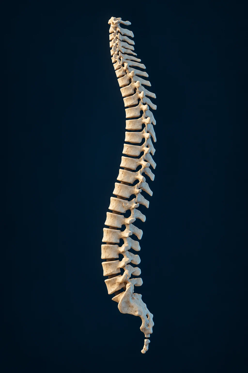
The sacrum is at the lower end of this retained whole-column illustration. The small scale is appropriate for position; it cannot establish nerve-root courses, exact segment counts or an individual cord ending.
Retained native-guided AI-assisted spine illustration · CC BY-SA4.0. BodyParts3D/DBCLS, Z-Anatomy and the credited FBX preparation inform the source-guided family; methods and licence context below.
In adults the spinal cord usually ends in the upper lumbar region, with individual variation. Descending lumbar, sacral and coccygeal nerve roots continue below it as the cauda equina. The bony sacral canal is a continuation of the vertebral canal, not evidence that spinal cord tissue reaches the coccyx.
Anterior and posterior roots join to form a spinal nerve. That nerve then divides into anterior and posterior primary rami. Anterior rami contribute to pelvic and lower-limb pathways; posterior rami supply posterior regional tissues. Root and ramus are not interchangeable labels.
The bone illustrations deliberately do not invent a complete hidden nerve map. The anterior and posterior foramina can be named from gross bone landmarks without claiming that a generated rootlet, vessel or individual clinical level has been verified.
Teaching references: UTHealth · Anatomy of the spinal cord · UAMS · Lower-limb bone anatomy table
DEEPER LEARNING / TISSUE AND VARIATION
A gross surface is one scale of a living structure.
A cortical shell and internal trabecular bone contribute to the sacrum’s mechanical behaviour. The surface texture in the teaching plates is illustrative; it is not a measurement of bone density, age or disease.
The usual five-segment description is an orientation, not a guarantee for every person. Transitional anatomy and fusion patterns can change the apparent boundary between lumbar spine and sacrum. Clinical level numbering requires appropriate individual imaging and professional interpretation.
Sex-labelled source models describe their particular source specimens. They do not establish an individual’s function, pelvic dimensions or reproductive capacity, and separate source datasets must not be treated as perfectly aligned specimens.
Teaching references: OpenStax · Vertebral column · OpenStax · Pelvic girdle and pelvis
CONNECTED LEARNING / PRESERVE THE ORIGINAL REFERENCE
Keep the preparation, selected component and surrounding teaching distinct.
A male sacrum representation and the separate female skeleton reference are different source specimens. A compound source mesh that includes the coccyx is labelled as related sacrum knowledge, not an exact sacrum-only match. No individual source opening is assigned a clinical label without verification.
The Return control above retains the originating reference and selection. The illustrations explain normal gross anatomy; they do not replace the selected mesh or add unseen nerves to it.
Typically five sacral vertebrae unite to form one adult sacrum. Its broad upper base meets L5; its lower apex meets the coccyx. The two lateral surfaces articulate with the ilia at the sacroiliac joints. Individual anatomy and fusion patterns vary.
Sacroiliac joint surfaces and strong ligaments help transfer load from the trunk into the pelvis. The joints allow limited movement; they are not freely mobile hinges, and describing them as completely motionless is misleading. Posture and loading involve the whole pelvic ring.
The sacral canal continues the vertebral canal. There are normally four pairs of anterior sacral foramina and four pairs of posterior sacral foramina, providing passages for sacral nerve branches and vessels. Anterior and posterior foramina should not simply be relabelled as anterior and posterior nerve roots.
The adult spinal cord usually ends in the upper lumbar region. Descending nerve roots continue below it, with sacral nerve branches reaching pelvic and lower-limb targets. Bone-only geometry cannot establish the position of those nerves or a person’s neurological condition.
A male sacrum representation and the separate female skeleton reference are different source specimens. A compound source mesh that includes the coccyx is labelled as related sacrum knowledge, not an exact sacrum-only match. No individual source opening is assigned a clinical label without verification.
Normal gross sacral anatomy, limited sacroiliac movement and the distinction between bone and neural structures. Existing reference-book inventories FB020–021 and saved spine/bone research guide topic organisation; a fresh raw-book page review has not been completed for this upgrade.
Adult cord typically ends near L1-L2; descending roots continue below it as the cauda equina. Targeted introduction and longitudinal organisation checked.
Gross sacral features and the vertebral-column context.
Anterior/posterior foramina, primary rami, canal, auricular surfaces and hiatus.
The sacrum within a ring formed with the hip bones; articular and ligament context.
Publisher abstract checked: limited joint movement and differences between measurement methods; no universal normal angle is inferred.
New isolated-bone and pelvic-ring plates are original AI-assisted medical teaching illustrations, not scans or specimen photographs. Medical labels, directional cues and short descriptions are separate web layers. Their gross landmarks are checked; fine generated microstructure and hidden attachments are not independently certified. The SI movement source was checked as a publisher abstract, not the full restricted paper.
The retained whole-column illustration belongs to the existing native-guided spine family, CC BY-SA4.0: BodyParts3D/DBCLS (CC BY-SA2.1Japan), Z-Anatomy (CC BY-SA4.0) and FBX preparation credited to Lluis Vinent Juanico. See CC BY-SA4.0 and Z-Anatomy. It is a teaching illustration, not a new scan. Original masters and provenance are preserved.
The original NCBI Bookshelf Sacrum source remains in the starting notes; its fresh direct page required a verification challenge and was not bypassed. Human medical review and physical-phone acceptance have not been performed. No diagnostic, injection, operative or patient-specific interpretation is provided.