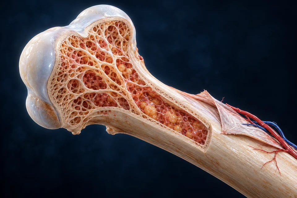
Retained original text-free adult bone illustration, not a scan. Generic structure, not an identified specimen or side. No open growth plate is shown. Anatomical names and explanations are editable layers.Structure notes · further context
Read the three scales
The whole organ carries loads. Matrix architecture distributes material. Living cells maintain and renew that matrix; the enlarged views below are generated teaching preparations, not acquired microscopy.
SupportA dense shell and inner struts share loads.
RenewDifferent cells remove, form and maintain matrix.
MoveCartilage, fluid and a capsule make a living interface.
30 seconds: locate the cooperating tissues. Three minutes: compare turnover, growth and the joint below. Deeper reading: retain the complete normal foundation and its sources. No playable video is presented.
02 / LONG-BONE
A bone is more than a dry mineral shape.
Dense cortex, internal trabeculae, marrow, vessels and connective coverings occupy different compartments. A smooth articular cap forms a different interface at a joint.
New original Golden-family text-free teaching preparation. Representative adult long-bone relationship, not an exact named bone, sex, side or individual scan. Broad articular, cortical, trabecular, marrow and vascular compartments are source-supported. Fine generated vessel branches, pores, marrow-cell identity and tissue thickness remain illustrative / not independently verified. No open growth plate is asserted. Human medical review: Not performed.Structure notes · further context
Articular cap
This surface is not covered by periosteum. Its thickness here is illustrative, not a measured cartilage layer.
Trabecular interior
Spongy bone is living bone, not soft sponge. Individual strut directions in this rendering are not a verified loading map.
Cortical shell
Thickness varies by site and individual. Cortical and trabecular organisation contribute together to support.
Marrow cavity
Fat-rich yellow marrow and blood-forming tissue have regional and age-related distributions; every adult bone end is not universally red marrow.
Periosteal covering
Its fibrous and cellular context contributes to attachment, nourishment and surface growth. The peeled flap is a teaching cutaway.
Bone blood vessels
Marrow, cortical and periosteal routes communicate in a more complex network than the visible branches. Blue is an educational venous convention.
The end of a long bone is an epiphysis; the shaft is the diaphysis. The metaphyseal region links them. The adult teaching model does not assert an open growth plate.
Endosteal tissue lines internal bone surfaces; periosteum covers non-articular outer surfaces. A joint cartilage cap should not be described as periosteum.
A skeleton is a connected framework of living organs.
The axial framework includes skull, vertebral column and thoracic cage. Limbs and their girdles form the appendicular framework; shape, tissue and joints together determine support and movement.
Axial context
Head → spine → rib cage
Supports and protects central structures while permitting selected movements.
Skull, spine and thorax each need their own regional atlas.
Appendicular context
Shoulder / pelvic girdles → limbs
Transfers loads and provides levers for movement.
A generic long-bone plate is not a complete limb or pelvis map.
Different bone shapes
Long, short, flat and irregular examples
Shape descriptions help explain different structural roles.
Long is a shape category, not a requirement that a bone be large.
This is a normal bone-and-joint foundation, not a complete whole-body skeletal atlas. Existing source-native atlas references retain their coverage and review limits.
Is every small bone a short bone?
Shape names do not simply describe size. A finger phalanx belongs to the long-bone family; a carpal bone illustrates the short-bone pattern. Flat bones may be curved, while vertebrae have an irregular form.
Sesamoid bones add a fifth category: they develop within tendons, with the patella as a familiar example. This matters because a small sesamoid and a small carpal do not have the same structural relationship to the surrounding tissues.
Collagen-rich organic matrix and mineral crystals contribute different mechanical properties. Living cells maintain the composite instead of letting it behave as one uniform piece of stone.
Organic matrix
Collagen-rich extracellular framework
Contributes tensile resilience and material organisation.
It is not a soft removable wrapping around all mineral.
Mineral component
Calcium-phosphate-rich crystals within matrix
Contributes stiffness and resistance to compression.
No simplistic calcium-carbonate reaction or exact generated crystal chemistry is claimed.
Architecture
Cortex + trabeculae + local orientation
Places material where support and internal tissue coexist.
Density alone does not completely describe strength or every loading response.
A hard shell: why call it living tissue?
The large preparation shows where material is placed; the enlarged surface view shows the cells that maintain it. Collagen and mineral belong to a shared matrix, rather than two removable shells. Mineral contributes hardness, while the collagen framework adds flexibility.
Marrow has another job: it houses blood-cell production in suitable regions. A bone therefore supports movement, stores mineral and contains living tissue at the same time. Its appearance alone cannot establish the quality of an individual person’s bone.
Mechanism references: NIAMS · What is bone? · Facts checked 2026-10-18. AI-assisted source check, not physician review.
Vascular routes supply bone cells and marrow. Blood-forming marrow and fat-rich marrow differ in their regional and age-related distribution; internal spaces are not normally empty holes.
Blood supply
Periosteal, nutrient and other regional vascular routes
Supports living cortex, internal tissue and marrow.
No complete vascular branching pattern is asserted by the image.
Marrow context
Selected axial and long-bone internal spaces
Blood-cell production occupies suitable marrow niches.
Every adult long-bone end is not guaranteed to contain only red marrow.
Nervous / connective context
Vascular routes and outer/internal surfaces
Living support includes more than mineral and one blood vessel.
Exact innervation is staged, not inferred from generated fibres.
An osteon puts a vascular route inside layered material.
Many cortical regions contain concentric lamellae around a central canal. Embedded osteocytes communicate through tiny canalicular surroundings, while larger channels carry blood.
New original Golden-family text-free teaching preparation. Source-supported osteon, lamellar, lacunar and canalicular relationships at an enlarged educational scale. Fine generated processes, lacunar size, mineral texture and exact cellular identity remain illustrative / not independently verified. Canaliculi are not blood vessels, and not all bone is built from complete osteons. Human medical review: Not performed.Structure notes · further context
Central canal
A central canal runs along the unit; the rendering shows selected vessels, not every nerve or exact individual lumen.
Concentric lamellae
Collagen and mineral arrangement supports the material. Individual generated rings are not measured histological lamellae.
Lacuna / osteocyte context
The enlarged profile is illustrative. Ground-bone preparations may show empty lacunae because organic material was removed.
Canalicular network
They support communication and exchange between embedded cells and nearby fluid environments; they do not carry red blood cells.
Perforating canal
These differ from the much smaller canaliculi. The selected channel is not a complete three-dimensional vascular reconstruction.
Between osteons
Remodelling changes cortical architecture. A neat block is not the exact appearance of every human bone region.
Cortical bone also includes other lamellar arrangements and remnants of remodelling. Trabecular bone is not simply a collection of miniature complete osteons.
A perforating canal connects vascular routes across cortical organisation. A canaliculus is much smaller and contains cell-process context, not a red blood vessel.
Formation, sensing and removal are different cellular jobs.
Several related and distinct cell populations coordinate matrix maintenance. The comparison below teaches selected roles rather than identifying cells from generated colour.
Osteoblasts
Bone-forming surfaces
Secrete organic matrix and support its mineralisation.
A mesenchymal-lineage cell; it does not fuse with an osteoclast precursor to create an osteoclast.
Osteocytes
Within mineralised bone matrix
Embedded cells communicate through processes and contribute to sensing and regulation.
An enlarged generated profile is not proof of a measured mechanosensory response.
Osteoclasts
Selected resorbing bone surfaces
Multinucleated cells remove bone during coordinated turnover.
Arise from haematopoietic monocyte/macrophage-related precursors, not osteoblast fusion.
Bone-lining cells
Selected resting bone surfaces
Participate in surface maintenance and communication.
Related to the osteoblast lineage; not a separate layer identical at every surface.
Chondrocytes
Cartilage matrix
Maintain specialised cartilage surroundings.
Cartilage and bone cells are not interchangeable simply because both support the skeleton.
Turnover is coordinated, not demolition without replacement.
Selected surfaces undergo resorption, preparation, matrix formation and mineralisation. The balance and timing depend on local and systemic signals rather than a fixed universal daily schedule.
Original generated 3D teaching preparation, not microscopy. Selected resorption and formation contexts are brought together for comparison, not a universal spatial conveyor or a measured time sequence. Fine processes, boundaries and proportions remain illustrative.Structure notes · further context
Two different lineages
Osteoclasts arise from haematopoietic monocyte/macrophage-related precursors. Osteoblast-lineage cells form bone; an osteoclast does not transform into an osteoblast.
Coupling is communication
Signals coordinate removal and formation across local contexts. The illustration does not show all signals or prove a fixed interval between the two jobs.
Remove selected material
The resorbing-cell context is at the left surface. An osteoclast removes bone locally; the entire bone is not being dissolved at once.
Gold outlines identify selected teaching locations in the full view. They do not measure activity, speed or a universal sequence; the narrow windows retain the same anatomical targets.
Mechanism comparison · all contexts
Resorption
Selected bone surface → removal by osteoclasts
Removal is a specialised cellular job, not a marrow cavity opening at random.
Preparation
Resorbed site → coordinated surface transition
Communication helps connect removal with new formation.
Formation
Osteoblast activity → organic matrix
New matrix is not identical to immediately mature mineralised bone.
Mineralisation
Matrix → mineral contribution over time
The cycle is not a universal fixed-duration clock.
Supporting mechanism comparison, not exact anatomical geometry or a measured time course. All explanations remain available without changing the selected context.
Supporting sequence beside the anatomy and cell comparisons. Steps overlap across sites; no cell count, turnover percentage or individual rate is measured.
Does a resorption bay mean the whole bone is failing?
A local removal surface is part of normal renewal. To judge the overall result, removal must be considered together with formation. The resorbing cell at the left and forming cells at the right illustrate different jobs, not a verdict about the strength of the entire bone.
The balance can change with age and biological context. The picture does not show an individual turnover rate, a fixed schedule or a guarantee that replacement always matches removal.
Mechanism references: NIAMS · What is bone? · Facts checked 2026-10-18. AI-assisted source check, not physician review.
Growth and adult remodelling are related but different.
Many bones begin through a cartilage template; others develop bone more directly within connective tissue. Longitudinal growth involves specialised growth-plate regions before maturation.
Original generated teaching views of a growing bone, not a mature adult bone or a measured tissue section. The larger panel explains broad regional order; cell fate, exact zone thickness and fine cartilage–bone boundaries are not certified by generated texture.Structure notes · further context
Growth is not adult turnover
Growth plates contribute to lengthening before maturation. Adult bone continues surface modelling and remodelling after plates have closed.
Not one universal fate
Lineage evidence is more complex than a claim that every hypertrophic chondrocyte always dies or always becomes a bone cell; much experimental evidence comes from animal models.
First locate the growth plate in the bone.
Original generated teaching views of a growing bone, not a mature adult bone or a measured tissue section. The larger panel explains broad regional order; cell fate, exact zone thickness and fine cartilage–bone boundaries are not certified by generated texture.Structure notes · further context
Growth is not adult turnover
Growth plates contribute to lengthening before maturation. Adult bone continues surface modelling and remodelling after plates have closed.
Not one universal fate
Lineage evidence is more complex than a claim that every hypertrophic chondrocyte always dies or always becomes a bone cell; much experimental evidence comes from animal models.
Then read the tissue from epiphyseal to metaphyseal side.
Original generated teaching views of a growing bone, not a mature adult bone or a measured tissue section. The larger panel explains broad regional order; cell fate, exact zone thickness and fine cartilage–bone boundaries are not certified by generated texture.Structure notes · further context
Growth is not adult turnover
Growth plates contribute to lengthening before maturation. Adult bone continues surface modelling and remodelling after plates have closed.
Not one universal fate
Lineage evidence is more complex than a claim that every hypertrophic chondrocyte always dies or always becomes a bone cell; much experimental evidence comes from animal models.
Find the growth region
The small organ view locates an open growth plate below the epiphysis. In the larger tissue view, epiphyseal-side cartilage lies above the metaphyseal replacement region.
Gold outlines identify selected teaching locations in the full view. They do not measure activity, speed or a universal sequence; the narrow windows retain the same anatomical targets.
Mechanism comparison · all contexts
Cartilage-template context
Developing cartilage → specialised growth regions
Many bones use endochondral development; not all bones begin identically.
Lengthening context
Proliferation / enlargement → matrix change and replacement
Human lineage outcomes are not reduced to one universal cell fate.
Maturation / shape
Growth regions change → adult modelling and remodelling
Closure ages vary. The adult main plate does not show an open growth plate.
Supporting mechanism comparison, not exact anatomical geometry or a measured time course. All explanations remain available without changing the selected context.
Growth-plate proliferation, enlargement and matrix changes support lengthening; bone replaces much of the cartilage template as vessels and bone-forming activity enter suitable regions. Surface apposition and modelling also change shape.
Closure timing varies among bones and people. Modern lineage research shows more complex chondrocyte outcomes than one universal fate, with much evidence from animal models; every human cartilage cell is not simply declared to die or turn into bone.
Loading, hormones and mineral balance influence living bone.
Local sensing interacts with systemic regulation. Calcium and phosphate exchange links bone with kidneys, intestine and endocrine signals; this is not a supplement or exercise prescription.
Mechanical context
Loading → local cell communication → structural adaptation
Osteocytes participate in sensing and coordination.
This chapter does not prescribe a universal safe load or claim every adaptation is identical.
Hormonal context
Growth, sex-hormone and calcium-regulating signals
PTH and other signals influence formation and resorption through cellular communication.
An osteoclast is not directly controlled by one arrow in every situation.
Mineral balance
Intestine ↔ blood ↔ kidneys ↔ bone
Organs coordinate availability and exchange of calcium and phosphate.
Bone is one participant, not a standalone thermostat.
Articular cartilage contains chondrocytes in hydrated matrix. Superficial, intermediate, deep and calcified-interface regions contribute different relationships to the smooth surface and bony support.
New original Golden-family text-free teaching preparation. A source-informed educational cartilage–bone interface, not a microscope section or pathology sample. Broad surface/middle/deep/calcified-interface/subchondral relationships are taught. Fine generated cell alignment, fibres, boundary thickness and exact tidemark remain illustrative / not independently verified. No blood vessel enters the uncalcified cartilage. Human medical review: Not performed.Structure notes · further context
Fluid-facing surface
Articular cartilage has no covering perichondrium. The bright upper film is an educational fluid context, not a measured fluid thickness.
Superficial region
Collagen organisation helps resist surface stresses. The illustration does not verify each fine fibre direction.
Intermediate region
Chondrocytes maintain the surrounding matrix. Their colour does not establish a molecular activity state.
Deep region
This broad orientation varies with site, age and cutting plane. Columns are not blood-vessel channels.
Calcified interface context
The exact tidemark and calcified-layer boundary are not independently verified in this generated image; use it for regional position, not measurement.
Subchondral bone
Vessels in the bone are distinct from the avascular uncalcified cartilage. Nutrient routes depend on region and tissue context.
Uncalcified articular cartilage is avascular and has no covering perichondrium. Diffusion and loading-related fluid movement contribute to exchange; exact nutrient routes vary with region and osteochondral context.
The enlarged plate separates broad regions for teaching. It cannot independently verify each cell, fine fibre, tidemark or boundary thickness and should not be read as a diagnostic tissue section.
A hydrated matrix combines collagen organisation with proteoglycan-related water interactions. Loading changes fluid and solid-matrix contributions over time rather than acting on a dry frictionless tile.
Low-friction interface
Opposing smooth surfaces + joint fluid
Helps surfaces move while load is distributed.
No exact friction coefficient is measured in the rendered plate.
Joint construction and movement are separate classifications.
Fibrous, cartilaginous and synovial describe how bones connect. Functional terms describe movement, so the two classification systems should not be treated as identical labels.
Fibrous
Connective tissue joins bones without a synovial cavity
Examples include skull sutures and selected ligamentous connections.
Movement varies; not every fibrous connection is equally immobile.
Cartilaginous
Cartilage participates in the connection
Examples include growth-related synchondroses and fibrocartilaginous symphyses.
Not every cartilage-containing structure is a synovial articular cap.
Synovial
Cartilage-capped surfaces within a capsule and cavity
Supports varied movements through different surface shapes.
An anatomical joint family does not alone give its exact range.
Synovium lines the envelope, not the contact cartilage.
A fibrous capsule surrounds the joint. Vascular synovial tissue occupies non-articular inner surfaces; smooth cartilage and a narrow fluid-containing cavity form the opposing interface.
New original Golden-family text-free teaching preparation. Generic synovial relationship, not an exact knee, hip, ligament map or measured joint gap. Broad cartilage/cavity/capsule/synovium/subchondral relationships are source-supported. Fine generated vessels, fibres, attachments and tissue thickness remain illustrative / not independently verified. A schematic process supplements rather than replaces this anatomy. Human medical review: Not performed.Structure notes · further context
Fibrous capsule
It contributes mechanical support together with surrounding tissues. Capsule thickness and attachment vary among joints.
Synovial lining
It does not coat the opposing articular cartilage. Synovial tissue helps maintain the fluid environment.
Opposing cartilage
Cartilage reduces friction and distributes loads with its hydrated matrix; it is not merely dry paint on bone.
Joint cavity / fluid
The cutaway separates surfaces for explanation. Real fluid volume and the three-dimensional space are not measured here.
Trabecular bone context
Trabecular spaces contain living tissue. The generated pattern is not a precise model of load-bearing alignment.
Subchondral support
Its interface with calcified cartilage links two different materials. The detailed boundary remains illustrative.
Capsular attachment context
This marks the broad capsule–bone relationship, not a verified enthesis, named ligament or exact attachment boundary. Reinforcing fibres remain illustrative.
Synovial fluid includes a plasma-derived component and substances secreted locally, including hyaluronan-related contributions. It is not simply blood or a cell-free layer painted over bone.
Ligaments, tendons, muscles and bone shape contribute to stability. The generic external band is not a named knee ligament, and the image does not claim every joint has identical internal structures.
Capsule, lining and cartilage: why keep them separate?
Follow the joint image from outside inward. The fibrous capsule contains the articulation. Synovial lining belongs on non-articular inner surfaces. Opposing cartilage caps face a fluid-containing space; they are not one bridge joining the bones.
Neighbouring structures can serve different jobs. A meniscus or articular disc is an additional feature of selected joints, not a universal layer. A bursa or tendon sheath reduces friction around moving tissues; neither is another name for the main joint cavity.
Surface shape guides movement without replacing soft-tissue control.
Plane, hinge, pivot, condyloid, saddle and ball-and-socket examples describe broad geometry. Real movements combine bone shape, capsule, connective tissues and muscle activity.
Mechanism comparison · all contexts
One dominant movement
Hinge / pivot examples → selected bending or rotation
These broad categories do not reconstruct every joint axis.
Two-plane context
Condyloid / saddle examples → combined motions
Surface form and soft tissues together guide movement.
Multi-directional context
Ball-and-socket example → varied movement directions
Freedom is constrained by stability and surrounding tissues.
Supporting mechanism comparison, not exact anatomical geometry or a measured time course. All explanations remain available without changing the selected context.
Supporting movement comparison; no exact joint-axis reconstruction or fixed degrees. The knee, for example, is not perfectly described as a simple uniaxial door hinge.
Can the name of a joint predict every movement?
A shape category describes useful possibilities, not a complete movement map. Compare the shoulder and hip: both are ball-and-socket joints, yet their sockets and surrounding support produce different mobility and stability.
Ligaments constrain movement; muscles and tendons also contribute to support. Use the generic preparation to identify the shared interface, then follow a regional lesson for the actual bones and attachments. No exact axis or range can be measured from this illustration.
Movement links bone with muscle, sensation and circulation.
Muscle force reaches bone through attachment systems. Sensory information helps coordinate position and movement, while vascular and marrow tissues connect skeletal organs with wider body functions.
Force transfer
Muscle → tendon / attachment → bone → joint
Levers and stabilising activity make movement possible.
A complete regional muscle atlas is separate work.
Sensory coordination
Joint / muscle / tendon context → nervous system
Position and movement depend on multiple sensory inputs.
No exact receptor map is inferred from generated texture.
Living tissue exchange
Vessels / marrow ↔ broader circulation and immune context
Support and movement coexist with tissue maintenance.
An isolated bone model does not teach the whole body alone.
Preparation changes what a microscope can actually show.
A ground section, decalcified section and developing-tissue section preserve different evidence. Species, age, cutting plane and processing must be known before assigning structures or cells.
Human ground bone
Duke0070_A transverse and0071_A longitudinal records
Useful descriptions for canals, lamellae and empty lacunar spaces.
Organic material is not preserved; this is not an intact living-cell view.
Compare different cartilage contexts and preparations.
Epiglottis, trachea and disc are not automatically articular cartilage.
Developmental records
UCSF61 human fetal finger; other records include animal examples
Development requires a known specimen and stage.
The0301_A fetal-finger description explicitly lacks an epiphyseal plate; guinea-pig joint examples are not human.
Historical source record: the following note concerns the Duke slide descriptions listed above. The independently acquired Berkshire photograph below is a separate source.
Record descriptions inspected; no slide pixels or patient images acquired. Original rendered plates are educational anatomy, not microscopy or clinical imaging.
The regional skeleton remains a set of distinct teaching jobs.
Skull, spine, thorax, pelvis, hand and foot need different views because their shapes and relationships solve different problems. This tissue-and-joint foundation does not pretend those branches are complete.
Skull / spine
Protection, openings and segmental support
Future regional plates need recognisable sutures, vertebral features and surrounding relationships.
Not replaced by repeating the generic long bone.
Thorax / pelvis
Protection, breathing framework and load transfer
Future plates should show region-specific connections and neighbouring organs.
No forced donor alignment or substituted sex-specific geometry.
Limbs / hands / feet
Lever systems, dexterity and stance
Future views should show distinct joints, attachments and architecture.
Existing partial or source-native 3D references retain their provenance and limits.
Structure, living cells, matrix turnover, growth, regulation and joint interfaces now connect across scales. Fracture management, arthritis, diagnosis and treatment remain later-phase work.
Now taught
Organ → bone tissue → selected cells → joint interface → normal mechanism
Four distinct realistic plates and visible supporting comparisons teach different jobs.
Source-supported stated scope; human medical review not performed.
Still staged
Regional atlases, licensed microscopy, exact biomechanics and clinical branches
These need their own sources, visual jobs and acceptance.
No invented completeness percentage or full skeletal-system claim.
Broad adult long-bone, osteon, cartilage and generic synovial-joint relationships; selected bone-cell roles, matrix turnover, growth, regulation, joint families and movement context. Authoritative references are used critically rather than repeating every simplified or historical claim.
Explicit limits
AI-assisted visual/content production. Fine generated cellular, vascular, canalicular and matrix details remain illustrative / not independently verified. Human medical review: not performed. Educational only; not diagnostic, clinical or surgical material.
Revision: bone-joints-english-2026-10-02-deep-v1. Stable local key: ahb_ch_bone_joints_english_v1. No invented canonical CMS/atlas entity or 3D dataset. Five retained qualified text-free preparations plus two new normal teaching masters use separate semantic anatomical targets and language copy. Original twenty-five labels and their detailed explanations are retained. Generated fine cellular, canalicular and matrix topology is not acquired microscopy, a measured growth plate or an exact joint reconstruction. Migration physical-phone acceptance: Pending, non-blocking.