Follow sound from air to fluid to a sensory signal. Then explore how neighbouring organs sense head motion and work with vision and body-position information.
Chapter overview film planned. No playable video is currently available.
Original anatomical exhibit · fine generated detail illustrative
02 / ORGAN IN CONTEXT
Locate the compartments before following the signal.
The canal ends at the tympanic membrane. An air-filled middle cavity couples to an inner-ear fluid system within the temporal bone.
Air-pressure changes reach a moving boundaryFollow the canal to the eardrum. The amber cue stops at this boundary; it is not a stream of air entering the cochlea.
Original Golden-family source-informed preparation. Representative combined educational view, not a patient side-specific section or surgical specimen. External canal, tympanic boundary, middle space, auditory-tube destination and inner-ear context are taught. Fine generated ossicle, nerve, vascular, bone-pore and duct details remain illustrative / not independently verified. No air stream enters the inner ear. Human medical review: Not performed.Structure notes · further context
Auricle
Its cartilaginous contours vary; this is not a measured patient pinna.
External canal
It is distinct from the deeper auditory tube.
Tympanic membrane
The attached mechanical chain transmits motion; the membrane is not a cochlear sensory epithelium.
Middle-ear space
An enlarged separate plate identifies the three selected bones and two windows.
Auditory tube
The distal pharyngeal opening is outside this view; this is not an inner-ear duct.
Cochlear context
The next section teaches compartments at a different scale; this surface does not prove cellular anatomy.
Air pressureCanal → moving eardrum
Coupled motionOssicles → inner-ear interface
Sensory responseCochlear tissue → separate afferents
Amber / cyan / green cues distinguish mechanical input, coupling and sensory context. Other outlines indicate anatomical location only, not measured flow, voltage, firing or a reconstructed tract.
Supporting relationship map · compare the connections
Supporting mechanism · educational relationships, not an anatomical plate or measured trace. All basic explanations stay visible.
Air pressure
Canal → tympanic movement
Pressure variation supplies the initial mechanical input.
Air is not sent through a nerve.
Mechanical coupling
Ossicles → stapes interface
Connected bones transmit movement between interfaces.
Air-pressure variation moves the eardrum and ossicles. Inner-ear fluid mechanics then drive a sensory response; air itself does not travel through the cochlear nerve.
The auditory tube links the middle ear with the nasopharynx. Ventilation and pressure relationships differ from the stapes-to-inner-ear mechanical interface.
Auditory tube
Middle cavity → nasopharynx
Provides a route for ventilation and pressure equalisation.
Not an inner-ear duct or open passage to brain tissue.
Tympanic boundary
Canal / middle cavity
Pressure on its two sides affects mechanical behaviour.
Three distinct bones bridge the membrane and window.
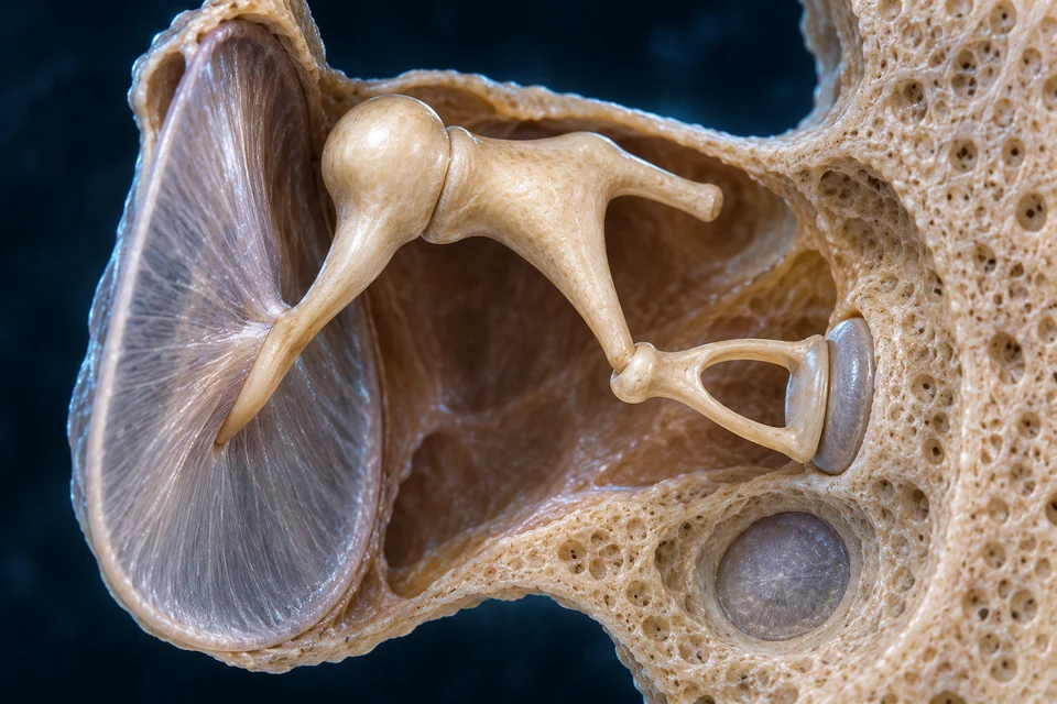
Malleus, incus and stapes form a linked mechanical chain. Their connections matter more than the familiar hammer, anvil and stirrup nicknames.
Original Golden-family source-informed preparation. Representative enlarged middle-ear teaching view. Three bone identities and separate mechanical window relationships are taught. Fine generated joints, ligaments, bone dimensions, membrane thickness and operative wall anatomy remain illustrative / not independently verified. No fixed amplification ratio or drain is depicted. Human medical review: Not performed.Structure notes · further context
Malleus
Its body articulates with the incus; the view does not verify every ligament or exact attachment extent.
Incus
A long process reaches the stapes head through the incudostapedial connection.
Stapes
Its two crura surround an opening; the footplate is at the oval window, not the round window.
Tympanic membrane
It separates the canal from the middle cavity; nerve impulses are not generated inside this membrane.
Oval-window interface
No open air channel through the footplate is taught.
Round-window membrane
It is not the next nerve station or a drainage hole.
The stapes footplate occupies the oval window. The separate round-window membrane permits displacement in the fluid system; it is not a drain or a neural relay.
The enlarged plate shows their separate positions. These are closed tissue interfaces; movement transmits mechanical energy without an air jet flowing into the inner ear.
No exact amplification ratio, displacement measurement or operative window anatomy is asserted.
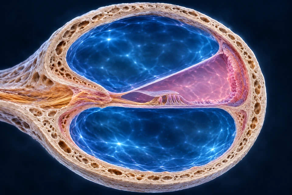
The cochlear duct lies between two perilymphatic spaces. Their membranes and ionic environments matter as much as their location.
A transverse turn separates three fluid spaces.
The cochlear duct lies between perilymphatic spaces. Membranes and lateral tissue define its environment; names cannot be assigned from blue colour alone.
Original Golden-family source-informed preparation. Representative enlarged turn; dimensions and individual shape are not measured. Three scala relationships and separating membranes are taught. The excessive generated sensory ridge was locally corrected to a representative one-inner/three-outer profile; exact bundle attachments and fine cell identity remain illustrative. The controlled supporting inset teaches selected cellular relationships. Fine generated bundle attachment, synapse, cell subtype, ganglion and vascular detail remain illustrative / not independently verified; the editable cellular inset clarifies selected relationships without claiming histology. Human medical review: Not performed.Structure notes · further context
Scala vestibuli
It is distinct from the endolymphatic cochlear duct.
Scala tympani
Fluid-space names do not imply that these are blood vessels.
Cochlear duct / scala media
The organ of Corti lies within it on the basilar membrane; the whole cochlea is not inside this duct.
Vestibular / Reissner membrane
Its exact thickness and cellular lining remain illustrative.
Basilar boundary
The controlled supporting inset distinguishes cell populations and the tectorial relationship.
Lateral-wall context
This location provides strial context, not a verified map of every generated epithelial or vascular element.
Different compartments sustain different conditions.
Endolymph in the cochlear duct differs from perilymph in the surrounding scalae. Lateral-wall tissue contributes to the environment supporting sensory transduction.
Scala vestibuli / tympani
Outside the cochlear duct
Perilymphatic compartments participate in cochlear mechanics.
Not veins supplying the sensory cells.
Scala media
Cochlear duct
Contains the endolymphatic environment.
Not an open mixing channel joining all three spaces.
Lateral wall
Stria / supporting tissue
Helps maintain the ionic and electrical setting.
The rendered fine cellular layers are illustrative.
Locate the sensory cells and supporting tunnel, then connect bundle movement to a receptor response and separate neuronal output.
A sensory epithelium sits on a mechanical foundation.
The organ of Corti rests on the basilar membrane within the cochlear duct. Hair cells, supporting cells and nearby membranes form a specialised unit.
Move the bundle, change the receptor membraneRelative mechanics act near the sensory apices. Deflection changes mechanically gated channels; the image does not identify individual channel proteins.
Original generated educational tissue preparation, not microscopy. Medial left, lateral right. The usual one inner row / three outer rows are represented across a transverse section, not a total cell count. Inner-bundle gap, outer-tip coupling and broad pillar/tunnel relationships are source checked. Fine membrane borders, proportions, individual fibres, synapses and molecular channels are not independently established; yellow endings cannot be assigned exact afferent/efferent identities from this image.Structure notes · further context
Tectorial membrane
It extends from medial tissue over the organ. Relative motion and the local fluid environment influence bundle stimulation; this is not a sheet of sensory neurons.
Inner hair cell
Inner hair cells provide the main sensory output to separate afferents. The displayed gap is a representative relationship, not proof that contact is impossible during every motion.
Outer hair-cell coupling
Three cells represent three usual outer rows. Voltage-dependent outer-cell motion contributes to cochlear amplification and tuning; no measured displacement or molecular motor is shown.
Tunnel of Corti
Supporting cells provide architecture around the sensory cells. This open space is not a blood vessel, nerve bundle or fourth fluid scala.
Basilar foundation
Movement of the basilar membrane interacts with supporting tissue and the sensory unit. Generated thickness and layered borders are not measurements.
Separate nerve endings
Hair-cell receptor potentials affect transmitter release; action potentials travel in separate afferent neurons. These gold profiles do not resolve synaptic ribbons or distinguish every feedback fibre.
Outlines locate the selected anatomical context; they do not measure firing, membrane current or a reconstructed neural tract.
Supporting relationship map · compare the connections
Supporting mechanism · educational relationships, not an anatomical plate or measured trace. All basic explanations stay visible.
The sensory apices face endolymph, while the basolateral cell environment is kept separate by the epithelial barrier. This separation supports the electrical driving force for transduction; the illustration does not display measured ion concentrations.
Bundle deflection changes mechanically gated channel activity. Potassium entry contributes to depolarisation, which can open basal voltage-gated calcium channels and change transmitter release onto separate sensory afferents. A hair-cell receptor potential is different from the action potentials propagated by those neurons.
Supporting cells provide the architecture around the receptors. Inner hair-cell bundles are not firmly embedded in the tectorial membrane; the tallest outer bundle tips couple to it. Outer-cell electromotility contributes active mechanics rather than duplicating the inner-cell output role.
Supporting relationship inset · further explanation
Source-supported supporting relationship inset, not primary anatomy, specimen histology, measured cell dimensions or exact membrane geometry. Labels and paths remain editable. Basic explanations stay visible.
Inner bundle / gap
Inner hair-cell stereocilia are not firmly embedded in the tectorial membrane. Local fluid and mechanical motion contribute to stimulation.
Outer bundle / coupling
Representative outer bundles contact the overlying membrane. One inner row and three outer rows describe the usual transverse organisation, not the total cochlear cell count.
Basal support / output
The unit rests on a basilar foundation. Separate afferents receive sensory output; no nerve action potential is drawn as travelling along a hair-cell axon.
The primary turn remains a realistic model. Its tiny generated bundle attachment cannot be independently established from the image; this controlled inset states the supported relationship. Fine tip links, synapses, cell packing and support-cell subtypes are not validated.
Sensory output and active mechanics are complementary.
Inner hair cells supply the main sensory output. Outer hair cells contribute to active cochlear mechanics; they are not simply another identical row of nerve cells.
Inner population
Medial side of Corti tunnel
Transmits sensory information to afferents.
Hair-cell receptor changes differ from afferent spikes.
Outer population
Lateral side
Electromechanical behaviour influences sensitivity and tuning.
Fine subtype, synapse and fibre counts are not verified here.
Mature cochlear bundles are stereocilia; no long kinocilium is added. The model is not an ion-current trace, synapse reconstruction or molecular animation.
Mechanical preference varies along the cochlea. Central comparisons combine information across frequencies, ears and experience.
Frequency preference changes along the spiral.
Higher-frequency peaks tend toward the basal region; lower-frequency peaks lie nearer the apex. Cochlear and neural processing preserve a frequency-related organisation.
Read position along the partition, not a change in the sound.
Basal · higher frequency preferenceApical · lower frequency preference
Near the base: the partition is relatively narrow and stiff.
Toward the apex: it becomes broader and more compliant.
This qualitative scientific comparison supplements the opened cochlear turn. It is not an anatomical cut surface, a calibrated curve or three exclusive receptor bins.
Supporting relationship map · compare the connections
Supporting mechanism · educational relationships, not an anatomical plate or measured trace. All basic explanations stay visible.
Uncoiled position comparison only: the direction does not mean that a higher sound frequency changes into a lower frequency.
Basal region
Higher-frequency preference
Mechanical properties influence where a response peaks.
Not a fixed frequency label for every cell.
Along the spiral
Position-dependent response
This is a coordinate comparison, not frequency becoming another frequency.
Real tuning is broader than three bins.
Apical region
Lower-frequency preference
The apical direction tends toward lower-frequency peaks.
No numerical audible range measured here.
An uncoiled coordinate supports the real cochlear context. No exact individual tuning curve, audible range or one-frequency-only receptor is asserted.
Cochlear nerve activity reaches brainstem processing and higher relays. Central auditory pathways have substantial bilateral organisation.
From cochlear afferents into brainstem processingCochlear nuclei receive the auditory nerve input. Other brainstem circuits compare information from both ears; this background locates the broad setting without inventing visible nuclei.
Original generated regional context, not MRI, a specimen or a traced auditory pathway. Two separate preparations: lateral cortex at one side; selected posterior exposure after cerebellar and other tissue removal at the other. Points identify broad regions only. Exact auditory nuclei, Heschl gyri, crossing fibres and radiations cannot be identified from these rendered surfaces.Structure notes · further context
Temporal cortical context
Connected cortical networks contribute to interpreting sound.
Exposed superior temporal plane
Auditory cortical regions lie within this broader anatomical setting.
Thalamic / diencephalic context
Auditory relay roles include the medial geniculate body, not resolved here.
Dorsal midbrain context
Collicular regions relate to ascending processing; exact nuclei are not outlined.
Lower brainstem context
Cochlear and binaural processing begins in brainstem circuits, not every point here.
A cortical setting for hearing
Original generated regional context, not MRI, a specimen or a traced auditory pathway. Two separate preparations: lateral cortex at one side; selected posterior exposure after cerebellar and other tissue removal at the other. Points identify broad regions only. Exact auditory nuclei, Heschl gyri, crossing fibres and radiations cannot be identified from these rendered surfaces.Structure notes · further context
Temporal cortical context
Connected cortical networks contribute to interpreting sound.
Exposed superior temporal plane
Auditory cortical regions lie within this broader anatomical setting.
Selected ascending regional context
Original generated regional context, not MRI, a specimen or a traced auditory pathway. Two separate preparations: lateral cortex at one side; selected posterior exposure after cerebellar and other tissue removal at the other. Points identify broad regions only. Exact auditory nuclei, Heschl gyri, crossing fibres and radiations cannot be identified from these rendered surfaces.Structure notes · further context
Thalamic / diencephalic context
Auditory relay roles include the medial geniculate body, not resolved here.
Dorsal midbrain context
Collicular regions relate to ascending processing; exact nuclei are not outlined.
Lower brainstem context
Cochlear and binaural processing begins in brainstem circuits, not every point here.
Outlines locate the selected anatomical context; they do not measure firing, membrane current or a reconstructed neural tract.
Supporting relationship map · compare the connections
Supporting mechanism · educational relationships, not an anatomical plate or measured trace. All basic explanations stay visible.
The left-to-right map organises the lesson; real population activity involves parallel and recurrent circuits, not exactly three serial cells.
Auditory organization includes parallel and bilateral relationships.
Three boxes are not a complete crossing map.
Interpretation
Auditory cortex / connected networks
Context and experience contribute to perception.
No one-ear-to-one-cortex exclusive cable.
Named relay roles and visible anatomy are different claims. The cochlear nuclei receive peripheral input; binaural circuits involve the superior olivary complex; ascending networks include the inferior colliculus and medial geniculate body before cortical processing. The regional preparation does not resolve those nuclei, show all crossings or trace auditory radiations.
Differences in timing and level between ears contribute to localisation. Outer-ear filtering and context also help; one cue is not a complete three-dimensional sound map.
Timing relationships
Two-ear input
Differences can contribute directional information.
No universal microsecond threshold measured here.
Level relationships
Head / ear context
Acoustic differences also help central interpretation.
No one-size-fits-all rule for every frequency and environment.
Different mechanical loads distinguish the sensors.
Start with the vestibular structures, then compare a crystal-loaded macula with a crystal-free cupula above a crista.
A neighbouring labyrinth responds to motion.
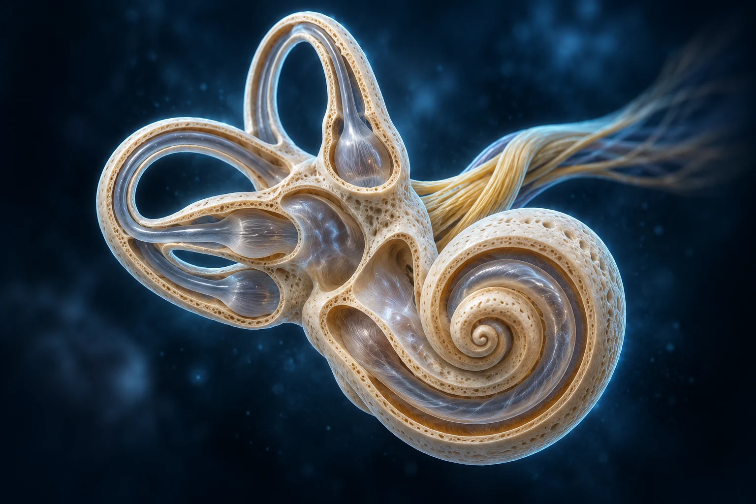
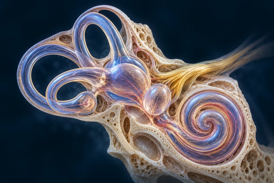
Semicircular ducts and otolithic organs are distinct vestibular components. Their sensory regions are enclosed within the inner ear.
Original Golden-family source-informed preparation. Representative opened labyrinth, not an exact individual duct-plane or surgical connection map. Selected duct, ampullary, utricular/saccular and adjacent cochlear context are taught. Fine generated common-crus/duct topology, sensory-patch extent, nerve branching and membrane detail remain illustrative / not independently verified. Controlled supporting insets separately show crista/cupula versus macula/otolithic relationships; no normal otoconia are placed in the cupula. Human medical review: Not performed.Structure notes · further context
Semicircular-duct context
Their three-dimensional orientations differ; this projection does not verify exact individual planes or all hidden connections.
Ampullary context
The supporting inset teaches these tissue interfaces; a generated bulge alone is not independently verified cellular anatomy.
Utricular context
Macular direction and exact shape vary; its tissue organisation is taught separately.
Saccular context
The teaching view is not an exact human polarity or connection map.
Adjacent cochlea
Sharing the inner-ear location does not make hearing and balance the same mechanism.
Neural-output context
No exact branch count, course or individual fibre is verified by these generated strands.
Cupular and otolithic interfaces carry different loads.
Canal cristae use a cupula; utricular and saccular maculas use an otolithic membrane with otoconia. Normal otoconia are not placed in the cupula.
Otolithic interface: dense otoconia load gel above a macula.
Rotation interface: a crystal-free cupula couples to a crista.
Tilt / translation · otolithic load
A dense layer gives the gel a load
Original generated vestibular tissue comparison, not a specimen or measured motion. Left and right are separate preparations at illustrative scales. Normal otoconia belong with the otolithic membrane, not the cupula. Broad gel/ridge/nerve relationships are source checked; fine receptor subtype, complete calyx, polarity map, crystal distribution, liquid motion and dimensions are not established by the image.Structure notes · further context
Otoconia
This load contributes to gravity-related and translational sensing. These representative grains are not a measured human crystal size or distribution.
Otolithic membrane
The utricle and saccule have differently oriented maculas. A single local preparation cannot represent the complete changing polarity across either organ.
Macular hair-cell region
Vestibular bundles include stereocilia and a kinocilium. Hair cells are interspersed with supporting cells; not every epithelial cell is a receptor.
Otoconia overlie the macular sensory epithelium. Gravity-related loading and translational acceleration can affect this interface, unlike the normal crystal-free cupula.
Supporting relationship map · compare the connections
Supporting mechanism · educational relationships, not an anatomical plate or measured trace. All basic explanations stay visible.
Tilt / translation
Mechanical loading changes
Gravity-related and translational effects influence the otolithic membrane.
A single peripheral input can be ambiguous.
Sensory pattern
Hair cells / afferents respond
Local directions and populations contribute information.
Not one universal bundle polarity.
Interpretation
Other cues / central processing
Combined inputs help distinguish movement context.
Animal evidence is not a measured human fibre map.
Changing rotation · cupula and crista
Changing rotation loads the cupular interface
Original generated vestibular tissue comparison, not a specimen or measured motion. Left and right are separate preparations at illustrative scales. Normal otoconia belong with the otolithic membrane, not the cupula. Broad gel/ridge/nerve relationships are source checked; fine receptor subtype, complete calyx, polarity map, crystal distribution, liquid motion and dimensions are not established by the image.Structure notes · further context
Cupula · no otoconia
Relative endolymph mechanics during changing rotation deflect the cupula and its sensory bundles. Normal cupulae do not carry the otoconial layer shown in the other preparation.
Crista ridge
Its role differs from the macular otolithic interface. The image is not a precise map of receptor orientation or Type I and Type II hair cells.
Separate basal nerves
Receptor changes alter signalling onto separate nerve fibres. Gold colour is an illustration cue, not evidence of a specific synapse, full calyx or measured neural activity.
Relative endolymph mechanics deflect the crystal-free cupula and sensory bundles at the crista. The ring locates tissue; it does not measure flow or deflection.
Supporting relationship map · compare the connections
Supporting mechanism · educational relationships, not an anatomical plate or measured trace. All basic explanations stay visible.
Motion changes
Relative duct / fluid mechanics
Changing rotation can displace cupular interfaces.
No calibrated head angle or flow.
Continued motion
Changing relative response
Mechanical and neural responses change during sustained rotation.
Not an unchanging rotation meter.
Stopping
A new relative change
Stopping can generate a different mechanical response.
No clinical balance test or timing promise.
Each sensor uses a native source window of the retained preparation. Its controls stay beside its tissue; no new microscopic detail or measured movement is invented. Numbers are local to each named view.
Both interfaces contain sensory hair cells and supporting tissue, but the material loading the bundles differs. Normal otoconia add mass to otolithic gel; normal cupulae are crystal-free. Motion of the surrounding endolymph can deflect the cupula during changing rotation.
Vestibular bundles include a kinocilium. Deflection toward or away from it changes receptor activity in opposite directions, but local orientations vary. Neither preparation is a complete polarity map or a calibrated simulation of head movement.
Supporting relationship inset · further explanation
Source-supported supporting relationship inset, not primary anatomy, specimen histology, measured cell dimensions or exact membrane geometry. Labels and paths remain editable. Basic explanations stay visible.
Otolithic interface
Dense otoconia load the otolithic membrane; gravity-related and translational effects change the sensory relationship.
Cupular interface
A cupula above crista hair cells is displaced by duct/fluid mechanics during changing rotation.
These sensory interfaces are not full labyrinths or exact human polarity maps. Vestibular hair bundles include a kinocilium; their uniformly drawn direction here is illustrative. Crystal distribution, three-dimensional support cells and response magnitudes are not measured. Normal cupula and otolithic membrane are not interchangeable.
Head motion is one input to a coordinated response.
Interpret rotation, gravity-related loading and changing sensory inputs together. Balance depends on more than a single organ.
Relative fluid motion helps signal head rotation.
During changing rotation, duct and fluid mechanics displace the cupula. Directional populations and paired organs contribute to the resulting activity.
Dense material gives a membrane a mechanical load.
Otoconia in the otolithic membrane contribute to gravity-related and translational responses. The utricle and saccule have differently oriented sensory regions.
Hair-cell orientations vary across a macula. A small teaching inset cannot stand for its exact polarity map or a measured human crystal distribution.
An otolith signal does not uniquely name its cause.
Tilt relative to gravity and translational acceleration can produce ambiguous peripheral input. Other sensory information helps the brain interpret movement.
Vestibular, visual and body-position information converge during orientation and movement. None becomes another sensory modality along a single serial chain.
Proprioception helps relate the body to its changing position.
These are parallel sensory contexts, not three links in a serial cable. Coordinated gaze and posture depend on central integration; the pictures do not reconstruct those connecting tracts.
Supporting relationship map · compare the connections
Supporting mechanism · educational relationships, not an anatomical plate or measured trace. All basic explanations stay visible.
The two left branches compare parallel inputs or alternatives. Their convergence groups a shared destination; one branch does not turn into the other.
Vestibular input
Motion / orientation relationships
The labyrinth contributes selected mechanical input.
Not the only balance sense.
Visual + body input
Visual + body-position input
Eyes, muscles and contact supply complementary information.
Connect anatomical teaching with tissue observations and functional evidence while keeping their methods and limitations distinct.
Different observations reveal different scales.
Anatomical sections locate compartments; tissue observation resolves epithelial units; physiological recordings examine responses. A generated rendering is none of these measurements.
Gross / section view
Regional organisation
Shows compartments and mechanical interfaces.
Not patient CT or an operative specimen.
Tissue / cell view
Sensory epithelium
Can reveal cell/support/bundle relationships.
No human micrograph is fabricated or relabelled.
Functional observation
Changing activity
Investigates mechanics or neural response.
Supporting model symbols are not recordings.
Deeper learning · when normal interfaces no longer work well
Hearing can change at different levels: inner-ear sensory structures, middle-ear mechanics and pathways toward the brain. Age-related hearing loss is often gradual and can involve several of these factors. The normal renderings here do not diagnose a cause or show a measured hearing threshold.
For balance, the normal crystal-bearing otolithic layer and crystal-free cupula matter. In benign paroxysmal positional vertigo, displaced otoconia can enter a semicircular canal and disturb its response. This is a functional connection to the normal anatomy, not an illustrated patient diagnosis or instructions for a treatment manoeuvre.
A bounded normal-function lesson, with explicit limits.
This chapter teaches normal relationships at organ, tissue and mechanism scales. Clinical disorders, diagnosis, treatment and detailed operative anatomy remain later-phase work.
Independent facts support the labelled teaching scope. Fine generated tissue, exact fibre paths, membrane dimensions, cell subtype and individual variation remain illustrative / not independently verified.
Human medical review: not performed. Real physical-phone acceptance: not performed. Educational only. No new working-database entity or publication state is created.
Does air travel from the ear canal into the nerve?
Pressure variation moves a membrane. A bone chain couples movement to a fluid interface; sensory tissue then changes activity in separate neurons. These are different forms of communication, not one material moving through every compartment.
A hair cell changes its membrane potential and communication with neuronal endings. Inner-cell sensory output and outer-cell active mechanics have different roles. Compare the tissue before following the central regions.
Why do crystals belong on one sensor but not the other?
The otolithic gel carries dense material; the normal cupula does not. Different mechanical loads help explain why the macula and crista contribute different information. Neither is a complete balance system in isolation.
Why can balance change when vision or body information changes?
The brain combines complementary inputs. A vestibular signal, visual scene and muscle-position signal can agree or conflict; their integration supports gaze and posture rather than converting one sense into another.
Normal ear compartments, mechanical coupling, selected cochlear and vestibular tissue relationships, transduction and integration. Current references are checked critically; historical numerical and oversimplified claims are not adopted.
Explicit limits
AI-assisted original teaching content. Five retained Golden-family preparations and two retained tissue illustrations now connect with a new regional central-auditory preparation. Original normal-function knowledge stays available; coarse central landmarks are distinct from unresolved exact nuclei and tracts. Visible broad relationships are source checked. Fine membrane boundaries, sensory-patch extent, duct topology, subtype, synaptic, nerve and vascular detail are illustrative / not independently verified. No specimen microscopy, physician approval or human medical review is claimed. Educational only.
Revision: hearing-balance-english-2026-10-17-eyes-standard-v1. Stable local key: ahb_ch_hearing_balance_english_v1. No new CMS entity or publication state. Anatomy concept keys are separate from representation files; this chapter identity is not a fabricated model mapping. Text-free masters and editable English labels remain separate. Human medical review and physical-phone acceptance have not been performed. Authentic microscopy and fine inner-ear interactive geometry remain separate gaps.




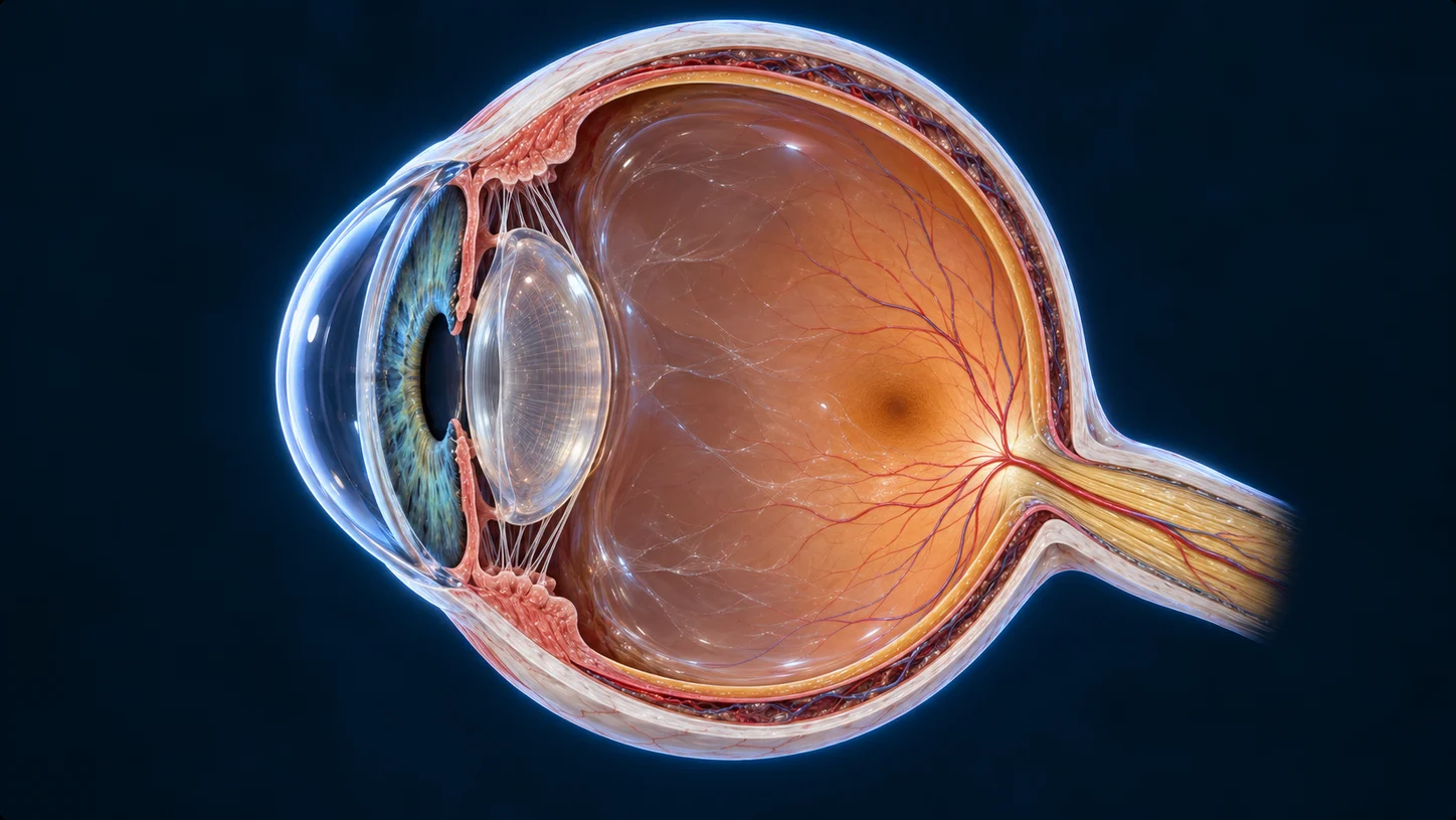 Eyes and visual context
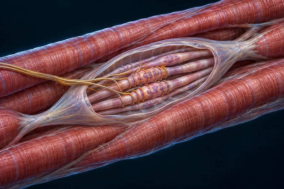 Muscle-position feedback