Special Senses / VISUAL EXPLAINER
Does Your Cochlea Contain Just One Kind of Fluid?
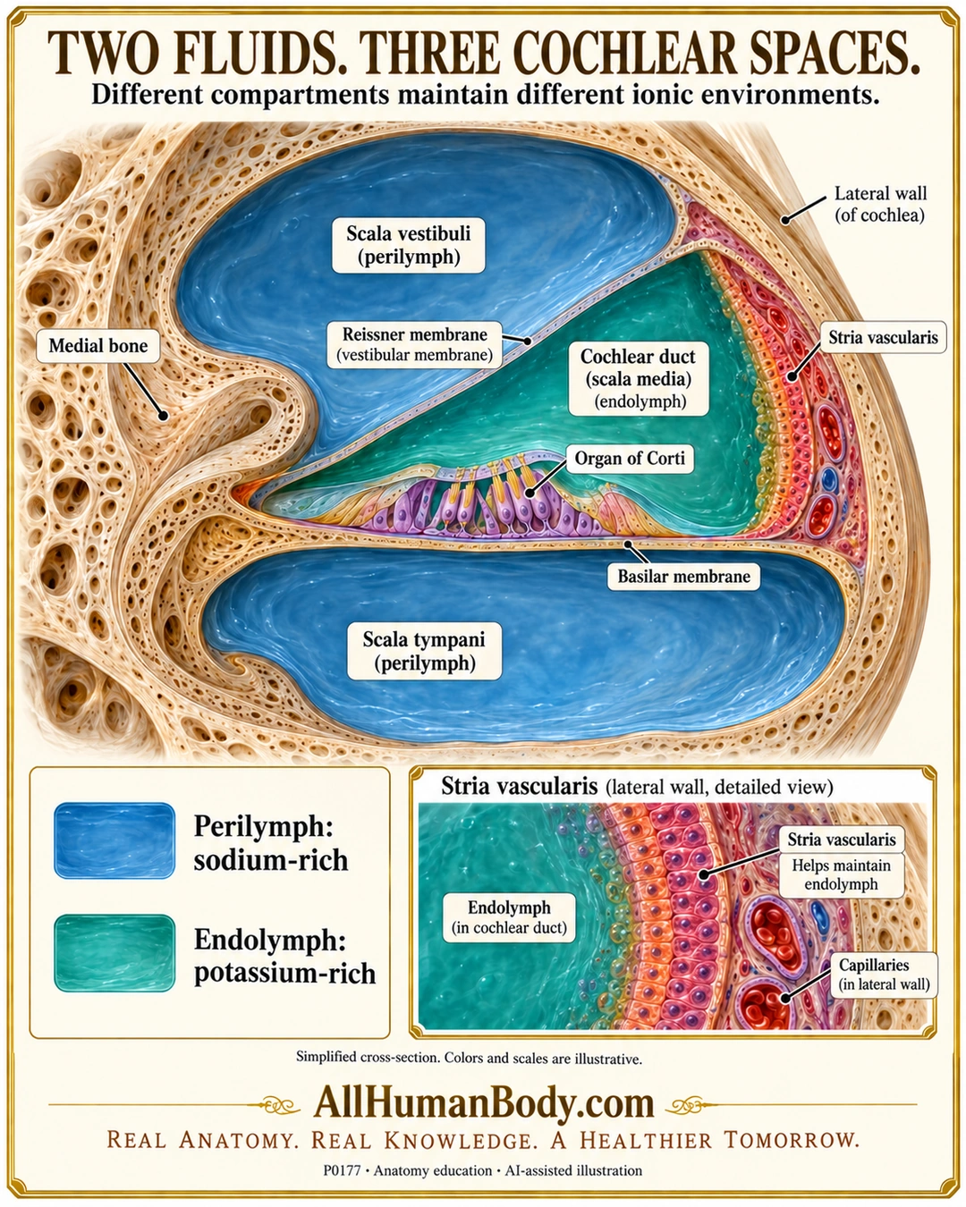
The cochlear duct contains potassium-rich endolymph, while the scala vestibuli and scala tympani contain sodium-rich perilymph. Separating tissues and the lateral-wall stria vascularis help maintain distinct fluid environments around the sensory apparatus.
READ THE IMAGE
What the illustration shows
Ivory-and-gold anatomy poster titled Two Fluids. Three Cochlear Spaces. A dimensional cross-section shows blue perilymph in the upper scala vestibuli and lower scala tympani. A triangular teal cochlear duct, or scala media, lies between them at the lateral wall and contains endolymph. Reissner membrane separates it from the upper space; the basilar membrane is below the simplified organ of Corti. Labels locate medial bone, lateral wall and stria vascularis. Below, blue and teal keys identify sodium-rich perilymph and potassium-rich endolymph. A lateral-wall inset shows strial tissue and capillaries adjacent to endolymph, without an open blood-to-fluid channel. The note says colors and scales are illustrative. Gold AllHumanBody.com and the complete brand slogan appear at the bottom.
How to interpret this poster
- Colors and thicknesses are schematic; fluid texture does not imply turbulence and exact concentrations or voltages are not shown.
- Organ of Corti is a locator, not validated cell counts, tectorial attachments or mechanotransduction. Its apical epithelial boundary matters: basilar membrane alone is not the complete ionic seal.
- No open blood-to-endolymph conduit, lymph-node fluid equivalence, energy-free maintenance or disease/diet advice. Apical scala communication is outside this section.
- AI-assisted educational illustration; not physician certification, microscopy, a diagnosis or individualized medical advice.
- Fine annotations need enlargement; the full description provides an accessible text alternative.
- Display the full uncropped illustration; do not trim titles, legends or boundary annotations.
AI-assisted educational illustration, adapted into web-sized images without changing its composition. This is general education, not an individual diagnosis or treatment recommendation.
EVIDENCE / CONTEXT
Sources and what they support
- Cochlea.eu: Cochlear fluids ↗
Three-space fluid allocation, strial maintenance and differing sensory apical/basal environments.