NORMAL PHYSIOLOGY / BOUNDARIES · QUANTITIES · COORDINATION
Fluid Balance & Osmoregulation
Locate the water. Distinguish amount from concentration. Follow the response.
Understand how water is distributed across cell boundaries, why an osmotic change differs from a volume change, and how drinking and renal transport work together.
30 SECONDS / FIND THE BOUNDARIES BEFORE FOLLOWING THE SIGNAL
Inside a cell. Around a cell. Within a vessel.
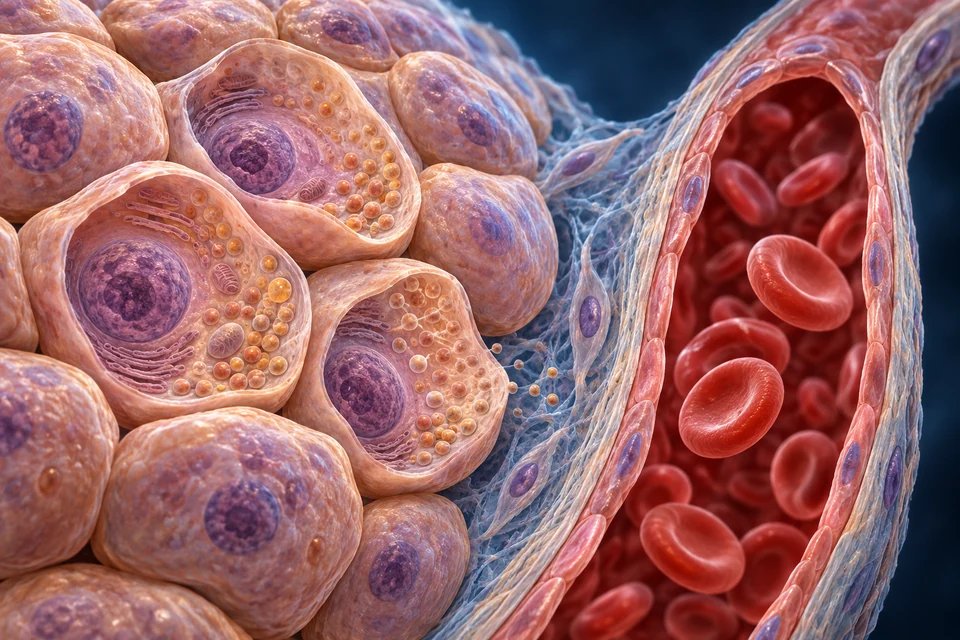
Structure notes · further context
- Cell interior
- No amount, solute concentration or specific organelle identity is inferred from colour. Blood cells also have intracellular fluid.
- Interstitial context
- The fibres are support, not a direct visual measurement of fluid. Cell membranes and vascular walls are different boundaries.
- Plasma context
- The red cells are not plasma. The visible blood region provides compartment context, not a quantitative image of proteins, solutes or water.
The cell membrane encloses intracellular water.
Find the opened tissue cell. Red blood cells also have intracellular fluid; plasma is outside them.
- LocateCell interior, tissue space and plasma are distinct.
- CompareWater amount and particle concentration are different quantities.
- CoordinateDrinking and renal transport help restore balance.
Rings locate a compartment, not its measured volume or permeability. The original tissue geometry is unchanged; names and meaning remain separate editable layers.
Sources for this relationship: OpenStax · Body fluids and fluid compartments
02 / INSIDE AND OUTSIDE
A cell membrane and a vascular wall make different boundaries.
Read the retained tissue cutaway before following any hormone: cell interior, tissue-side space and the fluid part of blood have different names.
Intracellular fluid is enclosed by cell membranes. Extracellular fluid includes interstitial fluid around tissue cells and plasma inside blood vessels. Blood cells contain intracellular fluid too; their red colour is not a picture of plasma.
The blood wall separates plasma from interstitial space; a tissue cell membrane separates interstitial from intracellular fluid. Lymph provides another return pathway. Cerebrospinal and other specialised extracellular fluids have additional barriers and are not all resolved in this one preparation.
A compartment is not a rigid container
The boundaries permit selected transfers, and water amounts change with conditions. The picture identifies locations; its coloured areas do not measure fluid fractions or an individual’s body-water content.
Sources for this relationship: OpenStax · Body fluids and fluid compartments
03 / AMOUNT AND CONCENTRATION
Count particles, then state what the denominator measures.
A water amount, a dissolved amount and a concentration answer different questions.
Osmolality expresses the amount of osmotically active particles per kilogram of solvent; osmolarity uses a litre of solution. The words are related, but their denominators are not interchangeable. Neither is simply the mass of salt in the body.
A given amount of particles becomes more concentrated when distributed in less water. Conversely, adding water can lower concentration without removing those particles. State the units before comparing two measurements or doing an accounting exercise.
An ion concentration is not total sodium content
A concentration is a ratio. It does not establish the total extracellular volume, total sodium inventory or amount of water lost. A normal-looking ratio can coexist with changed amounts on both sides of the ratio.
Sources for this relationship: Hypertonicity · physiological concept and experimental studies · Pressure natriuresis and renal blood-pressure control
04 / EFFECTIVE OSMOLES
Cell-volume effects depend on the boundary and the time.
Tonicity describes the sustained osmotic effect of the surrounding solution on a cell, rather than counting every measured particle equally.
Solutes that remain effectively separated across a particular membrane can sustain an osmotic water shift. Solutes that redistribute across it may raise measured osmolality without producing the same sustained change in cell water.
This is why a statement about osmolality alone cannot determine tonicity in every setting. Permeability and the observation interval matter. Urea illustrates that a solute can contribute to measured osmolality while behaving differently from effectively confined particles across many cell membranes.
Avoid “urea never matters”
Permeability is membrane- and time-dependent, and urea transport has an important renal medullary role. The two-compartment exercise below deliberately uses confined particles; it is not a universal model for every solute or nephron segment.
Sources for this relationship: Hypertonicity · physiological concept and experimental studies · Vasopressin-regulated renal water reabsorption
05 / FOLLOW WATER
A permeable route permits movement; a gradient supplies its direction.
Compare an idealised cell-water compartment with its surroundings while keeping the particle accounts explicit.
Auxiliary closed model: cell water starts at 2 kg with 600 mOsm confined particles; external water starts at 1 kg with 300 mOsm. Both begin at 300 mOsm/kg. These invented quantities are not personal reference values.
Reference balance
The two water spaces start at equal concentration. There is no net redistribution in the model.
- External water change
- 0 kg
- External particle change
- 0 mOsm
- Final total water
- 3 kg
- Final common concentration
- 300 mOsm/kg
- Final cell water
- 2 kg
- Final external water
- 1 kg
Cell-water change: 0 kg. Water shifts toward the side needing more solvent to balance the effectively confined particles.
Conservation and units, not a patient simulation: final concentration = total confined mOsm ÷ total kg water. Each side’s water = its confined mOsm ÷ the final concentration. No renal response, pressure force, permeating-solute kinetics, treatment fluid or individual volume prediction is included.
Water molecules move in both directions. A sustained difference in effective osmotic conditions produces a net transfer until the relevant forces balance. Aquaporins increase permeability; they do not actively pump water.
The exercise keeps particles on their original side and lets water redistribute. Real cells also regulate ions and organic solutes, possess non-water volume and face mechanical constraints. The model therefore explains a principle, not the precise swelling or shrinking of every cell.
What the calculation leaves out
No kidney, intake, ongoing metabolism, hydrostatic pressure, plasma protein distribution or timed permeability is simulated. Cell water plus extracellular water is conserved after the stated external change; this is a closed mathematical comparison, not a fluid-treatment calculator.
Sources for this relationship: OpenStax · Body fluids and fluid compartments · Hypertonicity · physiological concept and experimental studies
06 / VOLUME AND CONCENTRATION
Extracellular amount and osmotic concentration can change separately.
A proportional loss of water and effective particles differs from a loss of water alone.
In the constructed proportional-loss case, extracellular water falls but the final concentration stays at its reference value. In the water-only loss case, the final concentration rises and water redistributes across the cell boundary. These are different accounting outcomes.
Renal sodium handling is important for extracellular volume regulation. Effective circulation also depends on the pump and vascular distribution. Do not read “extracellular volume” as identical to plasma volume, arterial pressure or tissue perfusion.
Two control problems interact
Water balance affects concentration, while sodium balance strongly influences extracellular amount. The controls interact through circulation, thirst and renal transport; they are not two perfectly isolated switches.
Sources for this relationship: Pressure natriuresis and renal blood-pressure control · OpenStax · Electrolyte balance
07 / WHOLE-BODY BALANCE
A transfer within the body is different from a gain or a loss.
Water crosses gut, skin, respiratory and urinary boundaries as well as internal cell boundaries.
Water enters in drinks and food, and metabolic reactions also contribute. Urine, stool, skin evaporation and expired air provide output routes. Their contributions vary with conditions; this chapter supplies no fixed daily intake or loss target.
Reabsorption transfers water from tubular fluid toward the body’s internal circulation. It is not a new external water source. Similarly, movement from a cell into interstitial space changes distribution without itself changing total body water.
A balance needs an interval
For an explicitly defined interval, change in body water equals external inputs plus metabolic production minus outputs. A single urine volume cannot reveal every other term, and a momentary concentration cannot reconstruct the entire preceding balance.
Sources for this relationship: OpenStax · Water balance
08 / SENSING AND DRINKING
The motivation to drink integrates more than one signal.
Osmotic information, circulation-related inputs and drinking-related sensory cues contribute to regulation.
Brain networks receive information about effective osmotic conditions and circulatory state. These inputs help coordinate thirst with hormone release. Mouth and gut signals can begin reducing thirst before absorbed water has fully changed the circulating fluid.
The sensation and the eventual correction are therefore distinct events. A dry mouth, a thirst rating or relief after a sip is not by itself a quantitative measure of a person’s total water deficit.
Human evidence and detailed circuits
Studies of human sensation and brain activity complement detailed neural experiments in animals. The regional pituitary view below is not a fine map of human thirst neurons, and no single labelled nucleus is claimed to explain every drinking behaviour.
Sources for this relationship: Thirst · human physiology and animal-model primer · Thirst as an ingestive behavior · physiology and assessment
09 / A NEURAL HORMONE ROUTE
Synthesis in the hypothalamus. Release from posterior terminals.
The two pituitary lobes are neighbouring tissues with different delivery arrangements.

Structure notes · further context
- Hypothalamic floor
- Specific neuron nuclei are not assigned from this generated appearance. Neural and vascular delivery require different mechanisms.
- Infundibular stalk
- The detailed portal connections are explained separately in an editable supporting map; the red/blue surface branches are not a verified angiogram.
- Anterior endocrine tissue
- The pars distalis forms the major endocrine region. Its cells are not exocrine acini around a drainage duct; generated cell subtype colors are not diagnostic.
- Posterior neural tissue
- The pars nervosa stores and releases hypothalamic vasopressin and oxytocin. It is not the site where those neuronal cell bodies synthesize their hormone precursors.
- Vascular context
- Exact arterial, long/short portal and venous junctions are not established by this generated surface. The functional route map states the source-supported main distinction.
Vasopressin, also called antidiuretic hormone, is synthesised by hypothalamic neurons. Their axons pass through the stalk to posterior-pituitary terminals, where stored hormone can be released toward the circulation.
Osmotic and volume-related signals influence secretion. The posterior lobe is not the site of the neuronal cell bodies making the hormone, and the anterior endocrine lobe is not an interchangeable release route. The broad preparation shows this anatomical distinction without assigning individual nuclei or capillary junctions.
Feedback has more than one input
A fall in effective circulatory filling can alter the relationship between osmotic conditions and vasopressin output. The panel compares physiological priorities, not a universal secretion threshold or a complete clinical decision rule.
Sources for this relationship: Endotext · Neurohypophysis, vasopressin and oxytocin
10 / REGULATED PERMEABILITY
The collecting duct adjusts a route that still needs a driving gradient.
Identify tubular fluid, the apical surface, the cell, its tissue-facing surface and the separate blood vessel.
Vasopressin acts through V2 receptors on the basolateral side of principal cells. Intracellular signalling promotes availability of AQP2 at the lumen-facing membrane. AQP3 and AQP4 provide distinct basolateral pathways for water exit.
When conditions favour reabsorption, water can pass from tubular fluid through the epithelium toward interstitium and blood. Medullary osmotic conditions and adequate flow remain necessary context. Greater permeability does not mean that the hormone creates water or pumps it through a pore.
Channels, gradient and circulation are different jobs
Solute transport and renal architecture support the concentrating environment; blood flow returns recovered water. Individual channels are not resolved in the retained tissue illustration, and its vesicles must not be read as identified AQP2 carriers.
Sources for this relationship: Aquaporins in the kidney · molecules and water transport · Vasopressin-regulated renal water reabsorption
11 / SALT AND WATER CONTROL
Conserving sodium and changing water permeability are related, different responses.
Volume-linked hormonal and neural signals act alongside osmotic control.
Renal sodium excretion is adjusted with perfusion, neural and hormonal influences. Renin–angiotensin–aldosterone coordination helps regulate sodium handling and circulation; vasopressin has a separate prominent role in collecting-duct water permeability.
Aldosterone also participates in potassium handling, and potassium itself can influence its release. Do not reduce that hormone to a simple plasma-sodium concentration switch. Use the established adrenal lesson for the complete epithelial and potassium distinctions.
Retain the organ-level detail
Pressure-dependent excretion, local transport and endocrine responses interact over different timescales. This overview does not replace the mature kidney/adrenal mechanisms or infer a treatment from a hormone name.
Sources for this relationship: Pressure natriuresis and renal blood-pressure control · OpenStax · Electrolyte balance · Endotext · Neurohypophysis, vasopressin and oxytocin
12 / COMPARE PHYSIOLOGICAL PRIORITIES
Ask what changed before deciding which response matters.
A water-content disturbance and a proportional extracellular loss need not send the same set of signals.
The three comparisons below distinguish osmotic concentration, water excretion and circulatory priorities. They are teaching contexts rather than measurements of an individual. Inputs can oppose or reinforce one another.
Drinking and renal conservation complement one another. Reducing urinary water loss cannot replace external water indefinitely, and drinking-related relief can precede the completion of redistribution. Normal regulation is a coordinated process, not a fixed diagram in which every arrow changes by the same amount.

Increase water recovery when the integrated signal favours conservation.
Osmotic stimulation can promote thirst and vasopressin release. Increased apical permeability helps conserve water when an appropriate renal gradient exists. This describes a response tendency, not a measured hormone level or complete compensation.
Gold traces the previously checked overall reabsorptive route when its driving conditions apply; it stays a reference route in all three comparisons. A highlighted region is not a measured flux, an identified channel or a guarantee that reabsorption is occurring. Each membrane and blood boundary stays in its original location.
What remains outside this course
No symptom checklist, serum threshold, personal intake target, supplement, intravenous solution or correction rate is supplied. Disorders require clinical assessment; the normal comparison cannot establish a diagnosis or safe treatment.
Sources for this relationship: OpenStax · Water balance · Thirst as an ingestive behavior · physiology and assessment · Endotext · Neurohypophysis, vasopressin and oxytocin
13 / READING EVIDENCE
Name the measured quantity, site and interval.
A blood concentration, urine measurement, thirst report and anatomical image answer different questions.
Osmolality measures particle concentration relative to solvent mass; a thirst report measures a sensation. Urine amount and urine concentration describe output over an interval. Combining appropriate observations is more informative than treating any single one as a complete water inventory.
The illustrations identify normal compartments and broad tissue relationships. They do not acquire a person’s hydration status or measure hormones, channel abundance or osmotic gradients. The finite mathematical cases below are transparent examples, not validated physiological predictions.
Scope of the evidence check
Independent source checks support the stated mechanisms; generated fine anatomy and the whole chapter have not received physician certification. Existing FB134 guides the organisation without a new full-book reading or copied book prose/artwork.
Sources for this relationship: Thirst as an ingestive behavior · physiology and assessment · Hypertonicity · physiological concept and experimental studies · Aquaporins in the kidney · molecules and water transport
Check the boundary, the denominator and the direction.
Does red blood belong to extracellular fluid?
Plasma is extracellular, but each blood cell encloses intracellular fluid. Distinguish the fluid component from the cells it surrounds.
Can equal concentration prove equal water amount?
No. A ratio can remain unchanged when water and particles both change in proportion.
Does every measured osmole have the same cell-volume effect?
No. The relevant membrane permeability and timescale determine whether a sustained osmotic gradient is maintained.
Does AQP2 pump water?
No. It supplies a regulated passive pathway; the osmotic conditions supply the driving force.
Is the posterior pituitary the site of vasopressin synthesis?
The neuronal cell bodies synthesise it in the hypothalamus. Their posterior terminals store and release it.
Does a thirst rating establish total body-water deficit?
No. Sensory feedback, circulatory and osmotic inputs, behaviour and the observation time all matter.
Evidence, retained illustrations and scope
Directed source check: 22 October 2026. Existing FB134 print247/PDF249 and prior foundation research guide coverage; mature organ lessons remain linked. No book text or image is copied and no new full-book reading is claimed.
- OpenStax · Body fluids and fluid compartmentsReadable official educational text for compartment boundaries and unequal solute composition. No fixed body-water percentage or universal capillary-reabsorption cartoon adopted.
- OpenStax · Water balanceReadable official text for water inputs/losses and coordinated intake/output. No daily intake target, minimum urine rule or universal response timing adopted.
- Hypertonicity · physiological concept and experimental studiesOfficial indexed review passages distinguish osmolality, effective osmoles and cell-volume behaviour. Direct PMC retrieval met an access challenge; not claimed as a new full-text read. Clinical correction recommendations are outside this course.
- Thirst · human physiology and animal-model primerOfficial indexed primer text supports osmotic/volume input and anticipatory drinking signals. Fine neural circuit experiments include rodents, not a complete experimentally mapped human pathway.
- Thirst as an ingestive behavior · physiology and assessmentOfficial indexed review abstract distinguishes initiation, diminution and measurement of human thirst. Direct page met an access challenge; no full-methods or diagnostic validation claim.
- Endotext · Neurohypophysis, vasopressin and oxytocinOfficial indexed normal-physiology passages for hypothalamic synthesis, posterior release, osmotic/volume control and limits. Direct text request met an access challenge. No personal secretion threshold or dose is supplied.
- Aquaporins in the kidney · molecules and water transportOfficial publisher indexed review text supports apical AQP2 trafficking and distinct basolateral AQP3/AQP4. Molecular work includes animal/isolated preparations; the retained atlas does not resolve channels.
- Vasopressin-regulated renal water reabsorptionOfficial indexed review passages support basolateral V2 signalling and passive water transfer along an osmotic gradient. Direct PMC text met an access challenge; no fresh experimental replication is claimed.
- Pressure natriuresis and renal blood-pressure controlOfficial indexed research-review abstract for renal sodium balance, extracellular volume and pressure. Mechanisms differ by setting; clinical treatment sections are not used.
- OpenStax · Electrolyte balanceReadable official text for broad electrolyte distribution only; simplified sodium-trigger and potassium narratives are not adopted as a complete control mechanism. Existing source-qualified adrenal teaching supplies the detailed link.
Three qualified original generated preparations are retained with unchanged pixels and anatomical positions. They are not microscopy, a hormone assay or a measurement of fluid volumes. Default names, numbers, selected explanations, directions and accessible text remain independent of the medical base images.
Normal physiology and transparent mathematical examples only. No personal intake target, diagnosis, treatment solution or correction rate. AI-assisted source, structural and teaching checks are separate from physician review; fine generated anatomy and physical-phone use remain uncertified.