NORMAL PHYSIOLOGY / AIR → BLOOD → ACTIVE TISSUE
Oxygen Delivery & Active Muscle
Follow the supply. Separate the load from the flow. Find where it is used.
Connect lung exchange and circulation with living muscle tissue. Then compare delivery, extraction and uptake, and follow how ATP support changes when activity starts, continues and stops.
30 SECONDS / ONE FUNCTIONAL JOURNEY, DIFFERENT ANATOMICAL SCALES
Oxygen crosses a boundary, travels with blood and reaches a consuming cell.
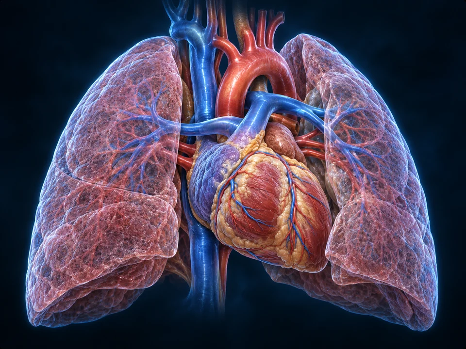
Organ scale · linked circulation
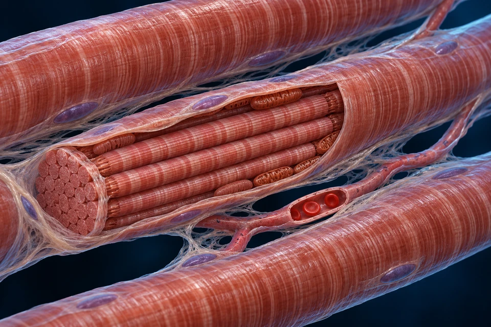
Cellular scale · blood beside muscle
Structure notes
- Skeletal muscle fibre
- A long striated cell lies beside other fibres; it is not a blood vessel.
- Mitochondrial context
- Oxidative ATP production is supported by mitochondria within the cell.
- Myofibril bundles
- Contractile structures use ATP and lie inside the opened fibre.
- Capillary lumen
- Red blood cells remain inside a vessel between the muscle cells.
- LoadAir is renewed; oxygen crosses into pulmonary blood.
- DeliverFlow carries a blood-borne oxygen load to the region.
- UseOxygen crosses into tissue; the returning load completes the balance.
Renewed alveolar gas meets pulmonary blood.
The alveolar-capillary boundary supports exchange; haemoglobin takes up much of the incoming oxygen. The broad lung highlight locates the organ, while the separate barrier view identifies the tissue scale.
Cyan short arrows: selected blood-flow directions in the organ view. Gold: qualitative net oxygen direction in the independent tissue views. Rings locate compartments; none of these cues is a measured flow, oxygen particle or continuous traced tube between the two scales.
Sources for this relationship: OpenStax · Transport of Gases · Oxygen flux from capillary to mitochondria · The pulmonary physiology of exercise
02 / AIR TO BLOOD
Ventilation renews air. Diffusion loads blood. Circulation delivers it.
Start inside an alveolus, cross its shared wall with a capillary, then follow the body circuit toward a muscle. Each step operates in a different compartment.

Air moves through conducting passages to exchange regions. Oxygen crosses a thin tissue interface and enters pulmonary blood; carbon dioxide moves toward alveolar gas during ordinary lung exchange. Red cells remain within blood, rather than entering the air space.
The heart links pulmonary exchange with systemic delivery. A breath, a beat and a mitochondrial reaction are connected events, but none can stand in for all the others. The retained regional image locates the pumps and lung tissue; the separate barrier view supplies the smaller exchange scale.
Why faster breathing does not specify tissue oxygen use
Breath size, rate and conducting-space ventilation affect fresh air reaching exchange regions. Perfusion, arterial content and the consuming tissue also matter. Observing chest movement cannot supply all the quantities in a tissue oxygen balance.
Sources for this relationship: OpenStax · Transport of Gases · The pulmonary physiology of exercise
03 / CONTENT AND PRESSURE
Identify the load before multiplying it by flow.
Arterial oxygen content describes how much oxygen is carried per volume of blood. Oxygen partial pressure and haemoglobin saturation describe related, different properties.
A large share of transported oxygen is reversibly associated with haemoglobin inside red cells. A smaller dissolved component contributes to blood oxygen tension. Binding and dissolved oxygen equilibrate; a red cell is not an air bubble.
A saturation percentage describes occupied binding capacity. Content also depends on how much haemoglobin is available, together with the dissolved contribution. Two saturation readings alone cannot establish identical oxygen loads or identical delivery to a region.
A pressure gradient and a transport reservoir do different jobs
Differences in oxygen partial pressure support diffusion. Haemoglobin provides a much larger transport reservoir than dissolved oxygen alone. Increasing a gradient, increasing binding capacity and increasing blood flow are separate changes, even though they interact in the living system.
Sources for this relationship: OpenStax · Transport of Gases · The pulmonary physiology of exercise
04 / DELIVERY BY BLOOD
Delivered oxygen depends on both blood flow and oxygen content.
Convective oxygen delivery to a selected region equals its incoming blood flow multiplied by incoming oxygen content. State the region and use compatible units.
More blood per minute can carry more oxygen into a region at unchanged content. The same flow can deliver a different oxygen amount when content changes. Neither observation alone states how much oxygen the cells actually use.
A flow measured for one leg is not automatically whole-body cardiac output. Likewise, a sample from a regional vein does not automatically represent mixed venous blood returning from all systemic tissues. Keep the anatomical territory of the flow and the samples matched.
Pressure supports flow, but is not the delivered volume
A pressure difference helps drive flow through a resistance. The amount delivered per minute still requires flow and content. The existing pressure course explains the hydraulic relationship; this chapter follows the oxygen carried by the resulting flow.
Sources for this relationship: Exercise oxygen-consumption measurements · MRI validation study · The pulmonary physiology of exercise · Regulation of exercise hyperaemia
05 / EXTRACTION AND USE
The returning oxygen load completes the balance.
For a suitable steady balance, tissue oxygen uptake equals flow multiplied by the arterial-minus-venous oxygen content difference. Delivery and uptake therefore need not change in the same proportion.
Regional delivery = flow × arterial content.
Regional uptake = flow × (arterial − venous content).
Reference balance
A constructed regional bed receives 200 mL O2/min and removes 50 mL O2/min from the incoming load.
- Blood flow
- 1 L/min
- Arterial content
- 200 mL O₂/L blood
- Venous content
- 150 mL O₂/L blood
- Delivery
- 200 mL O₂/min
- Returning oxygen
- 150 mL O₂/min
- Uptake
- 50 mL O₂/min
- Extraction fraction
- 25%
Auxiliary accounting examples at a constructed steady regional balance; values are invented. Contents use mL oxygen per litre of blood, so multiplying by L blood/min gives mL oxygen/min. No patient input, normal range, diagnostic threshold, exercise intensity or adaptation prediction is supplied.
The oxygen entering a region is partitioned between oxygen leaving in venous blood and net uptake by the tissue. A lower returning content can indicate a larger arteriovenous difference when incoming content is held constant. It does not mean that all returning blood has used up every oxygen molecule.
The extraction fraction divides the arteriovenous content difference by arterial content. It compares a used fraction with an incoming load. The illustrative cases below independently change flow, content and extraction; they are mathematical comparisons, not four obligatory stages of exercise.
The Fick balance has time and sampling conditions
During rapid transitions, changes in oxygen stores and the delay between locations complicate an instantaneous calculation. Regional flow must be paired with representative arterial and draining venous contents. A local muscle measurement and pulmonary oxygen uptake are not necessarily identical at every moment.
Sources for this relationship: Exercise oxygen-consumption measurements · MRI validation study · The pulmonary physiology of exercise · Human oxygen-uptake and phosphocreatine on/off kinetics
06 / BLOOD TO MITOCHONDRIA
Blood stays in a vessel; oxygen crosses into living tissue.
The muscle preparation places an extracellular red-cell capillary beside a striated fibre. Within the fibre, mitochondria and myofibrils have different roles.
Oxygen leaves the blood compartment and traverses vascular and tissue boundaries toward consuming cells. Flow transports oxygen along the capillary; diffusion contributes to movement across the local pathway. Capillary delivery and tissue diffusion must work together.
Mitochondria support oxidative ATP production, while myofibrils use ATP during contractile cycling. The drawn direction joins these compartments conceptually. It is not a tracked molecule, a measured gradient or a claim that red cells or haemoglobin move into the myofibrils.
Return to the extracellular capillary and intracellular organelles →
An oxygenated vessel beside a cell is necessary context, not a complete performance test
The surrounding tissue geometry, transport properties and cellular demand influence oxygen use. The atlas does not measure capillary density, diffusion distance, mitochondrial capacity or a person’s limiting step. Those quantities require appropriate methods rather than a count of painted organelles.
Sources for this relationship: Oxygen flux from capillary to mitochondria · OpenStax · Muscle Fiber Contraction and Relaxation
07 / DEMAND CHANGES
ATP demand changes while delivery and oxidative use adjust.
At activity onset, ATP turnover can increase before a new metabolic steady state is established. Existing ATP-regenerating processes contribute together, with changing proportions.
Demand rises while the system adjusts.
Rapid ATP regeneration and developing oxidative support coexist. Phosphocreatine changes and pulmonary oxygen uptake should not be drawn as identical instantaneous measurements.
A selected conceptual comparison, not a zero-oxygen stage or a fixed number of seconds.
These contexts compare explanations; they do not animate an individual’s metabolism or impose one fixed sequence on every fibre. Static onset and recovery lessons remain available below and through search.
Phosphocreatine supports rapid ATP regeneration; glycolytic reactions and oxidative metabolism also contribute. Oxidative metabolism was already operating before movement began. A drawing with three boxes should not imply that one energy system must finish before the next starts.
Pulmonary oxygen uptake and local muscle metabolism have their own dynamics. Human knee-extension experiments measured oxygen uptake alongside phosphocreatine changes and found that behaviour depends on intensity and on whether activity is starting or stopping. This does not establish one countdown for every task.
An oxygen deficit is a comparison, not proof of zero oxygen
In exercise physiology, the term can describe the difference between a required steady-state equivalent and the developing oxidative response. It does not by itself establish that the tissue had no oxygen or that all early ATP was regenerated by one pathway.
Sources for this relationship: Human oxygen-uptake and phosphocreatine on/off kinetics · OpenStax · Muscle Fiber Contraction and Relaxation
08 / COORDINATED SUPPLY
Local demand is served within a whole circulation.
Active muscle can receive increased flow, but regional vascular regulation also operates within a system that supports arterial pressure and other organs.
Local metabolic and endothelial influences interact with neural control, muscle contractions and the pressure driving flow. Rhythmic contraction can assist venous return while also mechanically affecting vessels in the contracting tissue. No single drawn substance accounts for every phase of exercise hyperaemia.
The recruited muscle mass and the task matter. A result from one small muscle group cannot be scaled directly to all-body activity. Maintaining pressure, supporting working tissue and exchanging heat at the skin create related demands on the circulation.
Supply and demand are not two independent knobs in a person
The arithmetic model deliberately holds selected quantities fixed to show their meanings. In a living system, changing activity also changes neural drive, local metabolism, distribution and cardiac responses. The model is an accounting lesson, not a forecast of the response to a flow change.
Sources for this relationship: Regulation of exercise hyperaemia · The pulmonary physiology of exercise
09 / LACTATE AND CARBON
A circulating metabolite can be produced and used at the same time.
Lactate can form when oxygen is present. Its concentration reflects the balance of appearance and removal, rather than reporting one tissue’s oxygen supply or ATP source by itself.
The lactate dehydrogenase reaction relates pyruvate, lactate and the cytosolic redox state. Regeneration of NAD+ supports continued glycolytic activity. This reaction does not require oxygen to disappear from the entire cell before lactate can be formed.
Lactate carbon can be used in oxidative metabolism or support glucose production in appropriate tissues. Transfer between tissues, oxidation and glucose synthesis are different processes. The blood concentration alone does not reveal the proportion following each route.
What a tracer experiment adds
The original graded-exercise study used a lactate tracer in six men to investigate appearance and disappearance alongside oxygen consumption. It distinguishes movement through a metabolic pool from the size of that pool. Its findings should not be turned into a universal percentage for every person or activity.
Why fatigue needs more than a lactate label
The existing muscle lesson describes neural drive, excitation, calcium handling and contractile/metabolic factors. Retain that wider explanation. Lactate should not become a universal waste product that single-handedly accounts for loss of force.
Sources for this relationship: Systemic lactate kinetics during graded exercise in man · The Science and Translation of Lactate Shuttle Theory
10 / RECOVERY METABOLISM
Stopping external work leaves physiological tasks to complete.
Energy reserves, ion distributions, oxygen stores and body conditions continue to change after activity. Restoring phosphocreatine and recovering performance are related, different outcomes.
Oxidative ATP production can support phosphocreatine restoration. Calcium handling and membrane-gradient maintenance also use energy. A stopped limb therefore does not imply that all associated energy turnover immediately returns to its prior level.
Post-activity oxygen consumption can remain elevated for multiple reasons. It should not be labelled as an exact payment for a single lactate debt. The human on/off kinetics experiment also cautions against drawing recovery as a simple mirrored copy of the starting response.
A return of one variable does not finish all recovery
An oxygen-uptake trace, phosphocreatine signal, heart rate, body temperature and force performance describe different aspects and timescales. The course does not supply a universal rest interval, a recovery score or a recommendation to continue activity.
Sources for this relationship: Human oxygen-uptake and phosphocreatine on/off kinetics · Metabolic bases of excess post-exercise oxygen consumption · Effects of exercise intensity and duration on EPOC · OpenStax · Muscle Fiber Contraction and Relaxation
11 / MEASUREMENTS AND LIMITS
Name the measurement site before interpreting the signal.
Expired-gas measurements, blood samples, regional flow and magnetic-resonance signals answer different questions. A visual atlas supplies anatomical context rather than an acquired recording.
Pulmonary oxygen uptake integrates exchange at the lungs. A regional Fick calculation uses the matching vascular territory and blood contents. Their relation depends on the conditions and timing. A single haemoglobin saturation percentage cannot replace the full set of quantities.
The cited MRI study investigates particular measurement methods during a defined exercise task. A method validation does not certify this illustration, every measurement setting or an individual’s fitness. All numerical cases in this chapter are invented and explicitly unit-labelled.
What remains outside the chapter
Clinical exercise testing, disease-related limitations, oxygen treatment, training prescriptions, supplements and individual diagnostic thresholds require separate evidence and safeguards. The normal explanations here do not authorise those extensions.
Sources for this relationship: Exercise oxygen-consumption measurements · MRI validation study · Human oxygen-uptake and phosphocreatine on/off kinetics · The pulmonary physiology of exercise
Check the compartment, the quantity and the time.
What additional quantity is needed when flow is known?
Incoming oxygen content is needed for delivery. Representative returning content is also needed to calculate uptake using the regional Fick balance.
Does a saturation percentage alone give oxygen content?
No. Haemoglobin quantity and the dissolved contribution also matter; pressure, saturation and content are related but different.
Do red cells cross into the muscle fibre to deliver oxygen?
No. They remain in the vascular lumen while oxygen crosses the local exchange pathway.
Does a positive lactate concentration prove no oxygen was present?
No. Lactate can form aerobically, and its concentration reflects production and removal across a pool.
Are onset and recovery necessarily mirror images?
No. Human task-specific measurements can have different on/off dynamics; variables also describe different compartments.
Are the model cases normal reference ranges or an exercise test?
No. They are constructed steady regional accounting examples, not diagnostic values, a simulation of adaptation or a personal performance prediction.
Sources, retained images and scope
Normal-physiology source check: 22 October 2026. The existing FB088 air-to-tissue book inventory and mature Muscle course guide organisation; no book prose or artwork is copied and no fresh full-book review is claimed. Independent human studies and reviews support the additional delivery, flux and transition explanations.
- OpenStax · Transport of GasesOfficial readable text for haemoglobin carriage, partial pressure and oxygen unloading. Existing Blood course supplies the deeper binding lesson.
- The pulmonary physiology of exerciseOfficial publisher indexed teaching passages for ventilation, oxygen content and Fick relations; no copied source graphics or individual exercise limits.
- Exercise oxygen-consumption measurements · MRI validation studyOfficial indexed primary-study passages identify flow and arteriovenous content difference. Direct full-text fetch returned403; no full-methods reading or universal clinical validation is claimed.
- Oxygen flux from capillary to mitochondriaRetained Muscle source check and current official indexed research record. Fresh direct page returned minimal content; broad anatomical relationships are reused, not fresh fine-structure certification.
- Regulation of exercise hyperaemiaIndependent research review, official indexed human circulation passages. Multiple mechanisms and competing needs; no universal single dilator or predicted exercise response.
- Human oxygen-uptake and phosphocreatine on/off kineticsOriginal human knee-extensor experiment, official indexed abstract. Task/intensity-dependent on/off behaviour; no universal seconds or compulsory phase trace.
- Systemic lactate kinetics during graded exercise in manOriginal tracer study of six men, official indexed abstract. Appearance/removal are fluxes distinct from one blood concentration; no all-population fraction is assigned.
- The Science and Translation of Lactate Shuttle TheoryOfficial indexed review abstract: lactate can form aerobically and participate in metabolism. Source prose, diagrams and therapeutic extensions are not reused.
- Effects of exercise intensity and duration on EPOCOfficial indexed 2006 review abstract distinguishes task/intensity/duration and measurement limits. No reported duration, percentage, weight-loss effect or exercise prescription is adopted.
- Metabolic bases of excess post-exercise oxygen consumptionFoundational review record, interpreted with the later human kinetics experiment. Its age alone does not invalidate the distinction between oxygen recovery and a single lactate-debt story.
- OpenStax · Muscle Fiber Contraction and RelaxationOriginal ATP-dependent contraction/relaxation foundation and energy-support framework, with lactate interpretation qualified by independent modern research.
All three main illustrations are retained qualified original generated project preparations. They are not acquired microscopy, MRI, metabolic recordings or clinical oxygen measurements. Native pixels and anatomical positions are unchanged; short names, directions, captions and accessible explanations are independent text/vector layers.
The numerical comparisons are constructed steady regional balances. They do not calculate fitness, diagnose a limitation, prescribe oxygen, supplements or training, or specify a recovery interval. Official indexed passages are distinguished from actually accessible full text. AI-assisted source and structural checks are not physician review; fine generated structures, physical-phone use and new WebGL performance are not certified here.