Two organs filter, recover and adjust. Urine carries away what the body no longer needs. The kidney is more than a waste outlet: it connects blood, working tubules, fluid and salt regulation, acid–base chemistry and endocrine signals.
Generated anatomical exhibit, not a specimen or scan. Anterior-oblique context: the person’s right kidney is image left. Fine hilar branch order is illustrative.
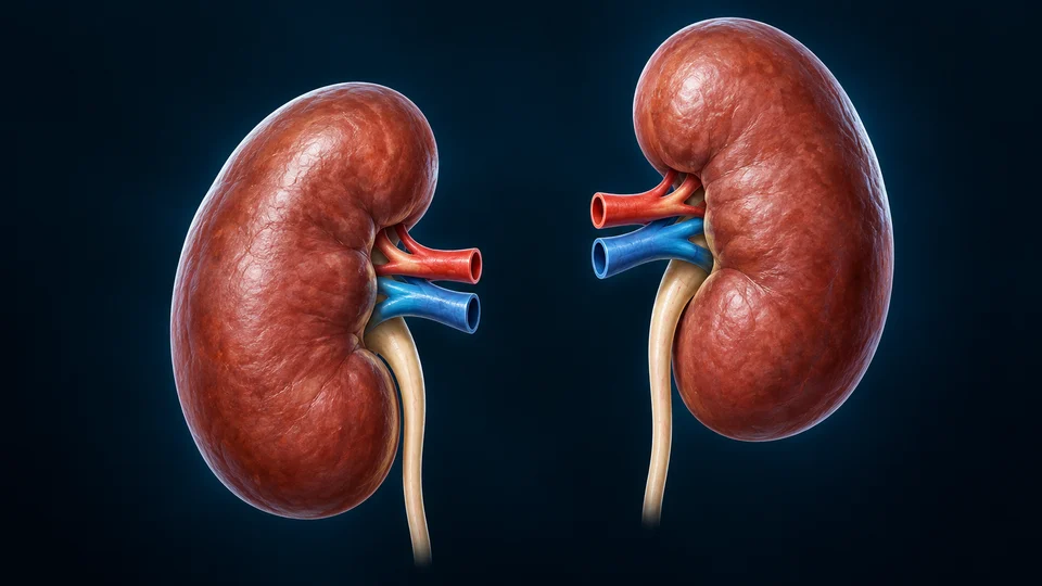
Selected kidney surface context. Short stubs distinguish blood from urine; their vertical or depth arrangement does not prove exact hilar anterior-posterior order. Both organs are separate, with different natural outlines.
Organ → functional unit
A filter followed by a working tube.
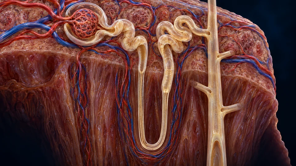
Each nephron begins in the cortex. Its tubular segments process the filtrate; a collecting duct receives fluid from several nephrons. The medulla provides a different environment from the cortex.
Generated expanded teaching preparation, not the appearance of one measured dissection. Fine segment boundaries, local vascular origins and physical distances are illustrative. Return of the distal limb to its own vascular pole/macula densa is not spatially resolved in this expanded image; the source-supported relationship is explained below.
Tubular-fluid direction · match the numbered names
2 Descending loop ↓Toward deeper medulla
3 Ascending loop ↑Back toward cortex
4 Collecting duct ↓Toward papillary drainage
Start a fluid stream. Change what remains.The corpuscle starts filtrate; the tubule recovers useful material and adds selected substances. The loop helps establish the medullary environment. Collecting ducts make further adjustments before urine drains onward. Blood keeps circulating alongside this separate fluid route.
These arrows describe regional travel along the tubes, not blood flow or water crossing a wall. Cyan rings locate selected contexts, not precise lumen boundaries, transport rates or a complete vascular map. Distinguish the three transfers →
Chapter overview video is planned; no unavailable playback or invented duration is shown.
Capsule → cortex → medulla → collecting spaces
Regions that work together.
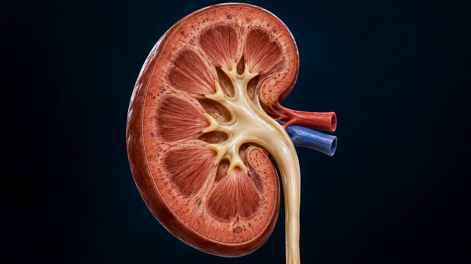
The kidneys sit below the ribs, one on each side of the spine. An outer cortex surrounds an inner medulla. Urine moves toward central collecting spaces.
Generated selected coronal section. Fine calyceal branching, pyramid counts, local texture and hilar anatomy are illustrative; a cortical column is tissue between pyramids, not a second ureter or a medullary air cavity.
Behind the abdominal lining
The kidneys lie retroperitoneally, against the posterior abdominal region rather than loose inside the abdominal cavity. Ribs, muscle, fat and fascial supports protect them. A fibrous capsule covers each organ. The right kidney commonly sits lower in relation to the liver; natural individual positions vary.
Cortex and medulla differ
The cortex contains renal corpuscles and proximal and distal convoluted tubular regions. The medulla contains longer tubular and collecting routes arranged within pyramidal regions. Cortical columns extend between pyramids. These are adjoining living tissues, not two unrelated organs or literal colour blocks.
Small collectors become one outlet
Collecting ducts drain toward papillae at pyramid tips. Minor calyces receive urine, larger collecting branches lead to the renal pelvis, and the pelvis narrows into the ureter. The ureter carries urine onward; it does not bring blood into glomerular filters.
Blood has its own hierarchy
The renal artery brings blood into the kidney. The renal vein carries blood away. Arterial branches distribute toward cortex and afferent arterioles. Glomerular capillaries lead through efferent arterioles to a further capillary network near tubules before venous return. The cutaway is not a verified map of each arterial generation.
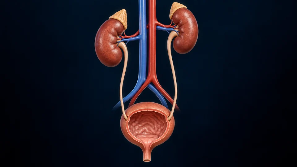
The kidneys make urine; the bladder stores it. Blood supply and urine drainage are different networks. Follow their position before looking inside an organ.
Generated anterior organ context, not a whole torso or a patient image. Adrenal appearances and exact ureteral wall entry are illustrative. This shortened generic urethral outlet does not portray sex-specific length, sphincters or pelvic-floor anatomy.
Ureters propel; bladder stores
Urine collects in the renal pelvis and passes through the ureter toward the bladder. Muscular waves in ureters propel it; drainage is not simply gravity acting on two rigid pipes. The ureters approach the bladder separately. Filling continues while the kidneys keep processing blood.
The wall accommodates and empties
Urothelial lining and the bladder wall accommodate changing volume. The detrusor muscle is involved in emptying, while neural coordination, outlet resistance and pelvic-floor support help govern storage. A folded empty-bladder rendering cannot supply a person’s capacity or diagnose a continence problem.
Shorter-loop cortical nephrons and longer-loop juxtamedullary nephrons share the same basic sequence. This expanded view teaches a long loop, not the average appearance of every nephron.
Blood stays alongside the fluid route.
After the glomerulus, efferent blood supplies nearby capillaries. Peritubular capillaries and, for long loops, vasa recta exchange with renal tissue. The rendered neighbouring vessels provide selected context rather than every connection or exact assignment.
Feedback close to the filter.
Macula densa cells sense salt delivery near the end of the thick ascending limb. Neighboring vascular cells participate in local filtration feedback and renin release. This juxtaglomerular relationship is real anatomy, but it cannot be located precisely from the expanded coiled profiles here.
Coiled regions return to the cortex
In this expanded preparation, the proximal coiled context lies just after the capsule at upper left; it begins bulk recovery. The later distal cortical context is upper right, leading toward the collecting system and further ion adjustment. These names and roles remain visible here; their exact return-to-vascular-pole contact is not resolved by the expanded image.
A receiver for more than one unit
Collecting ducts receive fluid from multiple nephrons. The tubular route and receiving system are related but not interchangeable. A single long loop does not mean one renal pyramid contains only one nephron, nor does its fluid connect directly to the renal vein.
LOOK CLOSER · THE JUXTAGLOMERULAR APPARATUS
The tubule returns to its own blood supply.
Find the meeting point beside the filter, then change scale to see the sensor and neighboring vascular wall. The tubule does not empty its fluid into an arteriole.
Original generated vascular-pole preparation, not microscopy. The returning tubular lumen remains separate from blood. Selected gross relationships are source-checked; exact cells, dimensions and capillary microstructure are not measured anatomy.
Fluid is sampled here. Blood is regulated nearby.The macula densa belongs to the returning tubule. Granular cells belong to the nearby vascular wall. Their close position supports local chemical communication without connecting the two lumens.
At the tissue interface
A closer window into the same preparation. Above the macula densa is tubular fluid; below is the basal tissue interface. These are adjacent tissues, not a connecting fluid channel. Generated granules and nuclei are explanatory detail, not a diagnostic micrograph.
Rings identify the selected tissues. A dashed cue indicates local chemical communication through tissue, not an open tube, exact molecular trajectory or quantified signal. No vessel is resized to pretend a measured response.
A return, not a shortcut
After the loop, the cortical thick ascending limb returns to the vascular pole of its own corpuscle, close to where the distal convoluted tubule begins. This local preparation shows the meeting point. The expanded nephron above supplies the larger route; the two are different illustrative preparations, not one continuous photographed specimen.
The apical surface faces tubular fluid. The basal side faces the extraglomerular tissue and neighboring vascular cells. Salt transport, including NKCC2 activity, contributes to sensing. This is not a direct blood-sodium meter or a way to read GFR from the picture.
Tubuloglomerular feedback adjusts local vascular resistance. Renin participates in a wider hormonal cascade. Related cells support both responses, but macula-densa signalling, renin secretion and the downstream renin–angiotensin system are not the same event.
Deeper learning · NaCl delivery does not directly measure filtration
What reaches the sensor depends on filtration and earlier tubular processing. Concentration, flow and salt transport are related, but are not interchangeable readings. A single simplified state cannot predict an individual’s kidney function.
Experiments in isolated rabbit juxtaglomerular preparations found that changing luminal chloride transport altered renin release. They support a transport-dependent signal; they are not a human clinical trial or evidence that every human response is controlled by chloride alone. The drawing does not resolve NKCC2 molecules or simulate a patient.
Directed source and gross-structure checks: 16 October 2026. Original generated teaching art with independent editable names, geometry and interaction. Fine cellular anatomy, physical-phone acceptance and human medical review remain unverified.
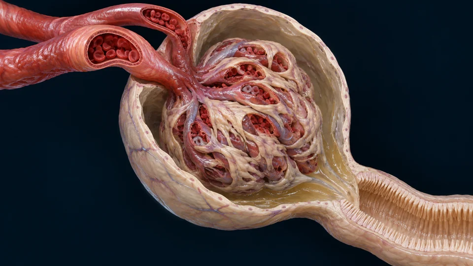
Filtration crosses a capillary wall. It does not pour whole blood into a tube. The renal corpuscle combines a glomerular tuft, its surrounding capsule and a space that receives the new filtrate.
Locate two fluids. Separate the force from the filter.
Blood continues through a capillary tuft while new filtrate enters the capsule. Compare this whole structure with its living wall, then ask which factor changes water filtration.
Generated selected corpuscle, not microscopy. Individual incoming/outgoing branch identities, podocyte outlines and loop counts are not inferred from the rendered weave. Blood cells are shown within blood-channel contexts, not within capsular or tubular fluid.Structure notes · further context
Arteriolar approaches
Afferent blood arrives; efferent blood leaves toward a second capillary network. Exact branch assignment is not inferred here.
Glomerular tuft
Opened capillary channels contain blood, separated from the surrounding filtrate space.
Capsular space
New filtrate is received outside the capillary tuft.
Proximal tubular lumen
The urinary pole leads onward into a working tubule.
Outer capsular wall
The capsule surrounds the tuft and its receiving space.
Gold locates the selected compartment or boundary on unchanged anatomy. It is not a pressure gauge, a pore map, a measured flow or a change in filtration area.
Hydrostatic differenceBlood-side pressure minus capsular pressure.
Osmotic oppositionProtein retention creates an opposing water-driving difference.
Filtering coefficientEffective surface area and water conductivity act together.
Corpuscle · two fluid routes · Hydrostatic difference
Capillary blood and capsular fluid exert hydrostatic pressure on opposite sides of the filter. Their difference favours outward filtration in the normal context. Blood still leaves by the efferent route; the urinary pole carries a separate fluid stream.
Two fluid pressures make one transcapillary difference.
Compare pressure inside the glomerular capillary with pressure in the receiving capsular space. Increasing the blood-to-space pressure difference tends to favour filtration when other factors are held constant. Increasing capsular pressure opposes it.
This is a local pressure comparison. An arterial cuff reading is not a direct measurement of glomerular capillary pressure, and no numerical value can be recovered from this drawing.
Retained protein changes the water-driving balance.
Plasma proteins contribute colloid osmotic pressure. Their much lower concentration in normal newly formed filtrate creates an osmotic difference that opposes outward water filtration. Small solutes that cross readily cannot sustain the same kind of difference.
Albumin is strongly restricted, not absolutely absent from every filtrate sample. Filtration and subsequent tubular protein handling are distinct processes; urine composition does not report only the glomerular step.
A driving pressure and its oppositions act together.
The blood-to-capsule hydrostatic difference and the opposing protein-related osmotic difference combine into an effective filtration pressure. Neither capillary pressure alone nor a count of visible openings gives the net result.
In a simple protein-retaining approximation, subtract the protein osmotic difference from the hydrostatic difference. Capsular protein osmotic pressure is often treated as small in normal teaching models; that is an assumption, not proof of a perfectly protein-free space.
Kf combines available area and water conductivity.
The ultrafiltration coefficient, Kf, combines the surface available for filtration with its effective hydraulic conductivity. Less functioning area or lower water conductivity can reduce filtration even if the driving pressure is unchanged.
The same observed filtration rate can result from different combinations of pressure, area and conductivity. A model term is not a structure one can point to in a single histological image.
Passing water and selecting molecules are different properties.
A highly water-conducting boundary need not freely pass albumin. Molecular movement also depends on size, shape, interactions with the barrier and the flow conditions.
The endothelial surface layer, extracellular matrix and slit-diaphragm relationships cannot be reduced to one row of identically sized holes. The relative contributions of these layers have been investigated with different experimental methods; the image assigns no universal pore size.
Blood composition can change along a filtering capillary.
As some water is filtered while most protein remains, the remaining plasma can become more protein-concentrated. Its increasing colloid osmotic opposition influences the pressure profile along the capillary.
The interplay with plasma flow was measured in classic rat micropuncture experiments. Those experimental conditions and coefficients are not universal human values; a single fixed-pressure cartoon cannot predict every nephron’s output.
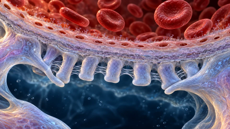
Living cells and extracellular material share the boundary.
Fenestrated endothelium and its surface layer face blood. Basement membrane lies between endothelial cells and the podocyte processes, whose slit diaphragms participate in the outer interface.
These components interact mechanically and biologically. They are not three independent inert screens. Modern native-human imaging resolves relationships far below the scale of this generated teaching preparation; its bridging strands are not certified molecular architecture.
Regulating a filter is different from raising systemic pressure.
Renal vascular resistance and local feedback influence glomerular pressures and plasma flow. Autoregulation can help limit the effect of changes in systemic pressure within a physiological range.
This does not mean more arterial pressure is always better, or that constricting one vessel has a single unconditional outcome. Use the existing juxtaglomerular lesson to follow local salt sensing and feedback rather than treating these choices as treatments.
Filtration starts a mixture; tubules determine what remains.
Glomerular filtration is a transfer from capillary plasma to capsular space. Reabsorption, secretion and later water adjustment change the fluid before final urine leaves the collecting system.
A change in final urine volume therefore does not, by itself, establish a change in filtration rate. Follow the existing tubular and ADH teaching for the next stages.
Experimental pressure and flow measurements, molecular transport studies and acquired human ultrastructure answer different questions. Combining them supports a physiological explanation; it does not make a generated image a measured specimen.
The choices here select an explanatory focus. They do not deform the filter, calculate GFR or simulate a patient. No diagnostic thresholds, fluid advice or medication rules are supplied.
Does higher blood pressure always mean more useful filtration?
No. Local capillary pressure, opposing pressures, functioning surface and renal regulation all matter. A systemic reading alone cannot determine them.
Is the basement membrane the only part that filters?
No. The endothelial surface, extracellular membrane and podocyte/slit relationships form an interacting living barrier.
Does strongly restricted albumin mean precisely zero protein is filtered?
No. Restriction is not an absolute zero, and subsequent tubular handling also affects what appears in urine.
Does a larger final urine volume prove a higher GFR?
No. Tubular recovery, secretion and collecting-duct water permeability also change the final volume.
Sources checked 21 October 2026. Normal educational scope. Animal measurements and human ultrastructure are distinguished; neither certifies the fine detail of generated anatomy. AI-assisted checks are not physician or physical-phone review. No personal GFR calculator or treatment advice.
Two poles do different work
At the vascular pole, an afferent arteriole supplies the glomerulus and an efferent arteriole drains it. At the urinary pole, capsular fluid proceeds into the proximal tubule. The blood route continues circulating rather than becoming the entire fluid route.
Pressure and selectivity both matter
Glomerular pressure favours outward filtration, while blood’s protein-related osmotic pressure and pressure in the receiving space oppose it. Net filtration depends on these forces and the available selective filtering surface. More blood pressure is not automatically healthier or better filtration.
Blood is above; receiving capsular fluid is below. Three interacting barrier components separate them.
Original generated cellular interpretation, not electron microscopy. Fenestrations, foot processes, bridging strands, surface coat and thicknesses are deliberately enlarged and not measured molecular structures. No hole size or literal pore-to-cell comparison can be inferred; whole blood cells normally remain inside the capillary.
Fenestrated endothelium
The blood-facing cell layer has small openings and a surface coat. Blood cells normally remain within the capillary.
Glomerular basement membrane
An extracellular layer between endothelial cells and podocytes contributes support and selective permeability.
Podocyte foot processes / slit diaphragms
Interlocking foot processes cover the outer capillary surface. Their slit diaphragms are part of the barrier, not open gaps in a simple sieve.
Barrier selectivity depends on the interacting endothelial surface, extracellular membrane and podocyte relationships, not one cartoon row of holes. Most large circulating proteins and blood cells are normally retained. Filtered water and small solutes enter the receiving space; the filtrate is not finished urine.
A nephron starts with a tiny blood-vessel filter and continues as a tubule. Filtration starts a separate fluid stream. Reabsorption returns material to blood; secretion moves selected substances into the tubule.
Blood is filtered and continues circulating. A second capillary network supports exchange and recovered material; filtration does not empty the whole circulation into the ureter.
Excretion concerns what finally leaves in urine. An increase in a filtered substance does not by itself prove the same amount will be excreted, because tubular recovery and secretion also change the final composition.
Filtration starts a separate stream; tubules recover some material and add selected substances. What finally leaves in urine is the result of all three transfers. Use the same anatomy to connect that journey with clearance.
Generated expanded teaching preparation, not the appearance of one measured dissection. Fine segment boundaries, local vascular origins and physical distances are illustrative. Return of the distal limb to its own vascular pole/macula densa is not spatially resolved in this expanded image; the source-supported relationship is explained below.
FilterBlood plasma → capsular fluid at the corpuscle.
RecoverTubular fluid → tissue → nearby blood.
Add by secretionBlood-side supply → tubular fluid.
ExcreteRemaining fluid → collecting system → urine.
Original qualified anatomy and label positions are retained. Read the structural names on the image; the four statements explain transfers between compartments rather than four precise molecular tracks.
One filtered load. Different final outputs.
Fixed illustration: plasma concentration 1 mass unit/mL, GFR 100 mL/min, renal plasma flow 500 mL/min. The solute is freely filtered, unchanged by renal metabolism, and has no renal production. These selected values are not patient data or normal targets.
Filtered reference
All of the filtered amount reaches urine. This ideal marker is freely filtered and has no tubular transport, renal production or breakdown.
Find the tissue: Start at the corpuscle. Follow the separate fluid route to its drainage; the blood route remains outside it.
The same solute output can travel in different amounts of water.
Urine flow1 mL/min
Urine concentration100 mass units/mL
Output stays the same100 × 1 = 100 mass units/min
Here the water choice changes concentration while keeping the selected solute balance fixed. It is a controlled comparison, not a simulation of all responses to drinking or a prediction of a person’s kidney function.
Two ratios, two questions
Urinary clearance: Cₓ = Uₓ × V̇ ÷ Pₓ. Equivalent plasma volume cleared of this solute per time; it is not urine volume.
Filtration fraction: GFR ÷ renal plasma flow. In this illustration 100 ÷ 500 = 20%. It describes plasma filtration before recovery, not the fraction of blood leaving as urine.
Read the assumptions behind the number.
Blood, plasma, filtrate and urine are four different streams.
Whole blood contains cells suspended in plasma. Renal blood flow counts both; renal plasma flow counts the plasma component. In a simple volume-accounting model, plasma flow equals blood flow multiplied by one minus the haematocrit, expressed as a fraction.
GFR describes fluid crossing from glomerular capillaries into capsular spaces per unit time. Most delivered plasma continues through the renal circulation rather than entering filtrate on that pass. Final urine flow is measured after tubular and collecting-duct handling. These volumes are not interchangeable.
A fluid rate becomes a solute load only after concentration is included.
For a freely filtered solute, multiply GFR by its plasma concentration to find the amount entering filtrate per unit time. A fluid rate in mL/min multiplied by concentration in mass/mL gives a mass rate, not another fluid rate. Higher concentration can raise a filtered load without changing GFR.
The simple expression assumes the solute concentration in newly formed filtrate matches the relevant plasma concentration. Protein binding or restricted passage changes that assumption. Total plasma concentration must not be used uncritically for a partly bound substance.
Count what crosses each boundary, then what leaves.
For an unchanged solute with no renal production or breakdown, urinary excretion equals filtration minus reabsorption plus secretion. The comparison uses amounts per unit time throughout. Concentration in one urine sample alone is not an excretion rate.
The difference between the filtered and excreted amounts identifies net tubular handling. It cannot reveal every opposing flux, every segment or a particular transport protein. A solute can be both recovered and secreted along its journey.
Clearance is an equivalent plasma volume, not a pocket of empty blood.
Divide urinary solute output per minute by the plasma concentration of that same solute. The resulting urinary clearance has units of volume per time. It asks how much plasma would contain the amount appearing in urine during that interval.
This is an accounting volume. The kidney does not produce a separate bottle of solute-free plasma, and clearance is not the volume of urine. Different solutes can have different clearances in the same kidneys at the same moment.
A filtration reference needs more than a matching number.
An ideal filtration marker crosses freely, is not bound to plasma proteins, and is neither reabsorbed, secreted, made nor broken down by the kidneys. Under suitable measurement conditions, its urinary clearance equals GFR. Inulin is the classic exogenous reference; its use requires controlled measurement rather than a visual inference.
For the freely filtered examples here, net recovery lowers clearance relative to GFR and net secretion raises it. Equality alone is not sufficient proof of absent transport: the opposing-transfer example gives the same net total. These comparisons change if free filterability or metabolic assumptions do not hold.
Changing water can change concentration without changing solute output.
Hold the solute excretion rate constant and double the urine flow. Its urine concentration halves; concentration times flow stays the same. With plasma concentration also fixed, calculated solute clearance remains unchanged.
This paired illustration deliberately holds other quantities constant to isolate the arithmetic. Real water regulation and solute transport can interact. The example does not assert that every change in urine volume leaves every solute clearance unchanged. Follow the collecting-duct lesson for the physiological water pathway.
Filtration fraction uses plasma delivery, not final urine.
Divide GFR by renal plasma flow to describe the fraction of delivered plasma volume that becomes glomerular filtrate. In this illustrative comparison, 100 divided by 500 gives 0.20, or 20%. These are selected teaching values, not a person’s measurements or universal normal targets.
A larger fraction can result from changes in its numerator, denominator or both. It does not state how much of the whole blood was filtered, how much solute was excreted, or how much final urine will form. Capillary protein concentration and downstream recovery add further physiology, explained in the existing filtration course.
Secretion helps a marker sample more than the filtered fraction.
At suitably low plasma concentrations, PAH is filtered and strongly secreted. Its urinary clearance can approximate effective renal plasma flow because much of the PAH delivered to functioning secretory tissue is removed during a pass. Extraction is high, not exactly complete.
True plasma flow requires accounting for the fraction extracted: in the appropriate steady-state balance, divide PAH clearance by its extraction ratio. Saturation, incomplete extraction and altered tissue function limit the approximation. PAH clearance is not simply another name for GFR. No infusion or testing instructions are provided here.
Creatinine arises from creatine metabolism, largely associated with muscle. It is filtered and also undergoes tubular secretion, so its measured urinary clearance need not equal an ideal filtration-marker clearance. Its plasma concentration also depends on production and the time taken to reach a new balance.
Laboratory estimated GFR, measured urinary clearance and directly measured filtration are related but distinct methods. Muscle mass, changing concentrations and collection quality matter. This normal-physiology lesson supplies no clinical cut-offs, personal estimate or diagnosis.
A stable concentration means inputs and outputs balance, not that nothing happens.
A substance can enter the body continuously and leave continuously while its plasma concentration stays fairly stable. After a change in clearance, concentration may change until generation and elimination reach another balance. The timing and other routes of elimination matter.
The simple tubular ledger excludes renal synthesis and destruction of the chosen solute. Glucose metabolism, ammonia production and other chemical transformations require additional terms; do not force them into the unchanged-marker example. The existing renal ammonia course explicitly follows those transformations.
A calculated relationship and a human experiment are different evidence.
The fixed values above demonstrate units and conservation; they are not a dataset. Historical human inulin work examined how concentration and collection affected the measured result. Timing, urine collection and the plasma value used for the interval influence a real clearance estimate.
The original human study was small and historical. It illustrates measurement constraints rather than supplying universal contemporary reference ranges. Generated anatomical preparations locate tissue compartments; neither their texture nor an emphasis ring measures clearance.
Does more concentrated urine necessarily remove more solute?
No. Compare concentration multiplied by urine flow over the same interval. Less water can concentrate the same output.
Can clearance exceed GFR without breaking conservation?
Yes. Secretion can add solute from the blood-side supply beyond the amount filtered. It does not create solute.
Does clearance equal to GFR prove that no tubular movement occurred?
No. Appropriate marker properties must be known; equal opposing transfers can cancel in a net measurement.
Does 20% filtration fraction mean 20% of the blood becomes urine?
No. Its denominator is renal plasma flow; most filtrate is subsequently recovered before urine leaves.
Can this unchanged-solute ledger describe renal ammonia production without extra terms?
No. Production and chemical conversion require additional accounting; follow the dedicated ammonia course.
Sources checked 22 October 2026. This is normal physiology teaching with fixed illustrative quantities. AI-assisted checks are not physician review or certification of generated fine tissue. Existing real-image provenance is unchanged; no clinical calculator, infusion procedure or treatment advice is supplied.
Tube wall → epithelial cell → transport
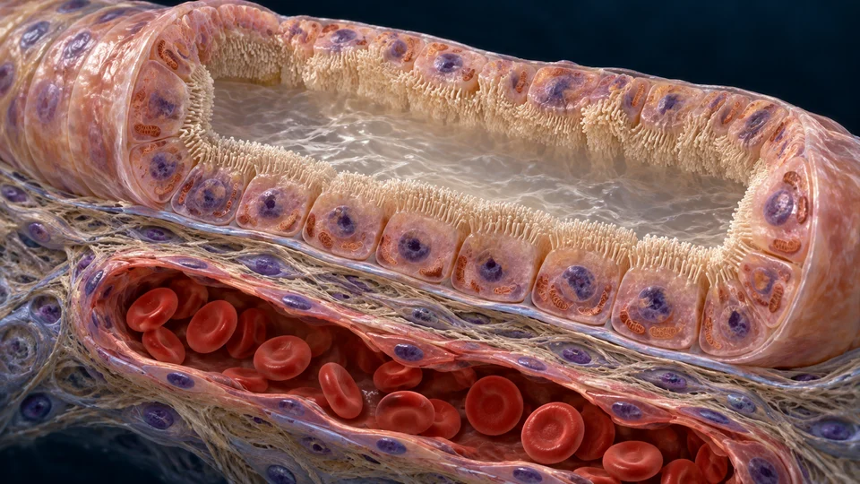
Two working faces. Different regional jobs.
Filtrate changes along the tube. Reabsorbed material returns to circulation through nearby capillaries. Secretion adds selected substances to tubular fluid; excretion is what finally leaves in urine.
Generated proximal epithelial enlargement, not authentic histology. Cell counts, organelle shapes and membrane thickness are illustrative. Short brush-border microvilli increase surface area; they are not the motile cilia of the respiratory clearance mechanism.
Lumen to surrounding tissue
Tubular fluid faces the luminal surface. The basal side faces supporting and interstitial tissue; recovered material then reaches nearby capillaries. Reabsorption moves from fluid toward circulation, while secretion has the opposite overall direction for selected substances.
Energy supports selective recovery
Pumps maintain ion gradients using energy. Other transporters can use those gradients; water follows osmotic conditions rather than an ADH water pump. The route and transport proteins vary by segment, and both through-cell and between-cell pathways can contribute.
Proximal tubule
Bulk recovery
Returns much of the water and sodium and normally recovers filtered glucose and amino acids. A brush border increases the surface available for transport.
Descending thin limb
Water leaves
Water moves toward the more concentrated medullary surroundings. The remaining tubular fluid becomes more concentrated.
Ascending limb
Salt leaves; water stays
Salt leaves while the wall is poorly permeable to water. Active salt transport in the thick limb helps maintain the medullary gradient.
Distal tubule
Ion adjustment
Further sodium and chloride recovery occurs. Later segments participate in hormone-sensitive sodium and potassium balance.
Collecting duct
Final adjustment
ADH increases water permeability. Principal and intercalated cells also contribute to ion and acid–base regulation before fluid drains toward a papilla.
A concentrated medulla provides the driving gradient. ADH controls how readily water can leave the collecting duct. Use the expanded anatomical loop and receiving duct above for location, then distinguish their mechanisms.
Multiplier ≠ exchanger
The nephron loop has unequal water and salt permeability. In the descending thin limb, water can leave toward a more concentrated environment. The ascending side transports salt while remaining poorly permeable to water; active transport in its thick portion contributes to the gradient. Opposing flows and regional transport cooperate across the medulla.
Vasa recta help preserve it
Vasa recta exchange is largely passive. Descending blood gains solute and loses water; returning blood reverses those exchanges. Circulation can remove recovered water while helping limit rapid washout of the medullary gradient. Drawing blood and filtrate as two U-shaped routes would not make their roles interchangeable.
Urea also contributes
Urea recycling adds to inner-medullary osmotic conditions. The concentrating mechanism is a tissue partnership, not simply one salt pump or one straight tube. A literal unchanging gradient or fixed collecting-duct water output is not implied.
The gradient and the gate.
ADH changes collecting-duct water permeability. It does not pump water or create the whole gradient on its own. Water can leave toward a more concentrated interstitium when the duct is sufficiently permeable; it returns to circulation through surrounding tissue and vessels.
LOOK CLOSER · FROM TUBULAR FLUID TO CIRCULATION
A permeable wall. A separate blood route.
Find the lumen-facing surface, cross the epithelial wall, then distinguish surrounding tissue from capillary blood. Water recovery requires both a permeable route and an osmotic driving gradient.
Original generated tissue preparation, not acquired microscopy. Principal-cell identity is the teaching context, not a diagnosis from cell appearance. Vesicles, membranes and organelles show gross compartments; individual aquaporins are not resolved. The tubular and blood lumens remain separate.
The luminal gate is on the apical side.
ADH (vasopressin) acts from the tissue-facing side through V2 receptors. Intracellular signalling increases the amount of AQP2 in the lumen-facing membrane. Water then enters down an osmotic gradient; the hormone does not pump it across.
Gold shows the selected overall transfer direction, assuming conditions favor reabsorption. It does not trace one molecule, identify a membrane pore or measure water flux. The ADH comparison below changes permeability; it does not change these anatomical compartments.
One signal, two time scales
Rapid regulation moves existing AQP2 between intracellular vesicles and the apical membrane. Longer-term regulation can change AQP2 abundance. A snapshot of one cell cannot measure its hormone level or water flux.
More water permeability alone does not guarantee concentrated urine. The surrounding osmotic environment depends on the renal tissue partnership, including loop transport, blood exchange and urea handling. Outer-medullary countercurrent multiplication is well established; the complete inner-medullary concentrating mechanism remains incompletely explained.
Osmotic sensing, thirst and vasopressin link water intake with renal water recovery. Vasopressin is synthesized in hypothalamic neurons and released from the posterior pituitary. Volume-related signals also affect its secretion; this is not a single-variable feedback switch.
Aquaporins provide a passive water pathway. Energy-dependent solute transport elsewhere helps establish conditions for water movement, while vasopressin signalling regulates the availability of apical AQP2. These are different roles; ATP is not consumed to push each water molecule through an aquaporin.
The tissue preparation has no measured osmolality scale or individually resolved channels. AQP2-bearing vesicles described in the mechanism are not identified from the illustrated round organelles. Fine molecular architecture and a complete inner-medullary exchange network require other evidence.
Directed source and selected gross-structure checks: 16 October 2026. Original generated teaching illustration with independent semantic positions, editable names and direction cues. Not acquired microscopy, a patient simulation or human medical review.
Kidneys adjust fluid and chemistry while taking part in endocrine signalling. Water, salts, acid–base balance, circulation and bone metabolism connect this chapter to the rest of the body.
Tubule ↔ blood
Sodium and potassium
Aldosterone promotes sodium recovery and potassium secretion in late distal and collecting segments. Water retention depends on water availability and permeability; aldosterone is not an interchangeable name for ADH.
Acid → urine / bicarbonate → blood
Acid–base balance
The kidneys reclaim filtered bicarbonate and generate new bicarbonate while excreting acid. This complements the lungs’ control of carbon dioxide; neither organ acts alone.
Reduced renal perfusion, sympathetic input and low salt delivery at the macula densa can stimulate renin. The resulting hormone network supports pressure and sodium balance; it is not simply an on/off blood-pressure switch.
Kidney → erythropoietin → bone marrow
Red blood cell support
The kidneys produce erythropoietin, a signal supporting red blood cell production. They do not manufacture the blood cells themselves.
Kidney → active vitamin D → calcium balance
Vitamin D activation
The kidneys help convert vitamin D to its active form, calcitriol. This links renal function with intestinal calcium uptake and bone health.
Local control is different from hormone signalling
Renal vessels and feedback around the glomerulus help adjust local delivery and filtration across ordinary changes. Tubuloglomerular feedback connects sensed distal salt delivery to vascular responses. Renin is released by specialised neighbouring vascular cells; macula densa sensing is not the same as manufacturing all downstream hormones.
Keep water and salts distinct
ADH-sensitive water permeability and aldosterone-sensitive sodium/potassium handling interact but are not interchangeable. Volume, osmolality, pressure and potassium are related variables with different signals. These are causal teaching links, not dosage advice, diagnostic targets or a complete hormone network.
These two epithelial preparations show different jobs. They are not a fixed sequence followed by every tubular cell, and the blood spaces remain separate from tubular fluid.
Original generated tissue preparation, not a micrograph. The magnified cell/brush-border setting identifies broad compartments. Transporters, carbonic-anhydrase molecules and measured dimensions are not resolved; arrows are selected functional directions.Structure notes · further context
Tubular fluid
Filtered bicarbonate is present in tubular fluid. This compartment is distinct from the blood below the epithelium.
Apical brush-border context
The luminal face has a dense microvillous brush border. Here secreted H⁺ can help convert luminal bicarbonate to CO₂ and water.
Proximal epithelial cell
CO₂ can enter the cell and participate in intracellular bicarbonate formation; this is not a direct apical bicarbonate hole.
Mitochondrial context
Numerous mitochondria and basal membrane organisation support an active transporting epithelium. Individual organelle counts are illustrative.
Blood-facing basal membrane
Bicarbonate exits across the basolateral cell membrane through transport mechanisms including NBCe1, not by opening the cell to the capillary.
Interstitium and support
Supporting tissue lies between tubular epithelium and a separate capillary. The two fluid lumens do not join.
Peritubular blood context
A reclaimed bicarbonate equivalent returns toward circulation. Red cells remain within the separate blood vessel.
Convert before crossing the luminal surface.
In this reclamation context, luminal H⁺ and filtered bicarbonate form CO₂ and water, aided by surface carbonic anhydrase. CO₂ can cross the epithelial membrane.
Apical tubular fluid and basolateral blood are separate compartments.
This view isolates filtered-bicarbonate recovery; it does not map every proximal-tubule process.
Original generated type-A intercalated-cell teaching context, not diagnostic histology. Transport polarity is explicitly supplied by the independently sourced overlay; morphology alone does not identify every cell subtype. Not all collecting-duct cells share this role.Structure notes · further context
Urinary fluid and buffers
Secreted H⁺ can bind urinary buffers. Ammonium and titratable-acid handling contribute to net acid excretion.
Luminal acid-secretory face
Type-A intercalated cells have apical proton-secretion mechanisms. The illustration does not resolve an individual H⁺-ATPase.
Selected intercalated-cell context
This is one acid-secretory epithelial role, not a model in which every collecting-duct cell performs the same transport.
Mitochondria-rich cytoplasm
Abundant mitochondria and irregular apical surface context differ from a uniform proximal brush border. The fine generated structures are not clinical subtype markers.
Basolateral exchange context
AE1 supports bicarbonate exit and chloride entry at this cell face. It must not be placed on the apical surface in this type-A example.
Interstitium
A separate supporting compartment lies between the epithelium and blood vessel; no open urine-to-blood pipe is present.
Blood-side bicarbonate return
Renal net acid excretion can generate new bicarbonate equivalents for the body. This differs from merely reclaiming filtered bicarbonate.
Direct H⁺ toward the lumen.
In this selected type-A context, apical proton secretion and basolateral bicarbonate exchange have different directions. The cell is not simply a reversed proximal tubule.
Type-A acid secretion is different from bicarbonate secretion by other intercalated-cell roles.
Renal acid–base balance is distributed; the two preparations are not a universal two-cell assembly line.
Why is recovering bicarbonate not the same as creating new bicarbonate?
Reclamation prevents filtered base from being lost. Net acid excretion, including ammonium and titratable-acid routes, replaces base consumed in the body. Proton secretion can participate in either setting; its presence alone does not establish net acid removal.
Do all collecting-duct cells secrete H⁺ in the direction shown?
No. This is a selected type-A acid-secretory context. Other intercalated-cell roles can secrete bicarbonate, while principal cells have different transport functions. Ammonia handling also spans multiple nephron segments; neither image is a complete kidney transport map.
Normal-physiology sources checked 20 October 2026. AI-assisted source and structural checks are not physician, ultrastructural or physical-phone certification. No diagnostic targets or treatment rules.
Make an acid carrier. Move it between segments. Count what actually leaves.
The preserved base-recovery and acid-secretory preparations above explain two epithelial jobs. This course connects their cellular setting to nitrogen metabolism and the final whole-body result.
Glutamine → proximal metabolismGenerate ammonia-related nitrogen and bicarbonate equivalents.
Loop ↔ tissue → collecting ductRedistribute nitrogen; NH3 and secreted H+ can meet in tubular fluid.
Final urine → net acid removalExcretion differs from early secretion or nitrogen returned to blood.
A circulating amino acid can supply an excretable acid carrier.
Glutamine metabolism generates ammonium together with bicarbonate equivalents.
Retained original generated proximal-cell illustration, not microscopy. The opened cell locates metabolism and separate urine-facing and blood-facing boundaries. It does not identify individual enzymes or NHE3 molecules. The membrane is cut open for teaching, not ruptured in life. Selected teaching context: Make the carrier. Glutamine metabolism generates ammonium together with bicarbonate equivalents. Gold rings locate discussed structures; they do not show measured flow or a direct intersegmental duct. Read the image: Locate the proximal epithelial cytoplasm and mitochondrial context. The preparation locates the metabolic setting; it does not identify glutaminase or reconstruct each chemical reaction.Structure notes · further context
Tubular fluid
Early secreted nitrogen can still be transported again before final excretion.
Brush-border face
An apical boundary supports secretion into tubular fluid; NHE3 is not visibly resolved.
Proximal cell
Glutamine nitrogen and carbon are processed through several metabolic pathways.
Mitochondrial context
Mitochondrial reactions participate in ammoniagenesis; texture does not identify an enzyme.
Basal boundary
Transport toward interstitial fluid is separate from transport into the lumen.
Outside the epithelium
Supporting interstitium lies between epithelial cells and surrounding blood vessels.
Separate blood space
Base equivalents can return to blood; nitrogen returned in venous blood has a different fate from urinary excretion.
Proximal tubular cells metabolise glutamine. Its nitrogen can enter the ammonia pool, while metabolism of the carbon skeleton contributes bicarbonate. This cellular production is different from simply filtering ammonia already present in blood.
Read the image: Locate the proximal epithelial cytoplasm and mitochondrial context. The preparation locates the metabolic setting; it does not identify glutaminase or reconstruct each chemical reaction.
Recovering existing base and adding new base are different
Filtered bicarbonate must normally be returned rather than lost. The existing proximal lesson shows how luminal conversion and epithelial transport recover a bicarbonate equivalent. H+ can be secreted and reused during this process.
Net acid excretion replaces base consumed elsewhere in the body. Ammonia metabolism contributes a major route. Identifying a proton pump or a bicarbonate transporter does not, by itself, distinguish reclamation from a net addition of base.
Glutamine supplies nitrogen and a metabolised carbon skeleton
The proximal tubule is the principal site of renal ammoniagenesis. Mitochondrial glutaminase and glutamate-dehydrogenase pathways participate in removing glutamine nitrogen; subsequent carbon metabolism contributes bicarbonate equivalents. Production involves several reactions and compartments.
The familiar net summary of two ammonium and two bicarbonate equivalents per fully metabolised glutamine is biochemical bookkeeping. It is not a count of particles in the picture, a universal rate for every cell or proof that every produced nitrogen equivalent will leave in urine.
NH3 and NH4+ interconvert, but do not cross membranes identically
Ammonia is often used as a collective term for NH3 and NH4+. NH3 is uncharged; accepting a proton forms NH4+, which is charged. At physiological conditions much of the total is present as ammonium, but the smaller NH3 pool can be important for transport.
Membrane proteins and acid–base equilibrium determine which forms move where. The older picture of ammonia freely diffusing through every membrane is incomplete. Protonation in the lumen can promote retention, but it does not make the entire nephron a perfect irreversible trap.
From cell production to an early tubular nitrogen supply
Ammonia-related nitrogen can reach proximal fluid through NH4+ transport and through NH3 movement coupled with proton secretion. NH4+ can substitute for H+ in NHE3 transport. The brush border locates a working membrane, not a visible opening that pours cell contents into urine.
Not all produced ammonia is immediately committed to excretion. Some enters the circulation; some is transported and recycled within the kidney. Early tubular secretion is therefore a stage in handling, not a direct measurement of final urinary output.
The loop and collecting duct cooperate through surrounding tissue
The thick ascending limb can take up NH4+ by using the potassium-binding position of NKCC2. Basolateral exit and other transport processes contribute to medullary accumulation and recycling. A substance can leave tubular fluid at one site and still support its later secretion into a different segment.
The medullary interstitium is a tissue compartment, not a hidden pipe joining two tubules. The anatomical nephron map gives locations; the exact three-dimensional recycling network, concentration gradient and transporter distribution need more detailed evidence than that overview supplies.
Collecting-duct NH3 transport and H+ secretion are separate jobs
Rh proteins, particularly Rhcg, contribute to collecting-duct ammonia movement. Acid-secretory machinery can supply H+ to the lumen independently. Bringing the two components together favours ammonium formation and lets substantial acid be carried without all of it remaining as free H+.
Species, segment and cell type matter when assigning individual transporters. The retained type-A intercalated-cell illustration teaches an acid-secretory context; it does not certify every hidden membrane protein or exclude contributions from other collecting-duct cells.
Final urine and renal venous return lead to different outcomes
Urinary ammonium removes nitrogen and an acid equivalent from the body. In contrast, ammonia returned to systemic blood can be metabolised in the liver. Hepatic urea synthesis consumes bicarbonate, offsetting the same kind of retained-base benefit.
Urea excretion and ammonium excretion are therefore not interchangeable terms. The liver nitrogen course explains urea synthesis; this course follows the renal ammonia route. Changing the final nitrogen destination changes the acid–base accounting even when proximal production has occurred.
Amount of excreted acid is not the same as urine pH
Net acid excretion includes ammonium and titratable acidity, with urinary bicarbonate loss counted in the opposite direction. Phosphate and other urinary buffers can carry acid. Free H+ concentration alone does not describe the total amount carried out.
Physiological adaptation can change glutamine handling, production, segmental transport and nitrogen partitioning. This supporting account is not a urine-test calculator, dietary recommendation or diagnostic rule. It complements the established blood and lung lessons without supplying treatment instructions.
Has nitrogen already left the body when it enters proximal tubular fluid?
No. Later transport can redistribute it. Final excretion in urine is different from early secretion into a tubule.
Does reclaiming filtered bicarbonate always create an additional base gain?
No. Reclamation prevents loss of base already present. New bicarbonate linked to net acid excretion is a different accounting step.
Does the nephron overview show a duct between the loop and collecting duct?
No. Medullary handling involves transport through cells and surrounding tissue. The two tubular lumens remain anatomically separate.
Can urine acidity alone tell how much acid was excreted?
No. Buffer-bound acid, ammonium and bicarbonate loss affect total net excretion. This lesson provides no clinical interpretation rule.
Normal physiology · source checks 21 October 2026. Three retained wordless generated anatomical preparations, with original structural positions and independently editable terminology. Not acquired microscopy, fine histological certification, physician review, a urine-test interpretation or treatment/dietary guidance.
Connect the opened kidney with its three-dimensional setting.
Use the native reference to locate the organ, its blood supply and the route toward the bladder. Keep the chapter preparations alongside it to understand the working tissues. Choose text view when you prefer source names and learning links without loading geometry.
Adult male whole-body source
Kidneys, renal vessels and urine transport remain separate source structures.
This partial female assembly includes named internal regions and collecting routes. It does not identify a separate urethral mesh; use the chapter preparation to follow the outlet.
Native model shapes are retained. Named surfaces do not demonstrate filtration, transport rates, muscle contraction or a complete fine vascular tree. Use the cutaways, real microscopy and mechanisms in this chapter for those other scales.
From a picture to a connected explanation
Connect the scales.
Filter → selective cell interfaces
Why is filtrate different from whole blood and from finished urine?
Genuine human cortex shows a rounded corpuscle beside tubular profiles. It cannot map every three-dimensional tubular connection.
A filtering corpuscle lies beside working epithelial tubes.
The original acquired cortex image is retained. Use its broad structures to connect two different tasks, while keeping the photograph separate from the opened nephron and cellular transport illustrations.
RECORDED · LIGHT MICROSCOPY
Tubular profilesCapsular space contextCorpuscle / capillary tuft
Authentic human cortex, bright-field microscopy, not generated detail. Source stain is undocumented. No individual tubular subtypes, podocyte slits or diagnosis are assigned; the embedded0.1mm scale bar is retained, and display magnification varies with the screen.
Josef Reischig, CSc. · exact source and rights · CC BY-SA 3.0. Full acquisition frame retained, proportionally encoded for display; no generated microscopy, recolouring, sharpening or artificial detail.
A corpuscle is a filtering unit, not a large tubular cell.
Read the recorded structure
Find the rounded region at lower left and the pale capsular-space context around its tuft. The photograph retains the previously checked broad observation targets.
Connect structure with normal function
Glomerular filtration transfers filterable water and solutes from capillary blood into capsular space. It is different from the later work of tubular epithelium.
Keep the inference within the evidence
The fine filtration layers, podocyte slits, filtration pressure and rate are not established by this image.
Gastric epithelium combines protection with specialised secretion. Intestinal epithelium receives material from the lumen, while renal tubules selectively exchange material with their surroundings after filtration. Shared epithelial organisation does not make these organs interchangeable or their transport mechanisms identical.
Sources checked 20 October 2026. Source-photo rights are unchanged; new independent labels and explanations are offered under CC BY-SA 4.0. Source identities and normal biology checks are distinct from physician, fine-histology and original-calibration certification.
Josef Reischig, CSc. · author’s archive / source image · CC BY-SA 3.0. Original source and colour retained. Proportional WebP resizing only, no crop, recolour or AI detail; photo derivatives retain CC BY-SA 3.0. Source600× refers to a26cm printed width, not an on-screen setting.
Imaging · Where and how large?
Ultrasound and other urinary imaging can show organ shape, collecting-space enlargement or an obstructed route. They do not directly show the molecular filtration barrier. In intravenous pyelography, injected contrast enters urine and can outline collecting spaces, ureters and bladder on X-ray. This describes an imaging principle; no patient radiograph is displayed here.
Microscopy · What tissue?
Light microscopy shows renal corpuscles and tubular profiles in tissue sections. Electron microscopy resolves much finer barrier relationships. The human cortex photograph supplies tissue evidence; the enlarged corpuscle and barrier images remain illustrations, with no generated picture presented as microscopy.
Tests · How is it working?
Blood-based estimated GFR and urine albumin measurements answer different questions: filtration performance and signs of kidney damage. Interpretation needs clinical context. This chapter provides no result calculator or personal diagnostic threshold.
When a pathway changes
Crystals can form solid stones. A stone may obstruct urine drainage, which differs from an injured filtration barrier. Kidney infection often develops when bacteria spread upward from the bladder, rather than following the blood-to-filtrate arrow. Diabetes and high blood pressure can damage kidneys over time. Neither organ shape nor a generated filter image diagnoses these problems.
The kidneys filter blood, return much of what the body needs and send remaining wastes and water toward the bladder. Original explanations retain existing renal knowledge and expand the anatomy-to-mechanism relationships. Independent sources are separate from the reference-book teaching benchmark.
Generated fine pore sizes, slit strands, branch paths, cell counts and dimensions remain illustrative. No exact incoming/outgoing vascular assignment, macula-densa location or sex-specific urethral anatomy is claimed from unresolved pixels. Source checks and AI visual inspection are not human medical review; physical-phone acceptance is separate from simulated narrow-screen checking.
Authentic electron microscopy, patient urinary imaging, full verified continence / pelvic-floor anatomy and detailed disease education remain deeper gaps. The prior NIDDK IVP explanatory artwork is preserved in project history, with its correct principle retained here; it is not relabelled a patient X-ray or used as the main anatomy image.
Educational normal-function chapter, not exhaustive renal medicine or individual diagnosis/treatment. No kidney-specific interactive3D model or unavailable video is claimed.
 Regions work together
 Two working faces
 Follow the exit route