Receive and accommodate
Entry → proximal reservoir
The proximal stomach relaxes as a meal arrives. Vagal reflexes and changes in tone create a reservoir without treating the organ as a rigid bag.
CHAPTER / DIGESTIVE SYSTEM
Receive. Mix. Move onward.
Follow a meal into a changing muscular chamber. Explore its regions, wall and glands, then connect secretion, protection and movement with the rest of digestion.
Follow the wall, glands and meal →Chapter overview film planned. No playable video is currently available.
02 / ORGAN TO REGIONS
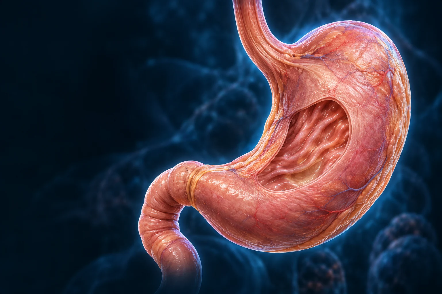
Below the diaphragm, mainly in the upper left abdomen, the stomach links the esophagus to the duodenum. Its shape changes with position and filling; a teaching cutaway is not a fixed outline for every person.
One chamber. Several overlapping jobs.The upper stomach receives a meal; the wall mixes it with secretions; the pyloric route delivers small portions to the duodenum. Receiving, mixing and emptying overlap rather than forming three sealed compartments.

Entry and reservoirEsophagus → cardia → proximal stomach
Mixing in one lumenBody → antrum ↔ return of contents
Controlled onward deliveryPyloric route → duodenum
Gold follows selected forward directions; cyan returns toward the wider chamber during mixing. The short duodenal cue does not trace the hidden pyloric opening. These local cues are not measured trajectories, rates or a contraction simulation. The arrows do not turn rugae into pipes. See how the wall supplies force →
Entry → proximal reservoir
The proximal stomach relaxes as a meal arrives. Vagal reflexes and changes in tone create a reservoir without treating the organ as a rigid bag.
Toward antrum ↔ retropulsion
Contractions carry contents toward the antrum. Resistance at the outlet sends some material back for further mixing and particle breakdown.
Small portions → duodenum
Fluid and sufficiently small particles pass into the duodenum. Gastric and duodenal signals regulate the delivery; digestion is not complete at this point.
These functions overlap during a meal. This is not a fixed transit clock or a simulation of pressure, particle size or emptying rate.
OpenStax · Stomach mechanical digestion · NIDDK · Digestive movement. Direction and normal-function check: 17 October 2026. Not physician review.
The lesser curvature is the shorter medial border; the greater curvature is the longer convex border. The cardia, fundus, body and pyloric region are broad anatomical areas, not four sealed rooms.
The liver lies nearby, and the pancreas lies behind the stomach. Arterial branches from the celiac circulation supply the wall; venous drainage reaches the portal system. This view does not map surgical vascular variants.
Follow the separate portal blood route →Continuous cavity, regional anatomy and controlled onward passage: OpenStax · Stomach structure and function · NIDDK · Digestive movement and controlDirected normal-function and claim correspondence check: 17 October 2026. Source checks are not physician review or a certificate for every generated pixel.
The reference shows a fixed stomach surface. Use the cutaway, wall and gland preparations here for relationships that this mesh cannot expose.
03 / FROM SURFACE TO MUSCLE
The mucosa faces the contents. Supporting submucosa lies beneath it, then the thick muscular wall and outer serosa. Rugae involve the inner layers and flatten as the organ expands.
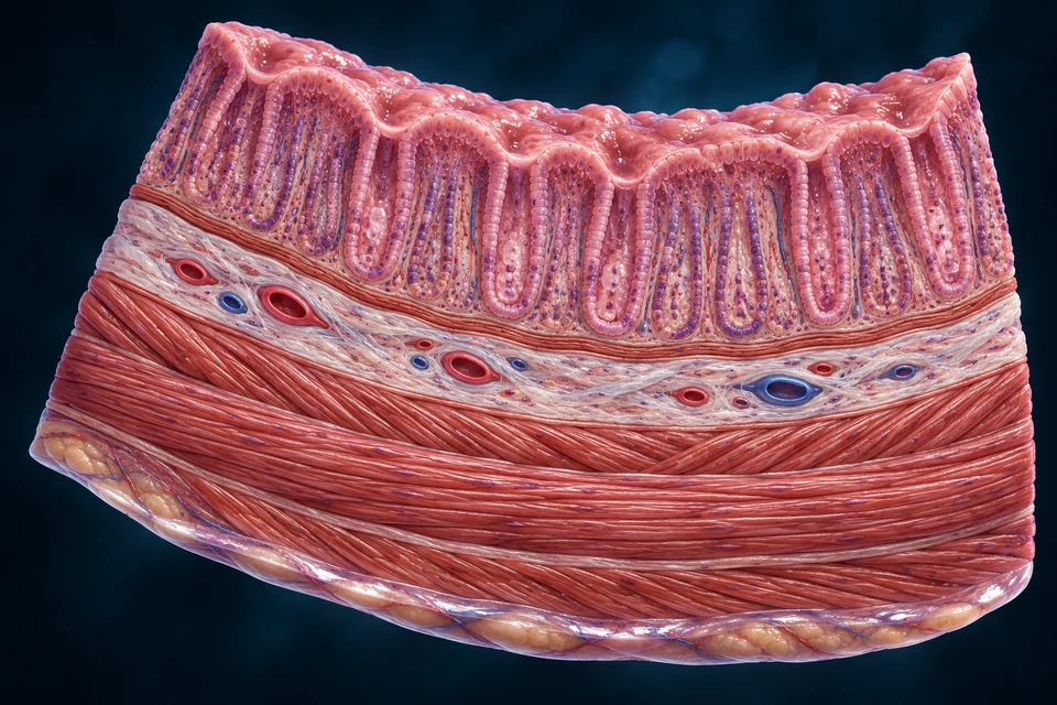
Oblique, circular and longitudinal smooth-muscle arrangements work together to change shape, mix and propel contents. They are not three independently timed pumps.
The submucosal and myenteric enteric plexuses help coordinate secretion and movement. Vagal pathways connect this local activity with wider digestive control. The generated wall does not resolve individual nerve fibers.
Explore neural signaling context →Wall layers, smooth muscle and enteric coordination: OpenStax · Stomach structure and function · NIDDK · Digestive movement and controlDirected normal-function and claim correspondence check: 17 October 2026. Source checks are not physician review or a certificate for every generated pixel.
04 / SMALL OPENINGS, DEEPER GLANDS
Gastric pits descend into the mucosa and communicate with glands. These are inward passages, not intestinal villi. The gland lumen and nearby blood vessels remain separate.
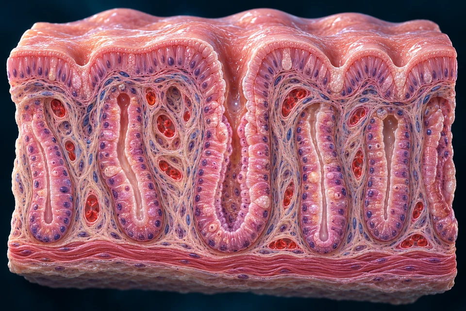
SUPPORTING CONNECTION INSET / SIMPLIFIED, NOT TO SCALE
Gland cells release into the digestive lumen. Blood stays in separate vessels outside the epithelium. Fine cell identity is explained below rather than guessed from generated texture.
Short pits lead to longer glands with prominent acid- and enzyme-producing cell populations. The gastric surface remains an epithelial barrier around those openings.
Mucous glands are prominent around the entry and outlet regions. Pyloric glands have deeper pits and include the main gastric G-cell population. One enlarged fundic view cannot represent every region.
Gastric pits lead into glands in the mucosa; regional glands differ: University of Leeds · Gastric tissue · University of Leeds · Cardiac and pyloric comparisonDirected normal-function and claim correspondence check: 17 October 2026. Source checks are not physician review or a certificate for every generated pixel.
05 / CELL ROLES
Some cells release products toward the digestive lumen; others signal through blood or within the tissue. Their identity requires appropriate evidence, not a guessed color in our illustration.
Mucus + bicarbonate
Surface and gastric pits
Help maintain a protective microenvironment over the epithelium.
Mucus
Neck of fundic glands
Contribute mucus within the glandular region; they differ from surface cells.
Acid + intrinsic factor
Fundic/body glands
Secrete acid into the lumen and a protein required later for vitamin B12 absorption.
Pepsinogen + gastric lipase
Deeper fundic/body glands
Supply a protease precursor and an enzyme that contributes to fat digestion.
Histamine
Fundic/body mucosa
A local signal stimulates neighboring parietal cells.
Gastrin
Mainly pyloric/antral mucosa
Release a hormone into blood that helps coordinate gastric secretion.
Somatostatin
Gastric mucosa
Provide inhibitory feedback within gastric secretion control.
Selected normal roles, not a cell census. G cells are not asserted to fill every fundic gland. Surface renewal and local signaling vary across the mucosa.
Selected secretory cell products and different tissue destinations: University of Leeds · Gastric tissue · OpenStax · Stomach structure and functionDirected normal-function and claim correspondence check: 17 October 2026. Source checks are not physician review or a certificate for every generated pixel.
06 / CHEMICAL WORK
Acid helps unfold dietary proteins and creates conditions for pepsin activity. Pepsin cuts protein chains into smaller peptides. Much further digestion and absorption take place in the intestine.
An inactive protease precursor enters the gland and stomach lumen.
Pepsinogen changes into active pepsin; pepsin can help activate more precursor.
Smaller peptides move onward for pancreatic and intestinal processing.
Parietal cells use a luminal H+/K+-ATPase to move hydrogen ions in exchange for potassium, using energy from ATP. Chloride transport contributes separately to the acidic secretion; acid is not packaged as a digestive-enzyme granule.
Gastric lipase contributes to fat digestion. Carbohydrate breakdown begins before the stomach and continues later. The stomach neither completes all nutrient processing nor absorbs most nutrients.
Continue the enzyme story in the pancreas →Acid conditions, pepsinogen activation and protein digestion: OpenStax · Stomach structure and function · NCBI · Physiology, PepsinDirected normal-function and claim correspondence check: 17 October 2026. Source checks are not physician review or a certificate for every generated pixel.
ONE QUESTION / CELL → MEAL → LIVING SURFACE
Acid-secreting cells export hydrogen ions toward the stomach’s contents rather than storing pre-made acid. Meanwhile, local buffering, connected surface cells, repair and blood supply protect the lining. Secretion and protection work together, but in different spaces.
A parietal cell is a specialized acid-secreting cell in a gastric gland. Follow its two membrane sides, then ask what happens to the acid after a meal arrives.
 Follow the open gland space into the cell cutaway ↓
3 MINUTES / FOLLOW THE BOUNDARIES
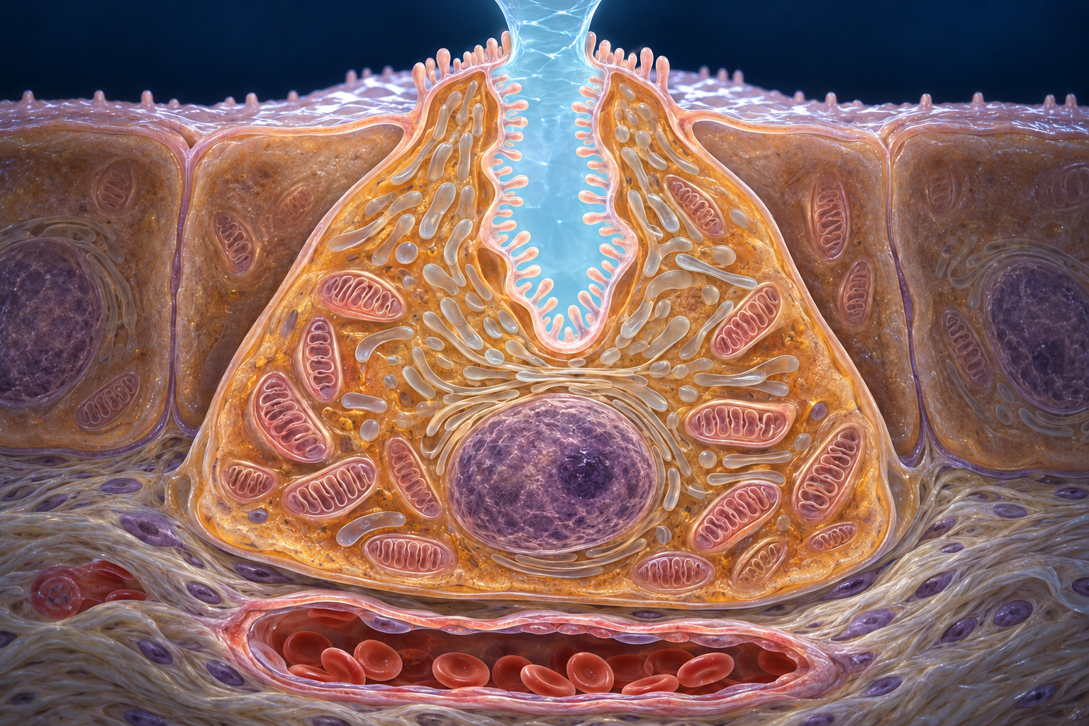
In glands of the stomach body and fundus, one cell connects a blood-facing supply side with a deeply folded lumen-facing secretory side. “Apical” means lumen-facing here; “basolateral” refers to the sides facing surrounding tissue.
A parietal cell is an acid-secreting cell in a gastric gland. Its canaliculus is a narrow, lumen-connected invagination of the apical membrane. The pale space is continuous with the gland lumen; it is not an acid reservoir inside the cytoplasm.
Gland lumen ↔ canalicular lumen. Blood remains in a separate vessel outside the cell.
The H+/K+-ATPase uses ATP, the cell’s usable energy supply, to move hydrogen ions from cytoplasm toward the lumen while taking potassium the opposite way. A separate potassium pathway returns K+ toward the lumen. These cooperating routes are not one open hole through the membrane.
H+: cell → lumen. K+: lumen → cell at the pump, then cell → lumen through a separate recycling pathway.
On the blood-facing side, a key exchanger moves bicarbonate out of the cell while bringing chloride in. Chloride then leaves across the lumen-facing membrane through a separate pathway. The proton pump itself does not carry chloride or bicarbonate.
Blood-facing exchange: bicarbonate out / chloride in. Apical chloride exit: cell → lumen.
Carbonic anhydrase speeds the reversible reaction of carbon dioxide and water that supplies hydrogen ions and bicarbonate. It does not pump them across membranes. Mitochondria support the cell’s energy demands; the image does not measure ATP production or identify individual transport proteins.
Cytosolic reaction supplies ions; membrane transport determines where they go.
The cytosolic reaction links CO₂ and water with H+ and HCO₃−. It is reversible; carbonic anhydrase accelerates it. This is not a complete quantitative membrane model, and the generic chloride exit does not identify a single exclusive human channel.
Evidence: Parietal-cell canalicular secretion ↗ · Human gastric AE2 localization ↗
SIGNAL → MEMBRANE → SECRETION
Parietal cells contain an internal pool of pump-bearing membranes. During stimulation, membrane can join the canalicular surface; retrieval helps reverse this redistribution. This moves secretory machinery, not little bags of stored hydrochloric acid.
Neural acetylcholine, circulating gastrin and local histamine cooperate. Gastrin promotes histamine release from nearby enterochromaffin-like (ECL) cells; histamine stimulates parietal cells. These are control signals, not digestive enzymes.
Recruitment can increase the secretory membrane available for pumping. ATP and the cooperating K+ and Cl− pathways still matter. Membrane area alone does not specify the output.
Acid-related signals participate in somatostatin feedback that restrains secretion. Somatostatin-producing D cells and their sensing differ by region; this is not a universal pH-triggered on/off switch.
Prediction: stronger coordinated stimulation can recruit more secretory machinery, but does not mean acid is already more concentrated in a buffered meal.
The cell image is an orientation plate, not two measured activation states. The membrane pool is illustrative; individual profiles are not molecularly identified.
Connect this mechanism to the retained whole-stomach signal lesson →Evidence: Parietal-cell canalicular secretion ↗ · Pump-bearing tubulovesicles and trafficking proteins ↗ · Cooperating secretagogues in isolated parietal cells ↗ · Luminal acidity and somatostatin release ↗
PREDICT / THEN EXPLAIN
A measure related to hydrogen-ion activity at the sampled place and moment. Lower pH means greater acidity.
An amount delivered per unit time. Concentration, fluid volume and time must not be treated as the same quantity.
Affected by food buffering, mixing, fluid and removal. It is not a direct meter of how many pumps are working.
Compare two hypothetical stomach samples after the same amount of acid is added to the same fluid volume. One sample contains more food-derived buffering. Mixing and removal are held equal for this comparison.
Hypothetical comparisons for reasoning, not a physiological simulator, personal measurement or diagnostic test. No numerical meal curves or clinical cutoffs are implied.
Evidence: Fordtran and Walsh · Meal buffering and gastric secretion ↗ · Richardson et al. · Meal-related acid secretion ↗
CELL → TISSUE / TWO BICARBONATE ROUTES
Bulk stomach contents can remain acidic while local mucus and bicarbonate buffering help protect the epithelial surface. The pattern is spatially variable: it is not a measured-looking color gradient or a surface that is always exactly neutral.
HCO₃− leaves toward surrounding tissue as Cl− enters. This is the blood-facing counterpart of secretion.
Parietal cell → interstitium/blood side
Surface epithelial bicarbonate contributes to the local mucus environment. This is a different cellular route, not a pipe from the parietal cell to the coating.
Surface epithelium → local mucus environment
Mucus is only one part of the defense. Closely joined epithelial cells, repair and restitution (rapid resurfacing by surviving cells), and underlying blood supply support living tissue. Blood remains in vessels; it does not wash directly through the gland lumen. Local defense does not neutralize the entire meal.
Inspect the retained surface-protection plate and its tissue layers →Evidence: Human gastric AE2 localization ↗ · Chu et al. · Gastric surface pH ↗ · Bicarbonate output in the normal human stomach ↗
Continue intestinal–pancreatic coordination → · Revisit controlled emptying → · Keep intrinsic factor and B12 distinct from acid →
No. Recruitment moves pump-bearing membrane to the secretory surface. Acidic secretion results from ion transport across that surface; it is not release of pre-made acid granules.
No. Parietal-cell bicarbonate exchange faces surrounding tissue and blood. Surface epithelial bicarbonate helps buffer the local mucus environment. They are different cells and routes, not a direct connecting pipe.
No. Local buffering, epithelial integrity and repair, and supporting perfusion cooperate. The surface environment varies; neither a fixed pH of 7 nor perfect protection is guaranteed.
No. Recruitment increases available machinery, but energy, signals and cooperating ion pathways also matter. This lesson does not calculate a patient’s acid output.
Source checks: 22 October 2026. Primary experiments include animal cells, tissues and human studies with distinct limits. They support the stated relationships, not a universal quantitative model or every generated detail. This is AI-assisted education, not physician review or medical certification.
Primary structural study; publisher abstract and figure descriptions. Atomic geometry is not reproduced.
Primary rabbit-cell evidence, indexed abstract; not a quantitative human membrane model.
Primary cellular study; supports membrane recruitment, not stored-acid granules.
Primary mouse study; physiological introduction supports carbonic-anhydrase and blood-facing transport context, not human quantitative output.
Primary mouse study; transport role, not a clinical or complete human channel model.
Primary human localization evidence for the blood-facing exchanger.
Primary mouse study; separate chloride pathway, not a unique universal human channel assignment.
Primary canine-cell experiments; qualitative cooperation, not human response percentages.
Primary isolated-mouse-stomach study; graded feedback, not a universal pH switch.
Primary rat study, full text checked; local conditions vary with preparation and physiology.
Primary healthy-volunteer study; local protection is active, not an impermeable coating.
Primary human experiments; normal measurement principles only, no patient numerical comparisons.
Primary human experiments; secretion and sampled acidity are different quantities.
Expert review supporting synthesis; not presented as an independent primary experiment.
Original generated parietal-cell image; no source figure copied. Nucleus, mitochondria, membrane profiles and microvilli are idealized. Existing qualified stomach anatomy, microscopy, protection, motility and B12 content remain separate and retained.
07 / DIGEST WITHOUT SELF-DIGESTION
Mucus, bicarbonate, epithelial integrity, repair and blood supply cooperate. Acidic contents do not mean every part of the tissue has the same chemical environment.
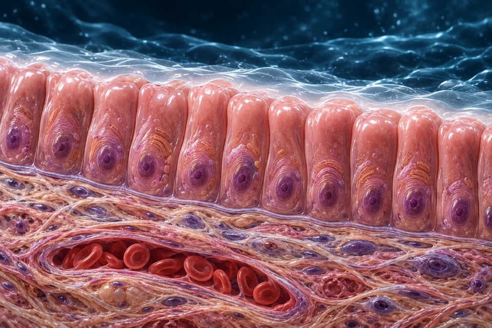
SUPPORTING DEFENSE CROSS SECTION / NOT TO SCALE
Multiple defenses cooperate. This is not an impermeable shield or a literal pH/pressure profile; generated textures do not demonstrate barrier function.
A peptic ulcer is a sore in the lining of the stomach or duodenum. Helicobacter pylori infection and nonsteroidal anti-inflammatory drugs are common causes; normal acid production alone does not explain every ulcer.
The protective lesson is the balance between luminal digestion and living tissue defenses. Our normal surface image does not diagnose an ulcer, show its depth or select treatment.
Cooperating surface defenses; ulcer definition and common causes: OpenStax · Stomach structure and function · NIDDK · Peptic ulcer definition and causesDirected normal-function and claim correspondence check: 17 October 2026. Source checks are not physician review or a certificate for every generated pixel.
08 / COORDINATED SIGNALS
Food-related sensory input, stomach distension and nutrients all contribute to control. Neural, hormonal and local signals overlap; they are not three isolated clockwork stages.
Vagal/enteric input + gastrin → ECL histamine and parietal-cell activity
Acetylcholine, gastrin and histamine act through different receptors and cooperate. Gastrin is a blood-borne hormone; histamine acts locally.
Somatostatin restrains gastrin, histamine and acid pathways. The response is a coordinated network, not one permanent on/off switch.
Supporting relationship map. Signals are not enzymes; neural acetylcholine, local histamine and circulating gastrin are different modes of communication.
Cephalic responses prepare secretion through neural pathways. Gastric responses then reflect distension and the chemical contents. The size of a response depends on the meal and physiological state.
Duodenal contents help coordinate gastric delivery with intestinal handling. Gut signals also recruit bicarbonate-rich pancreatic fluid and other digestive responses.
Follow intestinal–pancreatic coordination →Neural, endocrine and local regulation of secretion: NIDDK · Digestive movement and control · Schubert · Gastric secretionDirected normal-function and claim correspondence check: 17 October 2026. Source checks are not physician review or a certificate for every generated pixel.
09 / ACCOMMODATE, MIX, DELIVER
The proximal stomach accommodates a meal. Distal contractions mix and grind it. Movement and chemical digestion happen together while small portions leave through the pyloric outlet.
Coordinated contractions travel toward the distal stomach. The wall changes the space around its contents; the rugae are folds in that wall, not channels carrying a separate stream.
Compare the receiving, mixing and delivery regions on the cutaway →When onward passage is restricted, some contents return from the distal region toward the wider stomach. This helps mixing and grinding; it does not send food through the gastric glands or into blood vessels.
Distinguish the secretory gland openings →Interstitial cells of Cajal help organize electrical slow waves. Neural and hormonal inputs modify muscular activity; a slow wave is not automatically a measured emptying event.
Mixing, retropulsion and gastric–duodenal coordination: OpenStax · Stomach structure and function · NIDDK · Digestive movement and controlDirected normal-function and claim correspondence check: 17 October 2026. Source checks are not physician review or a certificate for every generated pixel.
10 / DELIVERY MATCHES DOWNSTREAM WORK
Meal composition, particle size and stomach–duodenum coordination affect emptying. There is no single illustrated time that applies to every meal.
Duodenal feedback, including responses to acid and fat, can slow gastric delivery. Neural pathways and hormones act together, rather than one hormone opening a simple tap.
Duodenal contents → feedback → adjusted gastric delivery
Buffering, bile, pancreatic enzymes and intestinal surface enzymes prepare molecules for absorption. The liver and pancreas supply different products; neither receives food through its tissue.
Follow bile delivery →Mixing, retropulsion and gastric–duodenal coordination: OpenStax · Stomach structure and function · NIDDK · Digestive movement and controlDirected normal-function and claim correspondence check: 17 October 2026. Source checks are not physician review or a certificate for every generated pixel.
11 / ONE STOMACH PRODUCT, A DISTANT DESTINATION
Parietal cells make intrinsic factor. Normal food-bound vitamin B12 handling then depends on gastric release, pancreatic processing and a specific absorptive route in the distal ileum.
Acid and proteolysis help release food-bound B12. It associates with haptocorrin in the acidic environment.
Pancreatic proteases break down haptocorrin. B12 can then bind intrinsic factor made by gastric parietal cells.
Specific intestinal receptors take up the intrinsic-factor–B12 complex. Making intrinsic factor does not mean the stomach absorbs that complex.
Selected: Stomach: release from food.
Normal food-bound B12 handling, not supplement or treatment advice. The intestine remains the main absorptive destination.
Connect the pancreatic enzyme contribution → · Continue to intestinal absorption → · Follow B12 and its changing escorts →
Food-bound B12 release, binding-partner transfer and distal ileal uptake: NIH ODS · Vitamin B12 handling · NCBI · Gastric intrinsic factorDirected normal-function and claim correspondence check: 17 October 2026. Source checks are not physician review or a certificate for every generated pixel.
12 / TISSUE EVIDENCE
A histological section shows tissue in one plane. Stain and sampling determine what is visible; our enlarged illustrations explain relationships without claiming to be actual microscopy.
The retained atlas opens the gastric wall. This real section adds another question: where does the epithelial surface continue inward, and where is glandular tissue arranged?
RECORDED · LIGHT MICROSCOPY

Nephron · exact source and rights · CC BY-SA 3.0. Full acquisition frame retained, proportionally encoded for display; no generated microscopy, recolouring, sharpening or artificial detail.
Gastric epithelium combines protection with specialised secretion. Intestinal epithelium receives material from the lumen, while renal tubules selectively exchange material with their surroundings after filtration. Shared epithelial organisation does not make these organs interchangeable or their transport mechanisms identical.
Sources checked 20 October 2026. Source-photo rights are unchanged; new independent labels and explanations are offered under CC BY-SA 4.0. Source identities and normal biology checks are distinct from physician, fine-histology and original-calibration certification.
The institutional MH 113 specimen shows pits, glandular tissue and supporting wall layers. Parietal and chief-cell appearances can be studied in their actual tissue context.
Open the human fundic specimen ↗External reference only. No specimen pixels are copied or embedded; redistribution rights were not established.
Cardiac, fundic/body and pyloric regions differ in pit depth and gland composition. A deep pit is not automatically an intestinal crypt, and a fold is not a villus.
Read the institutional regional comparison ↗This chapter teaches normal anatomy and physiology. It does not evaluate a symptom, diagnose disease or recommend treatment.
READ THE CONNECTION, THEN GO DEEPER
Pit and gland lumens open to the digestive contents. Blood remains in separate vessels outside the epithelium.
Acid changes the chemical environment. Pepsin cleaves protein chains, with further processing downstream.
Delivery has to coordinate with buffering, digestion and absorption beyond the stomach, rather than follow one universal transit time.
Intrinsic factor is made in gastric tissue but contributes to a later, specific intestinal uptake mechanism.
WHAT CONNECTS THE LESSON
Follow the organ into its wall and glands, then connect products and signals with the rest of digestion. The stomach begins important work; it does not finish the whole process.
Return to AllHumanBodyEDITORIAL RECORD
Source scope. Source-supported educational relationships: gross regions and route, wall order, pits and glands, selected cell roles, secretion and protection, mixing/emptying feedback and food-bound B12 handling. Sources support the teaching claims, not every generated pixel.
Review state. Public English normal-function teaching chapter. AI-assisted visual/content production. Fine generated cells, nuclei, gland branches, capillaries, fibers, hidden anatomy and proportions remain illustrative / not independently verified. Human medical review: not performed. Educational purpose only; not an exhaustive gastric or clinical guide.
English revision: stomach-english-2026-10-17-connected-quality. Stable chapter identity unchanged. The source-labelled 3D Stomach and its existing stable knowledge note show organ-scale context; neither resolves glands or simulates digestion. Physical-phone acceptance has not been performed. Editable labels and supporting diagrams remain separate from text-free anatomical masters.