Follicular cell
Thyroid hormone production
Works across a blood-facing side and a colloid-facing side; participates in thyroglobulin synthesis, hormone formation and release.
CHAPTER / LOCATION TO REGULATION
Nearby glands. Different messages.
The thyroid builds a hormone store inside tiny follicles. Separate parathyroids sense the mineral environment. Tissue organization connects these neighboring glands with different control systems and responsive organs.
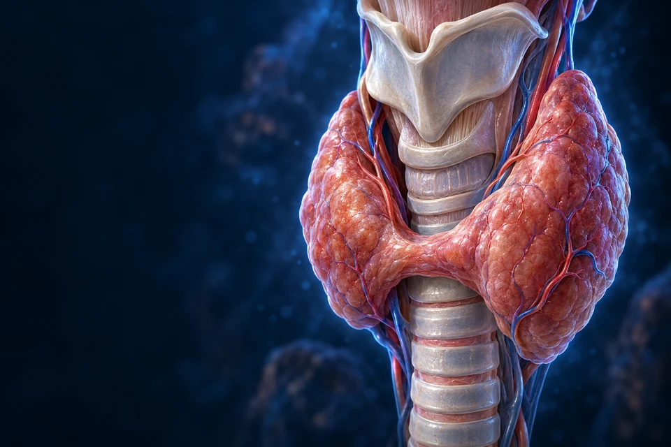
01 / LOWER NECK ORIENTATION
Two principal lobes lie beside the upper trachea. The isthmus crosses its front below the larynx. Natural proportions and anatomical variants mean a generic illustration cannot be an operative map.

The front view locates thyroid tissue beside the airway. It does not expose the parathyroids; use the separate posterior view below.
Teaching references: NCBI · Normal thyroid anatomy and tissue · Endotext · Thyroid anatomy and physiology
02 / A NEW VIEW, DIFFERENT GLANDS
Four is a common arrangement. Number and position vary, especially inferior glands. Their proximity does not mean that they make the same hormone or respond to the same control signal.

Teaching references: NCBI · Parathyroid anatomical variation · Duke · Endocrine tissue and specimen distinctions
03 / ORGAN → TISSUE → CELL
A follicular epithelial shell surrounds colloid rich in thyroglobulin. Apical surfaces face the colloid; basal surfaces face external support and capillaries. These two sides support different stages of the same process.

Iodide arrives from blood. Thyroglobulin and the apical enzyme environment support hormone formation at the colloid-facing side. The blood channel does not open into the central store.
Gold outlines locate the relevant compartment boundaries. Direction names explain function without inventing molecular transporters or a measured flow trace in this static preparation.
Teaching references: Leeds · Thyroid tissue organization · Endotext · Thyroid hormone synthesis and secretion · Duke · Endocrine tissue and specimen distinctions
04 / KEEP CELL ROLES DISTINCT
Follicular cells, C cells and parathyroid chief cells have distinct identities. An attractive generated tissue texture does not prove the hormone inside a particular cell.
Thyroid hormone production
Works across a blood-facing side and a colloid-facing side; participates in thyroglobulin synthesis, hormone formation and release.
Calcitonin
A distinct thyroid cell population; routine tissue appearance alone does not reliably prove its identity.
PTH
Mineral-sensitive endocrine control differs from the thyroid follicular TSH system.
A separate parathyroid population
Cellular function and identity need appropriate evidence. The generated main plate does not assign this subtype by color.
Teaching references: Duke · Endocrine tissue and specimen distinctions · Leeds · Thyroid tissue organization · NCBI · Normal PTH physiology
05 / BUILD → STORE → RETRIEVE → RELEASE
Iodide arrives through blood. Hormone formation involves tyrosine residues on thyroglobulin in the colloid-facing compartment. Later retrieval and cellular processing free T3 and T4 for release toward blood.
Different membrane sides and enzymes participate. The protein scaffold and iodine are inputs, not preformed free hormone flowing through a duct.
Synthesis into a luminal protein store and retrieval from that store are distinct directions. This is not a blood-filled cavity.
The whole thyroglobulin store is not emptied into blood as the normal hormone product. Exact rates and the complete molecular network are separate subjects.
These process comparisons supplement the detailed follicle preparation above. All three explanations remain visible; selected emphasis is not a measured transport trace.
The established route above is the summary. Use three scales below to connect the follicle, its polarised lining cell and a distant responding cell.
NIS brings iodide into the follicular cell; this is different from releasing finished hormone.
Iodide uptake: Blood-facing side → cell
The upward gold arrow marks ingredient entry across the basal boundary, supported by sodium-coupled transport. It does not depict T4 secretion.

At the basolateral surface, the sodium/iodide symporter couples iodide uptake to inward sodium movement. Na+/K+-ATPase uses ATP to help maintain the sodium gradient. NIS itself is not the ATP-driven sodium pump.
Read the image: Find the basal edge above the support tissue, then the separate blood channel. The supply arrow crosses only the cell boundary; it is not an open pipe from blood into colloid.
Mechanism evidence: Reactome · Sodium-coupled iodide uptake
Supporting pathway summaries beside the anatomical preparations; not a molecular shape reconstruction, measured flux or fixed time sequence.
This membrane step precedes hormone synthesis. ATP supports the gradient through a different protein; iodide uptake is not T4 uptake.
Mechanism evidence: Reactome · Sodium-coupled iodide uptake
MIT and DIT denote mono- and diiodinated tyrosine residues. Coupling appropriate iodinated residues forms T3 or T4 while the product is still attached to Tg.
Mechanism evidence: Reactome · Thyroxine biosynthesis · Endotext · Synthesis and secretion
Recovering iodine from unused iodotyrosines is not the same reaction as converting T4 to T3 or inactivating thyroid hormones in tissues.
Mechanism evidence: Reactome · Iodotyrosine dehalogenase · Endotext · Hormone metabolism
A plasma carrier, a membrane transporter and a nuclear receptor all bind or handle hormone, but perform different jobs in different places.
Mechanism evidence: Endotext · Serum binding proteins · Endotext · Cellular hormone uptake · NC-IUPHAR · Nuclear hormone receptors
An electrochemical gradient combines concentration and charge differences across a membrane. Sodium tends to enter under the conditions maintained by the cell. NIS couples that favourable movement to iodide transport, allowing the cell to concentrate an otherwise dilute ingredient.
Na+/K+-ATPase spends ATP while exporting sodium and importing potassium. Its work supports NIS indirectly. Calling both proteins an iodine pump would hide the distinction between energy use and coupled transport. The apical route toward colloid is a further membrane crossing, not the same NIS step.
Mechanism evidence: Reactome · Sodium-coupled iodide uptake · Cell biology · Angiofollicular units
Thyroglobulin is both a synthesis scaffold and a storage framework. Protein folding, processing and sorting occur along the ER–Golgi route before secretion at the apical face. The follicular cell therefore needs an organised membrane system, not merely a bag containing iodine.
The same epithelial polarity keeps colloid-facing secretion separate from blood-facing exchange. Apical means toward the lumen; basal and lateral surfaces face outside support and neighbouring cells. These are anatomical relationships, not fixed top and bottom directions in every possible drawing.
Mechanism evidence: Endotext · Synthesis and secretion · Cell biology · Angiofollicular units
Iodination adds iodine to selected tyrosines in thyroglobulin. TPO-dependent chemistry produces MIT or DIT residues. Coupling joins appropriate iodinated residues to produce hormone residues: two DIT contributors can form T4, while MIT and DIT contributors can form T3. This summary does not reconstruct the individual molecular transfer steps.
TPO works with hydrogen peroxide supplied by the apical oxidase system, including DUOX enzymes. The reactive chemistry is spatially organised at the colloid-facing boundary; peroxide is not simply poured into all of the cell. Surface texture in the illustration does not identify this enzyme complex.
Mechanism evidence: Reactome · Thyroxine biosynthesis · Primary study · DUOX and thyroglobulin · Cell biology · Angiofollicular units
The follicular lumen can retain hormone-containing thyroglobulin outside the epithelial cells. Endocytic retrieval brings selected material back into the cells; processing in endosomal/lysosomal compartments and protein breakdown liberate hormone. Storage and secretion can therefore change separately.
T4 and T3 subsequently leave the blood-facing side through transport processes. The small hormones are different from the large precursor protein. The drawing does not show a secretory tube through the epithelial layer, nor does it imply that all colloid is retrieved at once.
Mechanism evidence: Endotext · Synthesis and secretion · Reactome · Thyroxine biosynthesis
Protein breakdown also releases iodinated tyrosines that were not incorporated into hormone. Iodotyrosine dehalogenase, IYD, removes iodine from MIT and DIT so that iodide can be reused. Salvaging an ingredient avoids treating every iodinated residue as a secreted hormonal message.
D1, D2 and D3 act on iodothyronines such as T4 or T3. They belong to the hormone-metabolism story below. Similar names do not mean that IYD recycling and tissue activation are one universal enzyme reaction.
Mechanism evidence: Reactome · Iodotyrosine dehalogenase · Endotext · Hormone metabolism
Binding proteins maintain a large circulating pool that exchanges with the unbound hormone fraction. TBG, transthyretin and albumin differ in abundance and binding behaviour. A bound molecule does not need to remain attached for its whole circulation through the body.
Cell membranes have transporters involved in uptake and efflux; MCT8 is a well-established example. Transporter expression differs among tissues. The simple claim that all thyroid hormone freely diffuses into every cell would omit a biologically important control. This is normal physiology, not a guide to interpreting a thyroid blood test.
Mechanism evidence: Endotext · Serum binding proteins · Endotext · Cellular hormone uptake
T4 supplies an important precursor pool. D1 and D2 can remove an outer-ring iodine to generate T3; D3 can inactivate T4 to reverse T3 and T3 to T2. D1 also participates in hormone clearance, so a simple label of always activating would be incomplete. Enzyme distribution and regulation differ by tissue.
T3 interacts with thyroid-hormone receptors that regulate transcription through DNA-associated complexes and other regulatory proteins. Receptors do not turn every gene up, nor does hormone simply accelerate every cell reaction equally. Membrane entry, metabolism and receptor context make a coordinated response possible.
Mechanism evidence: Endotext · Hormone metabolism · Endotext · Cellular hormone uptake · NC-IUPHAR · Nuclear hormone receptors
The established TSH section below explains an upstream signal that influences follicular work. Synthetic capacity, stored precursor, release and target-tissue handling are related but distinct. An enlarged colloid compartment alone does not measure current secretion or whole-body hormone action.
Keep thyroid-hormone production separate from the PTH and calcium course later in this chapter. Nearby glands do not make one shared hormone. These mechanisms explain normal organisation and do not supply diagnosis, supplement advice or treatment instructions.
Mechanism evidence: Endotext · Synthesis and secretion · Cell biology · Angiofollicular units
No. It marks an ingredient entering the blood-facing cell boundary. Hormone formation, storage, retrieval and later blood-facing release remain separate.
No. It is outside the epithelial cells but enclosed by the follicle. Hormone-containing Tg must be retrieved and processed before hormone release.
No. IYD salvages iodide from MIT/DIT; D2 converts T4 to T3. Their substrates and physiological roles differ.
That is not the route taught here. Free hormone exchanges with tissues, membrane transporters handle cellular access, and local metabolism and receptors shape action.
Normal physiology · source checks 21 October 2026. One original wordless thyrocyte illustration and retained follicle/cell preparations. Generated tissue is not acquired microscopy, an atomic structure or a clinical measurement. Source-based Development checks are not physician review, physical-phone acceptance or diagnostic/supplement guidance.
The central compartment holds thyroglobulin-associated material. Uptake into a follicular cell and intracellular processing connect that store with release toward blood. These boundaries are visible in the follicular anatomy; the centre is not an outlet duct.
Thyroid synthesis and secretion sourceThyroglobulin supplies the protein framework. Thyroid peroxidase participates in iodination and coupling; this educational sequence does not reproduce every enzyme reaction or iodine-control mechanism.
Colloid storage differs from a granule packed with free hormone. Endocytosis and lysosomal processing connect stored material with a released product.
Teaching references: Endotext · Thyroid hormone synthesis and secretion · Endotext · Thyroid anatomy and physiology · OpenStax17.4 · Thyroid overview
06 / LINK TO THE PITUITARY
Hypothalamic TRH supports pituitary TSH release. TSH acts on follicular cells; thyroid hormones feed back to hypothalamic and pituitary regulation. Secretion and response also change over time and with physiological context.
A hypothalamic message reaches anterior-pituitary thyrotrophs.
Receptor signaling supports hormone synthesis and release.
Transport and local handling connect circulating hormone to response.
Thyroid-hormone availability helps restrain the axis; it is not a binary on/off thermostat.
Supporting endocrine-axis sequence, not a measured hormone trace.
Read the connected pituitary axes →Teaching references: Endotext · HPT axis physiology · Endotext · Thyroid anatomy and physiology
07 / BLOOD → CELL → RESPONSE
Most circulating thyroid hormone is protein-bound. Cellular transport, local deiodinases and receptor context shape access and action. T3 is an important receptor-active form; T4 supplies much of the circulating precursor pool.
Carrier-mediated cellular uptake matters. Lipid solubility does not mean universal unrestricted passive entry.
Deiodinases can activate or inactivate hormone forms; tissues need not handle the same blood signal identically.
Major thyroid effects involve nuclear receptors and altered gene expression. This is not the only possible cellular action.
Metabolism, growth and development depend on thyroid signaling alongside many other inputs, rather than a single universal speed dial.
Teaching references: Endotext · Cellular uptake of thyroid hormones · Endotext · Thyroid hormone metabolism · Endotext · Cellular thyroid hormone action
08 / SOLID CELL CORDS, DIFFERENT OUTPUT
Chief-cell-rich cords and clusters lie near vessels and connective partitions. Adult tissue may also contain adipose and oxyphil populations. Cell markers and specimens answer identity questions that a generated illustration cannot.

Cell cords lie beside a blood network. The illustration supplies tissue context; it does not independently identify every cell or locate a calcium-sensing receptor molecule.
Teaching references: Duke · Endocrine tissue and specimen distinctions · NCBI · Parathyroid cell regulation
09 / MINERAL ENVIRONMENT → CELL REGULATION
The calcium-sensing receptor, CaSR, connects extracellular ionized calcium to chief-cell control. Falling calcium generally permits increased PTH output; rising calcium strengthens restraint. Magnesium, phosphate, vitamin-D signaling and other context also matter.
Ionized calcium is not identical to total circulating calcium, which also includes protein-bound and complexed fractions. This distinction teaches physiology; it does not provide a laboratory interpretation rule.
PTH’s principal calcium-sensitive regulation is not the TRH–TSH pathway that controls thyroid follicles. Neighboring glands can therefore respond to different information.
Teaching references: NCBI · Parathyroid cell regulation · Endotext · Calcium and phosphate homeostasis
10 / BONE ↔ KIDNEY ↔ INTESTINE
PTH and vitamin-D metabolites connect mineral exchange, renal handling and intestinal absorption. Bone is living tissue; kidney filtration is not the same process as gut absorption.
Extracellular ionized calcium → chief-cell CaSR regulation → PTH output → coordinated organ responses

The nephron gives location to tubular handling. Calcium conservation, phosphate handling and calcitriol production are distinct responses; a long-loop overview does not identify each molecular transporter.
Continue in the existing tissue lesson →These qualified original illustrations are reused with their anatomical targets intact. They locate different responding tissues; they are not a new molecular transport or hormone-binding reconstruction.
The kidney conserves and exchanges minerals while also producing an endocrine signal. This differs from simply swallowing more calcium.
The major PTH–gut relationship here is indirect through vitamin-D activation. Absorption and renal reabsorption describe different boundaries.
PTH is not drawn as directly attaching to an osteoclast. Exposure pattern and tissue context matter; bone is not a passive mineral tank.
A fall in ionized calcium generally increases PTH; recovery of calcium strengthens restraint. Calcitriol and other signals also influence the system. This is a physiological relationship, not a clinical threshold tool.
Source-supported functional map. It supplements the organ/tissue plates and linked Kidney/Intestine lessons; it does not replace a precise skeletal cell atlas.
A CLOSER LOOK / FOOD, FILTRATE AND LIVING BONE
Taking a mineral in, recovering it and exchanging it are different jobs. Follow the boundary first. Then see how cells and signals coordinate the work.
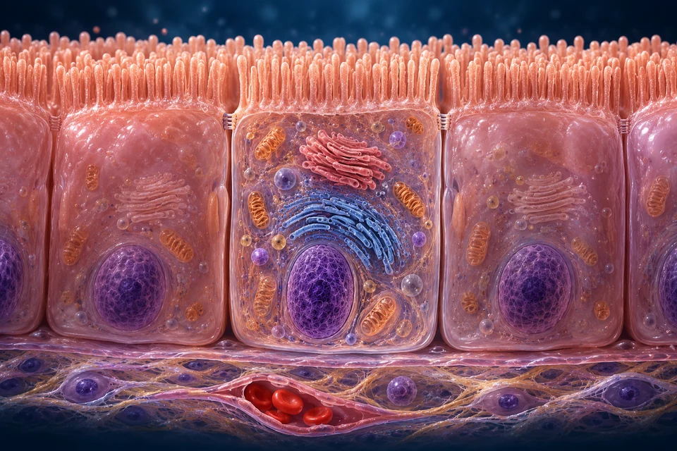
Read the anatomy: Brush border above; the blood-facing boundary and supporting tissue below. The separate vessel stays outside the epithelial cells.
EXPLANATORY COMPARISON / NOT MOLECULAR SHAPES OR MEASURED FLUX
TRPV6 / calbindin / PMCA1b–NCX1 is a useful model, not an exclusive chain or molecular image.
Read the evidence and limits for this comparison ↓Evidence: Biology of calcium homeostasis regulation in intestine and kidney (2025 issue) · Lieben et al. · Intestinal vitamin-D signaling and absorption in mice (2012) · Rodrat et al. · Calcium transport in a human-derived cell model (2022)
Open any explanation. The image above follows its anatomical context; each explanation keeps its own evidence and limits.
No. The starting compartment is different.
In the intestine, absorption moves material from the digestive lumen across the lining toward body fluids. In the kidney, reabsorption retrieves material that has already entered tubular filtrate. Filtration is not the final decision to excrete it.
In both settings, the lumen-facing membrane and the blood-facing membrane have different jobs. The blood vessel is not an open continuation of either lumen.
Keep this distinction: A route comparison, not a measured balance or a statement that every filtered substance is recovered.
Evidence: Renal Ca2+ wasting, hyperabsorption, and reduced bone thickness in mice lacking TRPV5 (2003) · NIH ODS Vitamin D Health Professional Fact Sheet
Yes. Epithelial junctions provide a selective route, not simply an empty gap.
The between-cell, or paracellular, pathway depends on permeability and electrochemical conditions. Individual movements can occur in both directions; net absorption is their balance.
Experiments in mice link junctional proteins with renal and intestinal calcium handling. They do not identify a universal calcium pore in every human intestinal region.
Keep this distinction: Do not infer a named junction protein from the decorative junctions in the illustration.
Evidence: Claudin-2 deficiency associates with hypercalciuria in mice and human kidney stone disease (2020)
A useful model separates entry, intracellular handling and exit.
TRPV6 participates at the lumen-facing side. Calcium-binding proteins such as calbindin help handle calcium within the cell. Blood-facing extrusion involves calcium pumps and exchangers, conventionally represented by PMCA1b and NCX1 in the intestinal model.
A pump uses ATP; a channel is not an ATP motor. The sodium gradient helps support exchange. The internal pathway is not a literal conveyor belt. Different proteins can have different importance across species and conditions.
Mouse intestinal vitamin-D-receptor experiments directly demonstrate altered absorption despite compensated blood calcium. Human-derived Caco-2 cells provide expression and epithelial-transport evidence, not proof of an exclusive normal-human chain.
Keep this distinction: The three-step model is not a claim that every absorbed calcium ion requires every named protein.
Evidence: Biology of calcium homeostasis regulation in intestine and kidney (2025 issue) · Lieben et al. · Intestinal vitamin-D signaling and absorption in mice (2012) · Rodrat et al. · Calcium transport in a human-derived cell model (2022)
It can pass between or through cells, but its transport machinery differs from calcium’s.
In a through-cell route, NPT2b couples phosphate entry to sodium at the lumen-facing membrane. It is distinct from the kidney’s NPT2a and NPT2c. Phosphate must subsequently leave toward body fluids; the molecular assignment of that intestinal exit is less firmly established.
Human donor-derived intestinal models show gradient-dependent, bidirectional phosphate passage between cells. Mouse studies also examine junctional permeability. Neither gives a single fraction for every healthy adult, intestinal region or diet.
Keep this distinction: The blood-facing exit is intentionally not assigned to XPR1, a calcium pump, or an invented transporter. No universal phosphate junction pore is shown.
Evidence: King et al. · Human intestinal models and phosphate permeability (2018) · Radványi et al. · Intestinal phosphate pathways in mice (2024)
The proximal tubule recovers calcium in association with sodium and water movement.
Much of this recovery uses a between-cell route. It is a different arrangement from later, through-cell calcium adjustment. A nephron outline helps locate the region but does not show every junction or capillary.
Keep this distinction: Qualitative regional comparison only; no fixed human segment fraction.
Evidence: Claudin-2 deficiency associates with hypercalciuria in mice and human kidney stone disease (2020) · Biology of calcium homeostasis regulation in intestine and kidney (2025 issue)
In the thick ascending limb, electrical conditions can favour calcium movement between cells.
NKCC2 carries sodium, potassium and chloride into the cell; it does not carry calcium. Salt handling, including potassium recycling, helps establish a lumen-positive electrical environment. This supports paracellular calcium recovery.
Claudin-associated junction selectivity matters. Cell-expression experiments inform this model, but do not reveal an exact atomic pore in an intact human tubule.
Keep this distinction: The thick ascending limb is a particular segment, not the entire ascending side of the loop.
Evidence: Claudin-16 and claudin-19 interact and form a cation-selective tight junction complex (2008) · Biology of calcium homeostasis regulation in intestine and kidney (2025 issue)
Distal convoluted and connecting-tubule regions use regulated through-cell transport.
TRPV5 participates in entry from filtrate. Intracellular calcium-binding proteins and blood-facing extrusion by NCX1 and calcium ATPases complete the model. TRPV5 is not responsible for all calcium recovery along the nephron.
Mouse knockout and micropuncture experiments directly demonstrate a distal transport defect with compensatory intestinal uptake. These are animal data, not measured healthy-human recovery percentages.
Keep this distinction: The retained nephron preparation does not resolve each distal cell subtype or transporter.
Evidence: Renal Ca2+ wasting, hyperabsorption, and reduced bone thickness in mice lacking TRPV5 (2003)
One mechanism is to reduce the uptake machinery available at the proximal surface.
PTH can promote removal of NPT2a from the lumen-facing membrane. Less phosphate is recovered from filtrate, leaving more available for urinary loss. This is reduced reabsorption, not active secretion of phosphate into the tubule.
NPT2a and NPT2c contribute to renal phosphate handling. The trafficking details established for NPT2a must not automatically be assigned to NPT2c.
Keep this distinction: The surface comparison is conceptual. It shows neither measured transporter counts nor a predicted urine result.
Evidence: Picard et al. · PTH and renal transporter trafficking (2010; publisher abstract) · Selective pharmacological inhibition of NPT2a promotes phosphate excretion (2020)
Different organs perform distinct chemical steps before the principal circulating active signal is formed.
The liver produces 25-hydroxyvitamin D. The kidney supplies the main endocrine activation step to calcitriol, or 1,25-dihydroxyvitamin D, involving CYP27B1. CYP24A1 contributes to metabolic inactivation.
The kidney is not the only tissue able to metabolize vitamin D. Local processing and the circulating endocrine supply are related but not identical contexts.
Keep this distinction: This is a normal-physiology pathway, not supplement, sunlight, dose or laboratory advice.
Evidence: NIH ODS Vitamin D Health Professional Fact Sheet · A PTH/salt-inducible kinase signaling axis controls renal vitamin D activation (2023) · Intestinal Cyp24a1 regulates vitamin D locally independent of renal Cyp24a1 in mice (2025 issue)
Yes. PTH and FGF23 can both reduce phosphate recovery while acting differently on calcitriol production.
PTH supports renal vitamin-D activation. FGF23 restrains activation and supports inactivation pathways. Comparing phosphate transport separately from vitamin-D processing prevents a misleading one-arrow summary.
Experimental evidence includes mice and human kidney organoids. An organoid is a research model, not a controlled whole-person measurement. The diagram does not predict a patient’s blood concentration.
Keep this distinction: Signal direction describes a regulated pathway, not a dose-response calculator.
Evidence: A PTH/salt-inducible kinase signaling axis controls renal vitamin D activation (2023) · Intestinal Cyp24a1 regulates vitamin D locally independent of renal Cyp24a1 in mice (2025 issue) · Selective pharmacological inhibition of NPT2a promotes phosphate excretion (2020)
Its calcium-sensing system responds to the ionized calcium environment.
The CaSR/PTH explanation above is the starting point. Hormonal messages change the behaviour of responding tissues; the message itself is not calcium traveling from one organ to another.
A solid parathyroid tissue preparation provides context. Individual receptors and secretory granules cannot be identified from the generated colours.
Keep this distinction: This links the retained sensing lesson to transport. It does not interpret total calcium or a laboratory report.
Evidence: Biology of calcium homeostasis regulation in intestine and kidney (2025 issue)
No. Exchangeable calcium and tissue remodelling describe different processes.
An exchangeable component of skeletal calcium can help buffer extracellular fluid. Remodelling, by contrast, involves osteoclast resorption and osteoblast formation of new matrix that subsequently mineralizes. An ion changing compartment does not mean an entire rebuilding cycle has occurred.
Animal research distinguishes rapid regulation from turnover, but does not establish a fixed normal-human pool, timing or a single controlling molecular gate. The precise contribution and control are not represented here.
Keep this distinction: The bone image supports remodelling context only. Exchangeable mineral is not a separately imaged sac or an instant osteoclast pump.
Evidence: MSD Manual · Calcium physiology overview (reviewed 2025) · Nordholm et al. · Rapid calcium regulation in rats (2015) · Osteocytes, not Osteoblasts or Lining Cells, are the Main Source of RANKL Required for Osteoclast Formation in Remodeling Bone (2015)
No. Tubular reabsorption can return it toward blood. Filtration and final urinary excretion are different stages.
No. A hormone carries information that changes tissue behaviour. Mineral movement is a separate pathway.
No. Phosphate and calcium use different machinery. A clearly stated evidence boundary is more accurate than an invented label.
Normal physiology · source checks 23 October 2026. Retained original illustrations with separate editable explanations. Animal and cell-model findings are not presented as measured normal-human fractions. AI-assisted production; human medical review not performed. No diagnosis, supplement dose or blood-test interpretation.
Intestinal absorption adds calcium from the gut; renal reabsorption recovers filtered calcium; bone turnover exchanges mineral within living matrix. PTH and vitamin-D metabolites help coordinate these different boundaries. They do not make every organ perform the same action.
Continue through renal regulation, intestinal absorption and living bone regulation.
Calcium and phosphate homeostasis sourceTeaching references: Endotext · Calcium and phosphate homeostasis · NCBI · Normal PTH physiology · NCBI · Bone-remodeling physiology
11 / A CONNECTED MINERAL NETWORK
PTH reduces proximal tubular phosphate reabsorption. Vitamin-D and FGF23 pathways connect bone and kidney with phosphate availability and vitamin-D metabolism. A calcium-only picture misses this connected regulation.
A substance can enter renal filtrate and later be reabsorbed. Calcium and phosphate responses need not move together through every regulated step.
FGF23 is a bone-derived signal that contributes to phosphate and vitamin-D regulation. It is part of the network, not a substitute name for PTH.
Teaching references: Endotext · Calcium and phosphate homeostasis · NCBI · Normal PTH physiology
12 / A DIFFERENT THYROID CELL POPULATION
C cells produce calcitonin, which can inhibit osteoclast-mediated bone resorption. Its usual role in adult human calcium homeostasis is smaller than that of PTH and vitamin D. A two-switch diagram would exaggerate its importance.
C cells are difficult to identify reliably in routine human light microscopy; no generated cell in the follicle plate is assigned a C-cell identity simply by pale color.
Teaching references: NCBI · Bone-remodeling physiology · Endotext · Calcium and phosphate homeostasis · Duke · Endocrine tissue and specimen distinctions
13 / KEEP THE LEARNING SCALES CONNECTED
Compare the input, cellular organization, control and target rather than grouping all nearby glands into one function.
| Learning scale | Thyroid follicular system | Parathyroid system |
|---|---|---|
| Tissue | Cells surround a colloid lumen | Solid cords and clusters beside vessels |
| Main output here | T3 / T4 | PTH |
| Principal control here | TSH with upstream feedback | Calcium-sensitive regulation |
| Connected response | Transport, local metabolism and receptor-specific action | Bone–kidney–gut mineral coordination |
Teaching references: Endotext · Thyroid anatomy and physiology · Endotext · Calcium and phosphate homeostasis · Duke · Endocrine tissue and specimen distinctions
14 / IMAGE VERSUS SPECIMEN
Follicle size, apparent cell height and colloid can vary with preparation and physiological state. These generated plates are explanatory anatomy, not a microscopy result.
Duke explicitly identifies parathyroid0312 as human H&E, thyroid0050 as monkey and0052 as young mammal. Its tissue notes also describe shrinkage and other preparation artifacts.
Read the external specimen record →Leeds provides external follicle/colloid teaching. Species or cell subtype should not be inferred when a displayed record lacks that information; staining alone has limits.
Read the external tissue teaching →Teaching references: Duke · Endocrine tissue and specimen distinctions · Leeds · Thyroid tissue organization
Real thyroid imaging and clinical disease/diagnosis/treatment are later-phase work. Their absence does not block normal anatomy and physiology.
EDITORIAL RECORD
Broad gland/airway orientation, separate parathyroids with variation, follicle polarity, selected cell outputs, hormone production/control and normal target/mineral coordination.
AI-assisted visual/content production. Fine generated vascular, cellular, cartilage and membrane details and hidden junctions remain illustrative / not independently verified. Human medical review: not performed. Educational only; not diagnostic or surgical material.
Revision: thyroid-calcium-english-2026-10-19-eyes-foundation-v1. Stable local key: ahb_ch_thyroid_calcium_english_v1. No new canonical CMS/atlas record or3D dataset. Migration physical-phone acceptance: Pending, non-blocking. Text-free masters,English labels and supporting paths remain separate and editable.