Read the structure, follow RNA and distinguish copying from expression. Look inside a nucleus, follow a selected RNA message, and connect experimental molecular views to the work of living cells.
▷
Watch chapter overviewFilm planned · not yet available
PDB1KX5 reconstituted construct · original controlled rendering
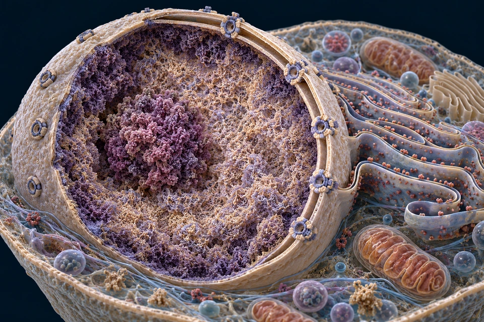
Original generated medical illustration, not microscopy. The cutaway locates broad cellular structures; it does not measure membrane thickness, pore composition or native chromatin spacing.Structure notes · further context
Nucleolus
A non-membrane-bound region where ribosomal RNA production and ribosomal subunit assembly are organised. It is not a second nucleus.
Chromatin
DNA associated with proteins occupies the nucleus in a changing, heterogeneous organisation; not all genes are active at once.
Nuclear envelope
Two membranes separate nucleoplasm from cytoplasm. The outer membrane connects with the endoplasmic reticulum.
Nuclear pore
A protein-lined gateway regulates exchange. Selected RNAs and proteins cross by organised transport, rather than DNA escaping through an open hole.
Rough ER
Membrane surfaces in cytoplasm carry ribosomes making selected secreted and membrane proteins. Many other proteins are made on free ribosomes.
DNA works within a cell, associated with proteins inside the nucleus.
The envelope and its pores separate nuclear and cytoplasmic work. Chromatin is heterogeneous; the nucleus is not normally filled with condensed mitotic X shapes.
A short B-DNA model shows two sugar–phosphate backbones and bases toward the interior. Blue and copper distinguish the strands; they do not mean oxygen-rich and oxygen-poor material.
New Golden-family controlled representation. Experimental-model coordinates, PDB 1BNA. Original controlled render; atom radii, chain colours and backbone guides are illustrative display choices. Missing atoms/water are not reconstructed. Not specimen microscopy or native-human molecular imaging. Human medical review: Not performed.Structure notes · further context
First backbone
The trace follows deposited phosphate coordinates; it is not an extra covalent bond.
Second backbone
Chemical direction is explained in a separate controlled map, not inferred from page position.
Interior bases
Not all atoms, ions or hydration sites are shown; no dynamic molecular claim.
Follow DNA from paired bases to protein-associated packaging, then distinguish a gene, a chromosome and a genome. Molecular views below use experimental coordinates; they are not photographs of a living human cell.
DNA
A molecule with a sequence
Its base order stores information that cells can copy and use.
A sequence is not an anatomical picture of the body.
Gene
A functional stretch of DNA
Its expression produces RNA; some RNA directs protein synthesis.
Not every gene produces a protein.
Genome
The genetic complement
Nuclear and mitochondrial genetic material contribute different parts.
Genome does not mean all genes are equally active.
A nucleotide combines a phosphate group, deoxyribose sugar and a base. The backbone joins successive nucleotides; complementary bases belong to opposing strands.
Phosphate
Backbone component
Connects sugars along a strand.
The smooth coloured trace is a guide, not an extra chemical bond.
Sugar
Deoxyribose
Connects the base to the backbone and gives strand direction its chemical meaning.
Atom sizes in the render are display choices.
Base
A, T, G or C
Base order supplies the readable sequence.
A letter represents a chemical group, not one atom.
This selected adenine–thymine pair comes from the same crystal model as the duplex. Covalent sticks show modeled connections within each nucleotide; hydrogen atoms and hydrogen-bond lines are not reconstructed.
New Golden-family controlled representation. Experimental-model coordinates, PDB 1BNA. Original controlled render; atom radii, chain colours and backbone guides are illustrative display choices. Missing atoms/water are not reconstructed. Not specimen microscopy or native-human molecular imaging. Human medical review: Not performed.Structure notes · further context
Adenine
Its experimental-model coordinates are retained without reshaping.
Thymine
Hydrogen bonds are explained separately; missing hydrogen atoms are not fabricated.
Sugar context
Covalent sticks are distance-based representations, not a validated bond-order map.
The two DNA strands are antiparallel. A written sequence needs an orientation: 5′ and 3′ refer to the sugar–phosphate chemistry, not the top and bottom of a page.
The sequence map is an original supporting mechanism, not a new molecular structure or a clinical sequence.
This reconstituted nucleosome model shows DNA associated with a histone assembly. It uses Xenopus histones and a deposited DNA construct; it is not a native human chromosome specimen.
New Golden-family controlled representation. Experimental-model coordinates, PDB 1KX5. Original controlled render; atom radii, chain colours and backbone guides are illustrative display choices. Missing atoms/water are not reconstructed. Not specimen microscopy or native-human molecular imaging. Human medical review: Not performed.Structure notes · further context
Associated DNA
Not a universal native human chromatin packing pattern.
Histone proteins
The construct contains Xenopus histones; visible modeled tails are retained.
DNA and associated proteins form chromatin. Local organisation can change with cellular work. A single nucleosome model does not establish a universal sequence of evenly spaced beads, a compulsory 30-nm fibre or one fixed chromosome shape.
Original generated medical illustration, not microscopy. The cutaway locates broad cellular structures; it does not measure membrane thickness, pore composition or native chromatin spacing.Structure notes · further context
Nucleolus
A non-membrane-bound region where ribosomal RNA production and ribosomal subunit assembly are organised. It is not a second nucleus.
Chromatin
DNA associated with proteins occupies the nucleus in a changing, heterogeneous organisation; not all genes are active at once.
Nuclear envelope
Two membranes separate nucleoplasm from cytoplasm. The outer membrane connects with the endoplasmic reticulum.
Nuclear pore
A protein-lined gateway regulates exchange. Selected RNAs and proteins cross by organised transport, rather than DNA escaping through an open hole.
Rough ER
Membrane surfaces in cytoplasm carry ribosomes making selected secreted and membrane proteins. Many other proteins are made on free ribosomes.
Compare the mechanism. All essential statements remain visible; selecting a focus changes emphasis rather than hiding the other steps.
Selected focus: DNA duplex · Two strands / local sequence
DNA duplex
Two strands / local sequence
The paired molecule is one level of organisation.
A short crystal fragment is not a chromosome.
Nucleosome
DNA / histone association
A protein assembly organises a local DNA segment.
A reconstituted particle does not establish native fibre spacing.
Chromatin
Context-dependent organisation
Many local interactions and cellular processes organise DNA.
No rigid universal 30-nm packing ladder.
Native-cell imaging studies reveal context-dependent organisation. Their results support caution about tidy textbook packing ladders; they do not prove that every cell has exactly the same arrangement.
How can the same nucleus contain both accessible and compact regions?
DNA is associated with histones and many other proteins. Local nucleosome arrangement, modifications, remodelling and interactions help influence which sequences are accessible to molecular machinery. Dense-looking regions in the illustration must not be read as a measured gene-activity map.
The envelope creates a boundary; nuclear pores support selective exchange. The nucleolus organises ribosome production without its own enclosing membrane. A single drawing therefore needs both a compartment explanation and a molecular explanation, rather than a universal ladder of thicker and thicker chromatin fibres.
A nuclear chromosome contains DNA organised with proteins. After replication, sister chromatids contain the copied DNA; during division they can condense into the familiar paired form. Before copying, a chromosome is not two identical sister chromatids.
Compare the mechanism. All essential statements remain visible; selecting a focus changes emphasis rather than hiding the other steps.
Original generated metaphase teaching preparation, not microscopy or a human chromosome count. Only selected exposed chromosomes are emphasised. The nuclear envelope has broken down; sister chromatids are still joined. Fine molecular attachment geometry is not a solved structural model.Structure notes · further context
Cell boundary
The plasma membrane encloses the cell. It must not be mistaken for a nuclear envelope around the spindle.
One spindle pole
Microtubules converge toward one centrosomal pole. The opposing pole lies across the same cell.
Opposing spindle pole
A bipolar spindle supports separation toward opposite poles; it is not a pipe carrying genetic material.
Joined sister chromatids
The textured replicated chromosome has two sister chromatids, still joined before anaphase. They are not a homologous pair.
Centromeric attachment context
Kinetochore protein assemblies at the centromeric region engage spindle microtubules. This expanded illustration locates the context, not individual binding molecules.
Spindle microtubules
Bundles connect the poles with centromeric attachment regions; other microtubules support spindle organisation. They do not copy DNA.
The image stays at metaphase while the cards compare chromosome states. DNA was copied earlier in S phase; sisters are still joined in this preparation. Anaphase separation is described below, not shown as already occurring.
Selected focus: Before copying · One chromatid per chromosome
Before copying
One chromatid per chromosome
A chromosome has not yet gained its copied sister.
Interphase chromatin is not normally a condensed mitotic rod.
After copying
Two sister chromatids
DNA copying produces sister DNA molecules within a replicated chromosome.
Copying DNA does not immediately double the chromosome count by centromeres.
During separation
Sisters become separate chromosomes
Mitosis separates the copies into daughter complements.
Homologous chromosomes are not the same as sister chromatids.
A gene and its control regions have different jobs.
Gene expression uses a DNA sequence to produce RNA. Regulatory sequences and associated factors help determine when and how much RNA is made. A protein-coding region is not the whole genome, and noncoding does not automatically mean useless.
RNA-producing sequence
Copied into an RNA product
Some products act directly as RNA; others supply protein instructions.
Do not equate every gene with one protein.
Regulation
Control of expression
Cell state and molecular interactions alter the use of a sequence.
Not a single permanent on/off switch.
Genomic context
Neighbourhood and packaging
Nearby sequences and chromatin contribute to access and control.
A diagram cannot predict an individual person’s traits.
RNA uses ribose and usually uses uracil where DNA uses thymine. RNA is often single-stranded, but it can fold and form paired regions. DNA is retained while selected information is transcribed into RNA.
Compare the mechanism. All essential statements remain visible; selecting a focus changes emphasis rather than hiding the other steps.
Selected focus: DNA · A retained sequence source
DNA
A retained sequence source
Selected regions are transcribed rather than used up.
Not every region is expressed at once.
RNA
Several working forms
RNA may fold, regulate or take part in translation.
Single-stranded does not mean unable to form paired regions.
Protein
Selected RNA-directed product
A coding message can be translated into an amino-acid chain.
RNA polymerase uses a DNA template to make a complementary RNA in the 5′→3′ direction. For this example, the template is read 3′→5′. The opposite coding strand has the corresponding sequence with T where RNA has U.
Compare the mechanism. All essential statements remain visible; selecting a focus changes emphasis rather than hiding the other steps.
Read three contexts: nuclear transcription · regulated export · cytoplasmic translation. Gold identifies RNA; coral identifies a growing polypeptide. The illustration locates working regions; use the sequence below to distinguish the individual template and coding strands.
Original generated teaching preparation with differently scaled molecular contexts. The nuclear transcription product, exported message and ribosome-bound message are separate exemplars, not one continuous RNA cable. Shapes are illustrative, not deposited atomic coordinates.Structure notes · further context
DNA template context
The blue/copper duplex remains on the nuclear side. Transcription locally separates DNA strands; it does not send DNA into a ribosome.
Transcription complex
RNA polymerase uses a DNA template to make complementary RNA. The underlying DNA remains available after transcription.
New RNA
A gold transcript emerges into the nucleus. Protein-coding transcripts undergo processing; this nascent example is distinct from the mature export example.
Export context
A selected mature message crosses the nuclear envelope through a pore. Many other RNAs remain nuclear or have non-protein-coding functions.
Cytoplasmic ribosome
Large and small ribosomal subunits read a mature message. tRNAs match codons with amino-acid delivery; fine binding sites are not resolved here.
Growing polypeptide
The coral chain represents a protein product emerging from the ribosome, not RNA passing through the pore. Folding and further processing determine the final working product.
Selected focus: Template DNA · Read this example left → right
Template read 3′→5′ → complementary RNA built 5′→3′. The coding strand is distinct from the template.
Template DNA
Read this example left → right
3′–TAC GGA CTT–5′
The template determines the complementary RNA.
Coding DNA
Opposite strand, same orientation as RNA
5′–ATG CCT GAA–3′
Coding is not another name for the template strand.
RNA product
Built in the 5′→3′ direction
5′–AUG CCU GAA–3′
U replaces T in the corresponding RNA sequence; this is an illustrative fragment.
Why is the RNA at the pore not the same stage as the emerging transcript?
A protein-coding nuclear transcript is made from a DNA template. Capping, splicing and cleavage with polyadenylation commonly contribute to maturation, with some processing occurring while transcription continues. An export-competent messenger ribonucleoprotein is a different molecular state from the short nascent RNA emerging at a transcription site.
The enlarged illustration places these contexts together to compare compartments, not to claim one RNA cable connects a gene to a cytoplasmic ribosome. Some RNAs remain nuclear; other RNAs function structurally, catalytically or in regulation rather than serving as protein templates.
For many human protein-coding genes, an initial transcript is processed: introns are removed and retained exon sequences are joined. Typical mature mRNAs also carry end modifications. These are not universal descriptions of every RNA molecule.
Compare the mechanism. All essential statements remain visible; selecting a focus changes emphasis rather than hiding the other steps.
Selected focus: Initial transcript · Retained / removed regions
Exon blocks are symbolic. An exon may contain coding or untranslated sequence. Alternative products require specific biological regulation; this is not a random mixing tool.
Initial transcript
Retained / removed regions
Selected exon and intron sequences are present before processing.
Box lengths are not measured sequence lengths.
Joined exons
Introns removed in this example
Retained sequences join in their original order.
Some retained sequence is untranslated.
Alternative combination
Selected exon choice
A different retained combination can change the mature product.
This schematic does not claim every combination occurs.
An exon is retained in the mature RNA. Protein-coding transcripts can include untranslated regions within exons, as well as a coding region. Alternative splicing can retain different combinations of exons; it does not randomly rearrange the DNA genome.
Exon
Retained sequence
May include coding sequence or untranslated sequence.
Not every exon entirely encodes amino acids.
Intron
Removed in the selected splicing example
Processing joins the surrounding retained sequences.
Not every gene has introns.
Alternative transcript
Different retained combination
Regulated processing can change a mature RNA product.
Not a guarantee that every combination is made or functional.
During translation, a ribosome reads an mRNA coding region in codons. tRNAs connect codons with amino acids; the growing chain becomes a protein product. The ribosome translates RNA, not the DNA strand.
Compare the mechanism. All essential statements remain visible; selecting a focus changes emphasis rather than hiding the other steps.
Read three contexts: nuclear transcription · regulated export · cytoplasmic translation. Gold identifies RNA; coral identifies a growing polypeptide. The illustration locates working regions; use the sequence below to distinguish the individual template and coding strands.
Original generated teaching preparation with differently scaled molecular contexts. The nuclear transcription product, exported message and ribosome-bound message are separate exemplars, not one continuous RNA cable. Shapes are illustrative, not deposited atomic coordinates.Structure notes · further context
DNA template context
The blue/copper duplex remains on the nuclear side. Transcription locally separates DNA strands; it does not send DNA into a ribosome.
Transcription complex
RNA polymerase uses a DNA template to make complementary RNA. The underlying DNA remains available after transcription.
New RNA
A gold transcript emerges into the nucleus. Protein-coding transcripts undergo processing; this nascent example is distinct from the mature export example.
Export context
A selected mature message crosses the nuclear envelope through a pore. Many other RNAs remain nuclear or have non-protein-coding functions.
Cytoplasmic ribosome
Large and small ribosomal subunits read a mature message. tRNAs match codons with amino-acid delivery; fine binding sites are not resolved here.
Growing polypeptide
The coral chain represents a protein product emerging from the ribosome, not RNA passing through the pore. Folding and further processing determine the final working product.
Selected focus: Message · An illustrative coding fragment
Coding-fragment example: AUG / CCU / GAA → methionine / proline / glutamate. A stop codon is not included in this short fragment.
Message
An illustrative coding fragment
5′–AUG CCU GAA–3′
The reading frame is specified here; untranslated regions are not drawn.
Delivery
tRNAs connect coding to amino acids
Codon recognition associates the message with appropriate amino-acid delivery.
The molecules are not invented anatomical machines.
Chain
Methionine → proline → glutamate
A growing amino-acid chain follows this specified standard-code example.
Not a complete protein or a prediction of its final fold.
Why do some proteins enter the ER while others stay in the cytoplasm?
Translation starts on cytoplasmic ribosomes. Targeting information in a growing protein can recruit the ribosome–nascent-chain complex to the endoplasmic reticulum for selected secreted or membrane proteins. A free cytoplasmic ribosome is not a different permanent ribosome species.
The protein product is an amino-acid chain, distinct from both its mRNA template and the tRNAs delivering amino acids. Further folding, modification, assembly and targeting help determine its working location. The short codon example below teaches code correspondence, not a complete protein or its final shape.
A chain is only the beginning of a working protein.
A new amino-acid chain must acquire a useful structure; many proteins also undergo processing or interact with partners. Delivery depends on the protein. The ER/Golgi route is not the destination of every protein.
Sequence
Order of amino acids
A coding region contributes to chain order.
This does not by itself display the folded structure.
Structure
Three-dimensional organisation
Folding and interactions support function.
No universal protein shape is shown.
Destination
Where the protein works
Signals and cellular pathways guide selected delivery.
rRNA contributes to ribosomal structure and function. tRNA carries amino acids into translation. Other RNAs participate in regulation or processing. An RNA product is not merely a temporary unused copy of DNA.
mRNA
Selected protein instructions
Its coding region is translated.
Not every part of an mRNA encodes amino acids.
rRNA / tRNA
Translation machinery
Different RNA classes organise translation and amino-acid delivery.
They are not translated into ribosomes or amino acids.
Other RNA
Regulation / processing
Some RNA products help regulate expression or process RNA.
Similar genetic material, different cellular work.
Many nucleated somatic cells share much of their nuclear genome but express different combinations and amounts of genes. Signals, chromatin, RNA handling and protein regulation contribute to changing cell states.
A message is prepared, transported, read and eventually removed.
DNA supplies selected RNA information. Cells then regulate access to that message and the fate of its products. Compare the jobs below; they are connected contexts, not a timed animation or a mandatory fate for every RNA.
An RNA transcript gains an export-ready organisation.
For a typical nuclear protein-coding message, a 5′ cap, splicing and 3′-end formation help make a usable mRNA. These activities can overlap transcription. Processing also changes the proteins associated with the RNA.
Read the image: Locate chromatin, the envelope and a pore. Processing happens in the nuclear compartment; the image does not resolve a spliceosome.
Original generated medical illustration, not microscopy. The cutaway locates broad cellular structures; it does not measure membrane thickness, pore composition or native chromatin spacing. Selected learning context: Prepare a message. Gold rings locate structures discussed in the explanation, not measured activity or molecular travel.Structure notes · further context
Nucleolus
A non-membrane-bound region where ribosomal RNA production and ribosomal subunit assembly are organised. It is not a second nucleus.
Chromatin
DNA associated with proteins occupies the nucleus in a changing, heterogeneous organisation; not all genes are active at once.
Nuclear envelope
Two membranes separate nucleoplasm from cytoplasm. The outer membrane connects with the endoplasmic reticulum.
Nuclear pore
A protein-lined gateway regulates exchange. Selected RNAs and proteins cross by organised transport, rather than DNA escaping through an open hole.
Rough ER
Membrane surfaces in cytoplasm carry ribosomes making selected secreted and membrane proteins. Many other proteins are made on free ribosomes.
Gold rings locate the structures being discussed. They do not show measured activity, a moving molecule or a fixed sequence followed by every RNA.
Not all genes contain introns, and not every human mRNA has a poly(A) tail. This is a common pathway, not a rule for every RNA.
Formation, maturation, export and removal influence which messages are available.
Protein production
Initiation, elongation and termination influence how a message is translated.
Working protein
Folding, partners, location and turnover help determine the functional result.
Follow the mechanism beyond a single arrow.
Cap, untranslated regions, coding region and tail do different jobs.
The 5′ cap is an end modification recognised by cellular machinery. The 5′ untranslated region precedes the translated coding region, and the 3′ untranslated region follows its stop codon. Both UTRs can carry regulatory information although they do not supply the amino-acid sequence of that coding region.
A poly(A) tail is added during 3′-end processing of most human mRNAs. It is not a DNA-encoded run that the ribosome translates into the end of the protein. Exons can contain untranslated as well as translated sequence.
Processing is coordinated work, not a compulsory three-stop conveyor.
Capping begins while a transcript is being made. Splicing and 3′-end formation are connected with transcription and RNA-associated proteins. A drawing that separates these jobs is organising the explanation, not asserting that every event waits until all the preceding work is finished.
Alternative splicing can generate different mature transcripts from the same gene. It changes the retained RNA sequence rather than shuffling the genomic DNA. Not every possible exon combination is produced or yields a functional protein.
The nuclear envelope separates nuclear transcription from cytoplasmic translation. A message associates with proteins during its formation; export competence depends on its processing and interactions, rather than merely on its size.
For bulk mRNA export, human NXF1–NXT1 is a major export-receptor pair. Other factors support assembly, pore interactions and remodelling. This description concerns a major pathway; different RNA classes and selected messages can use different machinery.
The mRNA coding region is read in a defined frame from 5′ toward 3′. Amino acids are added to the growing chain from its amino end toward its carboxyl end. Reading direction, frame and chain growth are related but distinct ideas.
Many nuclear mRNAs use cap-dependent initiation, with start-site selection influenced by surrounding sequence. Initiation controls entry into translation; elongation and termination have their own factors. Bacterial ribosome-binding rules should not be assigned wholesale to human cytoplasmic messages.
RNA quantity is a balance between production and loss.
Producing a transcript does not guarantee that it will be exported, translated repeatedly or retained for a long time. RNA-binding proteins and regulatory RNA can influence processing, localisation, translation and stability. Surveillance can remove selected faulty messages.
A reading of RNA abundance is a snapshot of that balance. It does not directly count how many ribosomes are translating each message or how much completed, active protein is present. The illustration is not a gene-expression dataset.
Why can similar RNA levels accompany different amounts of working protein?
Translation efficiency, protein folding, modification, assembly and protein removal operate beyond the RNA measurement. Even after a chain is synthesised, its function depends on structure, location and cellular context. More transcript therefore does not establish an equal increase in active protein.
For selected secreted or membrane proteins, targeting signals connect translation with ER entry; the later Golgi and vesicle route is taught in Cells. This is one destination pathway, not a universal finishing line for every protein.
Does removing an intron remove the same sequence from genomic DNA?
No. Splicing processes the RNA transcript. It does not edit the corresponding DNA out of the chromosome.
Is an untranslated region an unused part of the message?
No. It is outside that coding region but can contribute to translation control, localisation or RNA stability.
Does a message disappear as soon as a ribosome reads it?
No. One message can be read repeatedly; RNA degradation is a separate, regulated part of its lifecycle.
Does twice as much mRNA prove twice as much active protein?
No. Translation, maturation, targeting and removal also affect the amount and activity of the protein product.
Normal nuclear-encoded message teaching. Retained original generated compartment illustrations are distinct from the experimental molecular models elsewhere in this chapter. Fine molecular structure, human medical review and physical-phone acceptance are not certified.
Compare the mechanism. All essential statements remain visible; selecting a focus changes emphasis rather than hiding the other steps.
Selected focus: Shared information · Much of the nuclear genome
Shared information
Much of the nuclear genome
Many nucleated somatic cells have similar genetic sequence.
This is not universal for all cells or all DNA content.
Different use
Selected expression patterns
Different combinations and quantities of RNA/protein support different jobs.
No measured expression levels are plotted.
Changing state
Signals and cellular context
Expression and molecular activity can respond to the environment.
Not an irreversible one-gene fate switch.
The comparison is conceptual; it is not a measured gene-expression dataset. Some cells change DNA organisation or content, so “every cell has identical DNA” is not used as a universal rule.
How can related cells use similar DNA to do different jobs?
A neuron and a pancreatic secretory cell need different combinations and quantities of products. Regulatory DNA, transcription factors, chromatin organisation, RNA processing and degradation, translation and protein turnover all contribute. Regulation is not a single switch that irreversibly assigns one gene to one organ.
Many nucleated somatic cells share much of their genome, but this is not an assertion that all cells have identical DNA content: mature red cells lack a nucleus, gametes differ in chromosome complement, and specialised rearrangements or somatic changes can occur. Normal cell identity is better explained through coordinated cellular work than through a one-gene destiny.
Replication copies DNA before many forms of cell division. Each original strand can serve as a template for a new strand. DNA synthesis proceeds 5′→3′; fork geometry makes continuous and fragment-based synthesis necessary on different strands.
Compare the mechanism. All essential statements remain visible; selecting a focus changes emphasis rather than hiding the other steps.
Selected focus: Original duplex · Template strands
Original duplex
Template strands
Complementary original strands provide copying templates.
The fragment has no measured fork or polymerase.
New synthesis
5′→3′ addition
Continuous and fragment-based synthesis address antiparallel fork geometry.
Both new strands grow 5′→3′, not one growing 3′→5′.
Copied duplexes
Original + new strand
Each daughter duplex contains an original and a newly made strand.
Semiconservative copying is distinct from transcription into RNA.
No bacterial polymerase names, universal fragment length, timing or replication-fork speed is assigned to human cells.
A representative cycling cell passes through G1, S and G2 before mitosis and cell separation. DNA is copied in S phase. Cells do not all cycle continuously or at the same speed; some remain outside the cycling sequence.
Compare the mechanism. All essential statements remain visible; selecting a focus changes emphasis rather than hiding the other steps.
Selected focus: G1 · Growth / preparation
G1
Growth / preparation
A representative cycling cell prepares for copying.
Cells can leave the cycling sequence; duration varies.
S / G2
DNA copying / preparation
S phase copies DNA; subsequent preparation precedes division.
Not every cell cycles continuously or at one fixed speed.
M
Chromosome separation / cell division
Mitosis separates nuclear chromosomes; cytokinesis separates the cell.
The overview is not a timing chart or an account of meiosis.
What is copied, and what is actually separated?
S phase duplicates DNA before mitosis. The two sister chromatids of a replicated chromosome are distinct from the two homologous chromosomes inherited through the parents. DNA content can double without immediately doubling the chromosome count by centromeres.
During mitosis, condensed chromosomes become organised on a spindle. Microtubules engage kinetochores at the centromeric regions; after sister cohesion is released, the separated chromatids are chromosomes moving toward opposite poles. Nuclear division and division of the cytoplasm are related but distinct processes. Non-cycling cells do not continually repeat this sequence.
Most human nucleated somatic cells have a diploid nuclear chromosome complement. Gametes differ, and a mature human erythrocyte has no nucleus. Mitochondria carry a separate small genome; mitochondrial function also depends on many nuclear genes.
Nuclear complement
Typical diploid context
Commonly 22 autosomal pairs plus a sex-chromosome pair.
Not a universal count for every human cell.
Gametes
A different complement
Normal mature gametes carry one nuclear chromosome set.
This is a normal overview, not a fertility assessment.
Mitochondrial DNA
Separate genetic material
A small circular genome contributes selected mitochondrial products.
Mitochondria are not genetically independent of the nucleus.
A karyotype can show chromosomes isolated from a cell and arranged by features such as size and banding. It is not their position inside a living nucleus and does not reveal every base in a DNA sequence.
Compare the mechanism. All essential statements remain visible; selecting a focus changes emphasis rather than hiding the other steps.
Differentiation involves changes in gene expression and cellular organisation. Stem-cell populations differ in their renewal and differentiation potential. A tissue stem cell should not be shown as a universal source of every cell in the body.
Renewal
Maintaining a population
Selected cells can replenish cells within a tissue.
Capacity and division behaviour depend on context.
Specialisation
Changing cellular organisation
Signals and expression help produce a functional cell state.
Not explained by one gene switching alone.
Tissue context
Signals and surroundings
Neighbouring cells and the local environment contribute.
Not a treatment, cell-engineering recipe or fate prediction.
Different methods reveal different kinds of evidence.
Sequence analysis records base order. A karyotype compares chromosomes. Structural studies model molecules; microscopy examines cellular organisation. None alone gives a complete account of a cell’s current activity.
Molecular coordinates
Deposited structural model
The three primary views render selected experimental-model coordinates.
Not an electron-density map or a live human molecular movie.
Cellular imaging
Observed organisation at a method-dependent scale
Native chromatin research tests simplified packing assumptions.
No licensed micrograph is reproduced here.
Expression measurements
RNA / protein activity context
Different methods assess abundance, location or state.
This chapter has no patient-specific dataset.
Which picture can support which conclusion?
The retained PDB 1BNA DNA and 1KX5 nucleosome views are original renderings of deposited experimental constructs, with their source identity and coordinate-linked labels preserved. They are neither an entire human chromosome nor live chromatin footage.
Generated cellular preparations provide an interpretable spatial context. Their realistic texture is not experimental evidence for a particular protein conformation, pore composition or human chromosome count. A karyotype, a base sequence and a structural model answer different questions; none substitutes for the others.
Use molecular explanations to understand organ work.
Connect cell products and regulation to specialised tissues. These links add an explanation layer rather than replacing anatomy with a sequence diagram.
This chapter covers selected DNA structure, chromatin, RNA/protein expression and genome concepts. It does not provide genetic diagnosis, treatment, a complete catalogue of genes or a deterministic explanation of human traits.
AllHumanBody explanations and overlays are original. CC0 deposited coordinates are credited separately from the original rendering method. No reference-book prose, artwork or page layout is reproduced.
Human medical review: not performed. Real physical-phone acceptance: not performed. Source-supported teaching does not imply physician approval, complete molecular validation or a frozen flagship chapter.
Selected DNA/chromatin structure, RNA expression, protein production and genome distinctions. Reference claims are checked critically; gene/protein, exon/coding and universal chromosome-count oversimplifications are not adopted.
Methods and limits
AI-assisted original content and editable overlays. Four retained controlled Three.js renders use CC0 PDB coordinates, without coordinate deformation. New text-free cellular and expression-context preparations are original generated teaching illustrations, clearly separate from the experimental models; they are not microscopy or measured protein structures. Display radii, colours and backbone guides are illustrative; inferred covalent sticks are not a bond-order validation. Model provenance is not live-human microscopy or a complete molecular reconstruction. Human medical review: not performed. Educational only.
Revision: dna-genes-genome-eyes-context-2026-10-19-v2. Stable local identity: ahb_ch_dna_genes_genome_english_v1; not a new CMS entity. Private text-free masters and English editable concept labels stay separate. Migration physical-phone acceptance: Pending, non-blocking. No clinical advice, physician approval or independently validated human molecular simulation.