Contractile cells meet a continuous extracellular load path.
At a myotendinous junction, the muscle-cell membrane and its supporting matrix form a specialised interface. Intracellular contractile force is coupled through membrane-associated attachments into extracellular collagen. The muscle fibre does not simply turn into a collagen fibre.
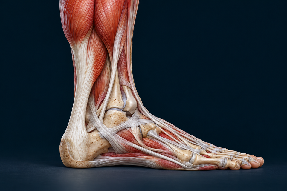
An aponeurosis spreads attachment over a sheet, while a cord-like tendon concentrates the visible route. Muscles can transmit force through both arrangements. The calf illustration locates this connection at organ scale; it does not resolve membrane proteins.
Sources for this relationship: Benjamin et al. · Tendon structure and function · OpenStax · Muscle attachments and lever systems
Collagen has several levels of organisation.
Collagen molecules assemble into fibrils; larger organised structures form fibres and fascicles within a tendon. Supporting matrix and interfaces connect these levels. Terminology and the relative prominence of levels vary between tendons and methods.
The retained cutaway shows a grouped fibrous architecture and elongated tendon-cell context. Its visible bundle ends are not counts of collagen molecules. A long-axis view and a cross-section answer different questions about the same material.
Sources for this relationship: Wang et al. · Tendon biomechanics and mechanobiology · Columbia University · Human muscle and tendon, H&E
An attachment must manage a change of material.
Soft tendon and mineralised bone deform differently. In a fibrocartilaginous attachment, a transition through nonmineralised and mineralised fibrocartilage helps distribute load across that mismatch. The local direction of pull and bending around the insertion matter as well as tension along the tendon.
Fibrous attachments are different: collagen connects with bone or periosteal regions without requiring the same four-zone fibrocartilaginous sequence. Do not apply the Achilles window to every tendon, ligament or point on a bone.
Sources for this relationship: Benjamin et al. · Where tendons and ligaments meet bone · Apostolakos et al. · The tendon-to-bone insertion
Guiding a tendon is different from attaching its end.
A synovial tendon sheath provides a specialised gliding environment where a tendon travels through a constrained route. A retinaculum or pulley helps hold that route near the skeleton; it is not another muscle and does not propel the tendon.
The Achilles has a surrounding paratenon rather than the same true synovial sheath used by many finger flexors. Follow the established hand lesson for actual finger courses and attachments; this chapter does not infer those fine routes from a generic tendon texture.
Trace the actual long-finger flexor routes →Sources for this relationship: Benjamin et al. · Tendon structure and function
A turning effect needs force and a moment arm.
Torque about a selected axis is force multiplied by the perpendicular distance from that axis to the force’s line of action. That distance is not automatically the tendon length, the bone length or the distance to an insertion.
As a joint moves, the force direction and moment arm can change. Two equal muscle forces may therefore have different turning effects. The small calculation here isolates one relation; real movement also includes multiple muscles, gravity, joint reactions and acceleration.
Sources for this relationship: OpenStax · Torque and rotational equilibrium
A short effort arm can favour excursion rather than force.
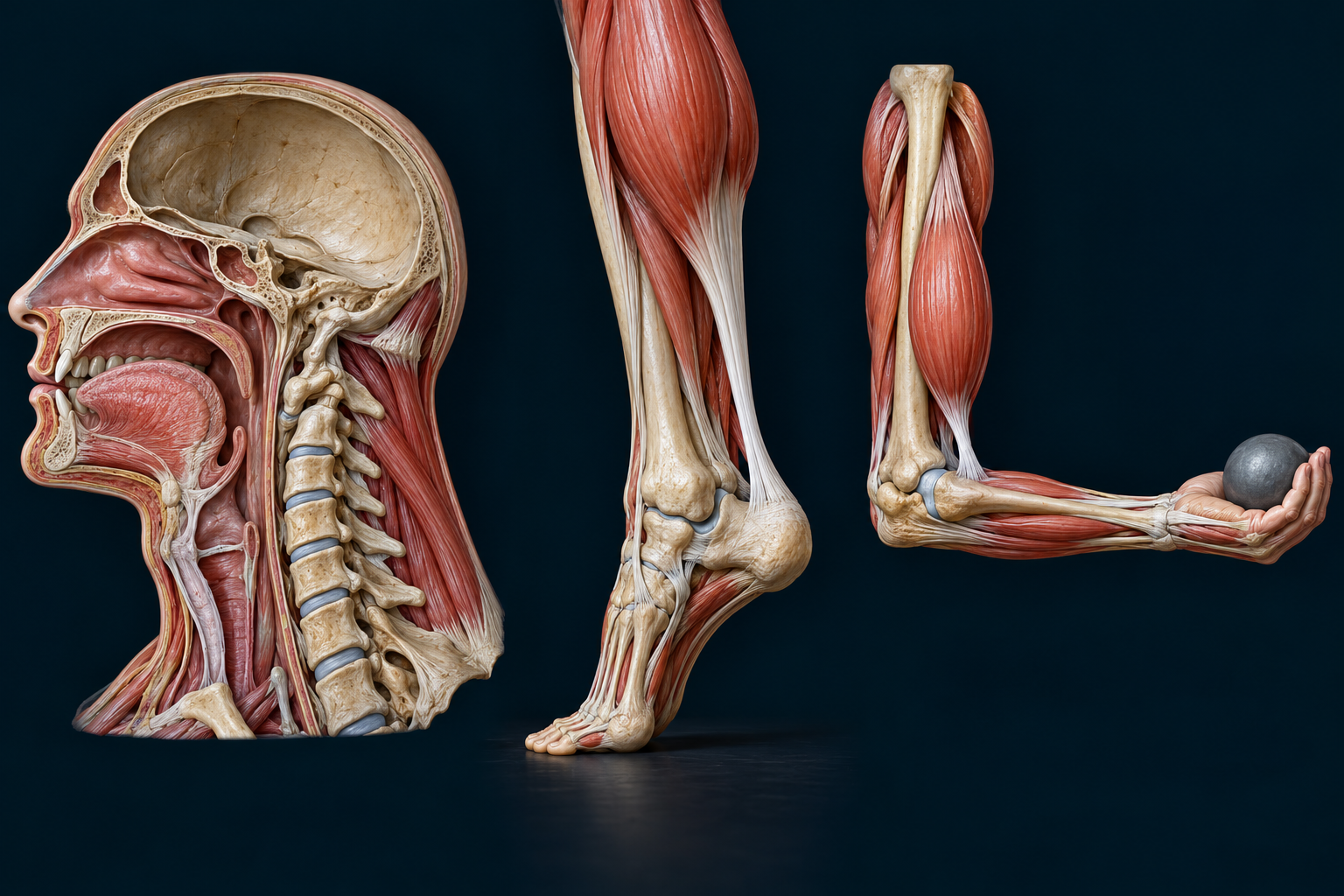
In a common third-class arrangement, muscle effort acts close to the joint while the hand moves much farther away. A comparatively small tendon excursion can accompany a larger distal movement. This can support range and speed, while requiring substantial muscle force to balance a distant resistance.
Lever class describes the relative order of effort, axis and resistance in the selected task. It does not rank how good a joint is, establish its efficiency, or guarantee a fixed mechanical advantage at every angle.
Sources for this relationship: OpenStax · Muscle attachments and lever systems · OpenStax · Torque and rotational equilibrium
A tendon can store and return some mechanical energy.
Tensile loading can deform the tendon while a muscle–tendon unit works. Part of that stored elastic energy can be returned as loading changes; some is dissipated. Energy-storing and positional tendons do not have identical material organisation or demands.
Returning stored energy does not mean the tendon generates active muscle force. Neither the static illustration nor the controls measure energy, stiffness or performance in a particular person.
Sources for this relationship: Wang et al. · Tendon biomechanics and mechanobiology · Lavagnino et al. · Tendon mechanobiology
The response depends on time and loading history.
In a creep experiment, deformation can increase while load is held. In stress relaxation, the force needed to maintain a fixed deformation can decline. Hysteresis describes the difference between loading and unloading paths. These terms ask different experimental questions.
A tendon is not a perfect steel cable or a lossless spring. Its nonlinear response reflects structure, water, matrix interactions and loading conditions. This chapter deliberately avoids a universal safe strain, exercise dose or injury threshold.
Sources for this relationship: Wang et al. · Tendon biomechanics and mechanobiology
Cells maintain the load-bearing matrix.
Tendon cells interact with their extracellular environment through several sensing and signalling routes. Changes in mechanical conditions can influence matrix turnover and gene expression. A single highlighted nucleus does not reveal which pathway is active.
Maintenance and adaptation occur over time. They depend on more than the presence of force, and experimental cell or animal results are not a personalised training or rehabilitation plan.
Sources for this relationship: Lavagnino et al. · Tendon mechanobiology
A pale tendon still belongs to living circulation and neural control.
Blood reaches tendon through regional connections, including its muscle and bone ends and surrounding connective tissues. The Achilles vascular study linked here examined cadaveric specimens; it does not establish identical living perfusion in every tendon or individual.
Sensory structures contribute information about the muscle–tendon system. Golgi tendon organs respond to tension-related deformation in series with muscle fibres. Muscle spindles lie within muscle and provide information related to length and change in length. Spinal and descending circuits shape the resulting response.
Continue with position and force-related feedback →Sources for this relationship: Ahmed et al. · Blood supply of the Achilles tendon · NCBI Neuroscience · Mechanoreceptors for proprioception · NCBI Neuroscience · Force-related afferent feedback
Tendon and ligament names describe different relationships.
Tendons generally connect muscle to bone and transmit muscular force. Ligaments connect skeletal structures and help guide or restrain joint motion. Their collagen-rich tissue can share features while its orientation, attachments and regional loading differ.
Neither is an inert fastening strap. A joint works through coordinated bone surfaces, cartilage, capsule, muscles, connective tissues and nervous control. Continue to the existing tissue and joint lessons instead of treating this distinction as the whole story.
Connect the tendon with joint organisation →Sources for this relationship: Benjamin et al. · Tendon structure and function · OpenStax · Muscle attachments and lever systems