Neurons integrate body-state information. Secretory cells make different products. Distinct neural and vascular routes connect this brain–gland bridge with responsive tissues.
▷
Watch chapter overviewFilm planned · not yet available
Neural–endocrine bridge · fine generated detail illustrative
01 / A NEURAL–ENDOCRINE LOCATION
The gland has a brain connection and a bony seat.
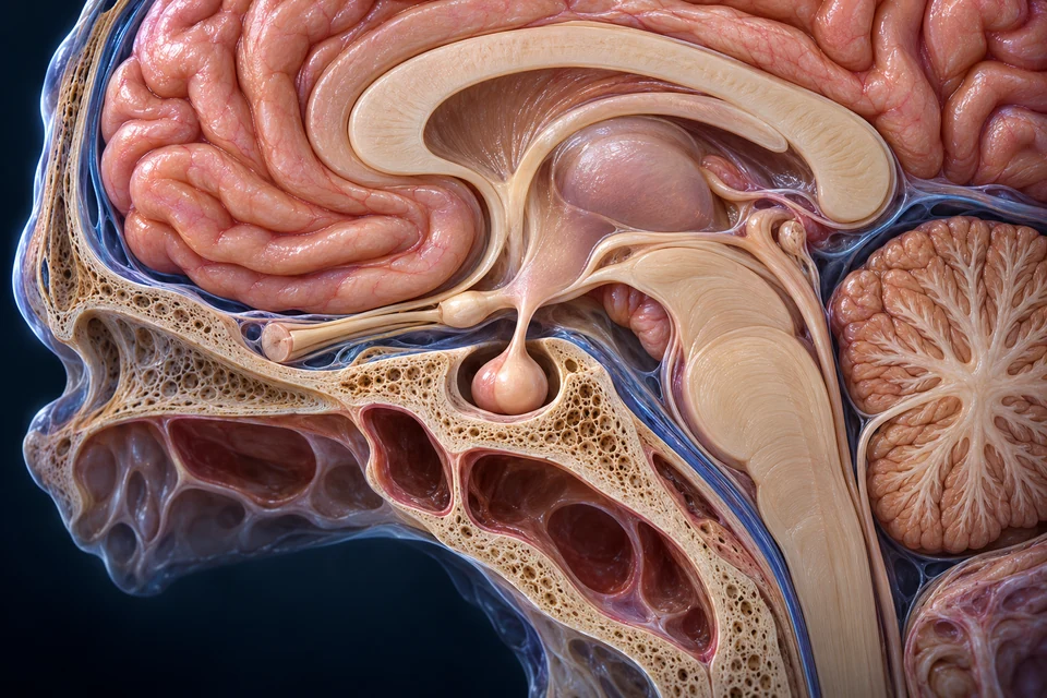
The pituitary lies below the hypothalamus in the sphenoid sella turcica. Nearby optic pathways are neighbors, not hormone ducts. The regional view establishes context; the next view enlarges tissue organization.
New original Golden-family text-free teaching preparation. Expanded near-midline regional preparation; anterior is image left. The exposed brain and skull contours, hidden optic relations and exact dimensions remain illustrative. This is not a surgical map or patient scan. Human medical review: Not performed.Structure notes · further context
Hypothalamic region
The region belongs to the base of the diencephalon. Individual nuclei and the generated ventricle/optic contours are not identified or claimed verified here.
Pituitary stalk
The infundibulum carries neural processes and vessels. It is not a duct carrying a brain fluid into the pituitary.
Pituitary gland
The anterior endocrine and posterior neural regions are enlarged in the next distinct view. This regional view establishes location, not all vascular junctions.
Bony seat
The pituitary lies in this bony depression below the brain. Bone and adjacent cavity contours are illustrative, not operative boundaries or a measured cross-section.
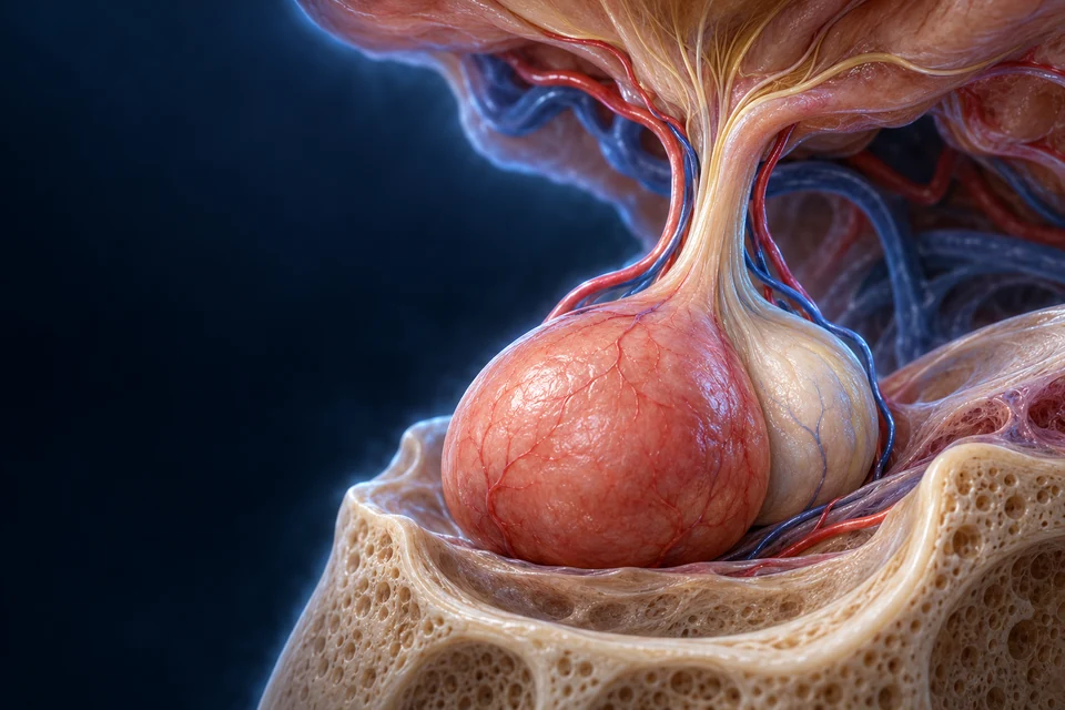
The stalk is a neural and vascular connection, not a hormone drainage duct. The tissue view below separates the two jobs.
The anterior glandular region and posterior neural region are connected parts of one organ. Neither is a drain from a ventricle. Their distinct developmental origins help explain their different tissue arrangements.
New original Golden-family text-free teaching preparation. Focused lobe/stalk view adds tissue continuity rather than repeating the skull context. Anterior is image left. Solid endocrine tissue is distinct from neural texture; exact cell/vessel detail and scale remain illustrative. Human medical review: Not performed.Structure notes · further context
Hypothalamic floor
Specific neuron nuclei are not assigned from this generated appearance. Neural and vascular delivery require different mechanisms.
Infundibular stalk
The detailed portal connections are explained separately in an editable supporting map; the red/blue surface branches are not a verified angiogram.
Anterior endocrine tissue
The pars distalis forms the major endocrine region. Its cells are not exocrine acini around a drainage duct; generated cell subtype colors are not diagnostic.
Posterior neural tissue
The pars nervosa stores and releases hypothalamic vasopressin and oxytocin. It is not the site where those neuronal cell bodies synthesize their hormone precursors.
Vascular context
Exact arterial, long/short portal and venous junctions are not established by this generated surface. The functional route map states the source-supported main distinction.
A blood-borne control signal reaches another secretory cell
Anterior cells manufacture their own products after receiving blood-borne control signals. The gold contexts locate tissue and vessels, not a reconstructed portal angiogram.
A glandular front
The adenohypophysis includes the pars distalis and tissue around the stalk. A thin intermediate remnant is not a large separate functional lobe in the adult human model presented here.
A neural back
Hypothalamic axons extend into the pars nervosa. Supporting glia, called pituicytes, belong to this tissue; they are not the main neuronal source of vasopressin or oxytocin.
Anterior control hormones reach endocrine cells through portal blood. Posterior hormone products arrive along axons before terminal release into blood. A red or blue branch in the artwork cannot by itself establish every connection.
Anterior · portal blood
A blood-borne control signal reaches another secretory cell
Hypothalamic releaseReleasing or inhibiting signals enter capillaries at the median eminence.
Portal transportPortal veins connect that first bed with anterior-pituitary capillaries.
Anterior synthesis and secretionEndocrine cells make their own hormone products; control signals can stimulate or restrain output.
Systemic deliveryReleased pituitary hormones reach the general circulation and responsive targets.
These ordered routes explain direction; both remain readable. The anatomical views above and below provide the tissue context. Neither list reconstructs exact vascular branching, hormone quantities or transport timing.
Why is the posterior pituitary a release site rather than a second gland making the same hormones?
Vasopressin and oxytocin precursors are synthesized in hypothalamic neurons. Axons carry packaged products to posterior terminals. Electrical activity and calcium-dependent secretion control release there; storage does not mean the terminal is continuously secreting.
Anterior endocrine cells instead make their own products after receiving releasing and inhibiting signals through portal blood. Neither pathway is an open hormone duct from the brain.
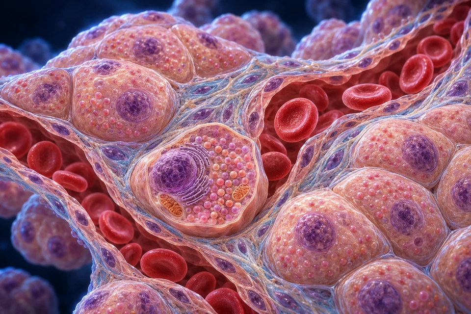
Anterior-pituitary endocrine cells lie in groups beside sinusoidal capillaries. Release first reaches the surrounding interface and then blood. Cell identity is determined by biology and appropriate markers, not a generated color.
New original Golden-family text-free teaching preparation. Representative tissue illustration, not H&E microscopy, a subtype stain or a measured cell model. Broad endocrine-cell/capillary relationships are taught; fine generated granules, membranes and cell identities remain illustrative. Human medical review: Not performed.Structure notes · further context
Endocrine-cell cords
The cell types have different hormone products. This unstained illustration cannot independently identify a somatotroph, lactotroph or other subtype by color.
Cell nucleus
Hormone synthesis depends on the cell’s molecular machinery. Nuclear size and chromatin appearance here are decorative, not a diagnostic observation.
Granule context
The exposed dots illustrate secretory storage, not an electron-microscope count, exact composition or universal granule geometry.
Capillary channel
Anterior-pituitary hormones enter capillary blood after secretion into the surrounding tissue interface. This lumen is not a gland duct.
Red blood cells
Their generated number, diameter and spacing are not physiological measurements or evidence of flow speed.
Vessel boundary
Fenestrated pituitary capillaries support exchange. Individual pores, basement membrane and molecular passage are not resolved or independently verified in this artwork.
A secretory cell prepares its own product
The cell nucleus and synthetic machinery support production. Stored granules are not evidence that secretion is occurring at this instant.
Gold outlines locate contexts in a static original teaching illustration. They do not animate release, quantify flow or establish molecular identity.
These selected anterior cell classes explain the principal outputs in this lesson. They are not every pituitary cell population or a literal count of hormones in all species and life stages.
Somatotroph
GH
Growth and metabolic effects; direct receptor action and tissue IGF-I pathways cooperate.
Lactotroph
Prolactin
Supports mammary milk production; hypothalamic dopamine provides an important inhibitory influence.
Thyrotroph
TSH
Signals thyroid follicular tissue; its output is distinct from thyroid T4/T3.
Corticotroph
ACTH
Signals adrenal cortical cortisol production; it is not the medullary catecholamine pathway.
Gonadotroph
FSH and LH
Supports reproductive tissue functions and hormone production; one cell class contributes two different outputs.
Ordinary H&E appearance cannot independently identify all hormone-producing subtypes. Generated nuclei/granules are illustrative. Support cells and cell-state variation are not reduced to five colored populations.
A neuron’s cell body can be far from its endocrine terminal.
Hypothalamic neurosecretory neurons synthesize precursors and send products along axons. Posterior terminals store and release them. Both supraoptic and paraventricular regions contain vasopressin- and oxytocin-related neurons; this is not a rigid one-nucleus/one-hormone map.
New original Golden-family text-free teaching preparation. Neurovascular tissue view, not a synapse onto a second neuron or exact electron microscopy. Terminals remain outside the blood lumen. Fine granules, support-cell identities and membrane thickness remain illustrative. Human medical review: Not performed.Structure notes · further context
Neurosecretory axon
The hormone precursor is made in the hypothalamic neuron and transported along its axon. This posterior region is not a cluster of new magnocellular cell bodies.
Axon terminal
Electrical activity can trigger calcium-dependent exocytosis. Release reaches the tissue-blood interface; the axon does not open directly into the blood lumen.
Terminal storage
Vasopressin and oxytocin products occupy neurosecretory granules. Counts, identities and packaging cannot be proved from generated dots.
Blood channel
This is the main posterior endocrine delivery relationship. It is distinct from hypothalamic control hormones traveling to anterior-pituitary cells through portal blood.
Tissue–blood interface
Interstitium and capillary endothelium are separate from the axonal membrane. Exact nanoscale spacing, fenestrae and supporting pituicytes are not independently verified by this image.
The neuronal cell body is upstream in the hypothalamus
The visible axon reaches a posterior release region. It is not a new hormone-producing neuronal cell body inside the posterior lobe.
Gold outlines locate contexts in a static original teaching illustration. They do not animate release, quantify flow or establish molecular identity.
Storage and release are different steps
Electrical activity can raise terminal calcium and trigger vesicle fusion. A stored product does not prove that release is happening at that moment.
Neurovascular, not a second-neuron synapse
Hormone products reach tissue capillaries. The illustrated terminal lies outside the endothelial boundary; it is not an open tube pouring directly into the lumen.
Vasopressin influences permeability, not a water faucet.
Vasopressin, also called ADH, helps regulate water conservation. Osmotic information interacts with volume/pressure-related inputs and other influences. Release is not governed only by a fixed blood-salt threshold.
Neural integration
Body-state inputs influence hypothalamic neuronal activity and posterior release.
V2-receptor signalling helps move AQP2 water channels to the apical membrane of principal cells.
Supporting cause-and-response chain. Water movement depends on the renal gradient and complementary pathways; ADH is not an active pump moving every water molecule, and it does not completely stop water loss.
The anterior prolactin pathway and posterior oxytocin pathway contribute different actions. This comparison teaches normal physiology, not feeding advice or a complete reproductive chapter.
Prolactin · production
Lactotroph output supports milk synthesis in mammary epithelium. Hypothalamic dopamine supplies an important tonic inhibitory influence; suckling can alter that regulation. It is not simply one universal releasing hormone switched on only during pregnancy.
Oxytocin · ejection
Posterior release supports contraction of mammary myoepithelial cells, helping move milk toward ducts. Oxytocin also participates in uterine contraction; neither function makes it a single proven “love hormone” explaining complex behavior.
Pregnancy-associated tissue preparation and the changing steroid environment matter. Hormone availability alone is not a guarantee of an individual output.
A hypothalamic signal influences pituitary output; selected peripheral glands then change their own secretion. Feedback affects the upstream response. This organization does not mean the pituitary controls every endocrine tissue.
Thyroid axis
1 · Hypothalamus · TRH
2 · Anterior pituitary · TSH
3 · Thyroid tissue · T4 and T3
Return influence Thyroid-hormone availability feeds back at hypothalamic and pituitary levels.
Target thyroid tissue changes its output; TSH is not itself thyroid hormone.
Adrenal cortical axis
1 · Hypothalamus · CRH
2 · Anterior pituitary · ACTH
3 · Adrenal cortex · cortisol
Return influence Cortisol provides negative feedback on hypothalamic and pituitary activity.
This cortical pathway is not the neural catecholamine route of the adrenal medulla. Aldosterone is not governed chiefly by this ACTH chain.
Gonadal axis
1 · Hypothalamus · pulsatile GnRH
2 · Anterior pituitary · FSH and LH
3 · Gonads · tissue responses and hormone output
Return influence Sex-steroid and inhibin feedback depends on cell, sex and reproductive context.
FSH/LH support gamete-related and steroid-producing tissue functions. Sustained preovulatory estradiol can provide positive feedback; not every phase is negative.
Supporting causal comparisons; no clinical concentration, dosing, fixed threshold or universal timing is predicted. Feedback changes secretion rather than circulating backward through a hormone duct. Basic pathways stay visible.
Growth hormone acts through its own receptors and stimulates IGF-I production in liver and other tissues. Local tissue action matters alongside circulating factors; not every growth effect is a single liver-only chain.
GH has direct targets
Its receptor-mediated actions influence metabolism and tissue function. The response depends on tissue, nutrition, developmental stage and other signals.
IGF-I adds a related pathway
Liver-derived circulating and locally produced IGF-I contribute to growth-related tissue responses. Feedback influences hypothalamic/pituitary secretion; GHRH and somatostatin provide opposing regulatory influences.
An original supporting comparison, not a treatment model, growth prediction, fixed hormone percentage or claim that adult and growing tissues respond identically.
Blood carries the message; receptors help determine its meaning.
Cells need the relevant receptors and signalling machinery. Receptor availability, local hormone handling and cellular state shape the response. A gland does not send a private hormone duct to every target.
Original educational cell illustration. These are selected surface and intracellular receptor contexts, not every hormone pathway or measured protein structures. Thyroid cellular entry involves transporters; it is not represented by the steroid-access example. Human medical review not performed.Structure notes · further context
Extracellular signal
The blue signal is approaching, not visibly bound; the picture does not prove receptor activation.
Cell-surface receptor
This is not a pore for the whole hormone. Its shape does not establish a particular receptor family.
Intracellular machinery
These representative proteins are not a measured cAMP cascade. Surface receptors use different downstream pathways.
Intracellular receptor
The small amber signal and protein are explanatory symbols, not an atomic reconstruction or a specified hormone.
Nuclear compartment
The static image locates the nucleus; it does not demonstrate completed gene expression or show a universal receptor movement sequence.
Plasma membrane
The right illustration is a steroid-access example. Thyroid hormones require cellular transport mechanisms; they are not depicted here.
A peptide signal need not enter the cell to change its activity. The receptor couples binding to internal signalling; different receptors use different machinery.
Intracellular receptor
A steroid signal reaches the cell interior → receptor-associated regulation
Receptor complexes can influence gene expression. Cortisol is one example. This is not a claim that all hormone effects are slow or use only one route.
Thyroid hormones are not simply interchangeable with peptides or steroids. Entry transporters and local conversion help shape the response; rapid additional actions are not fully mapped.
Original cell cutaways locate selected receptor contexts; no molecular structure or thyroid transporter identity is inferred from the image. Hormone identity, receptor availability and tissue state determine the response; one blood signal does not make every cell do the same thing.
Why can a circulating hormone affect one cell more than another?
Blood delivery gives tissues access to a signal, but access is not the whole response. Receptor availability, receptor coupling, local hormone conversion and the cell’s state can all change the effect. A similar blood concentration therefore does not imply an identical response in every tissue.
Neural, blood-level and hormone inputs can differ.
Endocrine regulation includes different sensing and release mechanisms. These routes cooperate; the distinctions prevent the false idea that every secretion must first pass through the pituitary.
Neural input
Hypothalamic integration and axonal release connect nervous activity with endocrine delivery. An electrical event and the subsequent blood-borne product are different signals.
Blood-level sensing
Pancreatic islets respond to metabolic inputs with local/neural influences. Insulin and glucagon are not synthesized by the pituitary.
Selected gland axes involve sequential signals and return influences. A thyroid or adrenal output is distinct from the pituitary signal that helped regulate it.
Pulses, circadian influences and physiological context can change secretion. GnRH’s pulsatile pattern matters to gonadotropin regulation. ACTH/cortisol and GH show temporal organization that one static diagram cannot predict.
Supporting qualitative pattern only · no measured patient trace, universal hormone timetable, fixed concentration or clinical interpretation. Feedback and temporal pattern are related but different questions.
A coordinating bridge is not the whole endocrine system.
Dedicated glands and endocrine cells inside other organs contribute different messages. Regional thyroid/parathyroid, adrenal and gonadal tissue lessons remain separate depth dependencies rather than being claimed complete here.
Brain
Hypothalamic integration connects autonomic, behavioral and endocrine functions without reducing the brain to one switch.
Tissue evidence asks a different question from an atlas render.
Real section staining reveals tissue arrangement under a method-dependent appearance. A generated cell image is not a real specimen, subtype stain or proof of a granule’s contents.
External specimen reference
MH149 records pituitary tissue and H&E staining, with anterior/posterior regions, capillaries and axonal storage context. The page does not identify the donor/species in its displayed metadata; do not label it as a verified human specimen.
Subtype hormone identity may require specific markers. Neither stain color nor AI-generated texture proves a somatotroph or lactotroph. Exact human microstructure, cell-state populations and molecular distances are not independently validated by these illustrations.
No specimen pixels or clinical scan were copied. Detailed sellar MRI/clinical comparisons, disease, diagnosis and treatment remain later phases; these do not block the normal-function foundation.
Broad gland location and lobe relationships; anterior endocrine versus posterior neurosecretory delivery; selected cell outputs; renal water response; production versus ejection; selected target axes; GH/IGF-I; receptor principles and temporal/feedback distinctions.
Explicit limits
AI-assisted visual/content production. Generated fine brain/bone contours, exact vessels, nuclei, cell identities, granules, membrane dimensions and hidden connections remain illustrative / not independently verified. Human medical review: not performed. Educational purpose only; not a surgical map, clinical guide or exhaustive endocrine atlas.
English revision: hypothalamus-pituitary-english-2026-10-18-eyes-foundation-v1. Chapter identity: ahb_ch_hypothalamus_pituitary_english_v1. No canonical pituitary CMS entity is asserted. Native-source links provide related regional learning, not certification of a complete pituitary model or its fine pathways. Migration physical-phone acceptance: Pending, non-blocking. Anatomy masters are text-free; labels, supporting paths and explanations remain editable English code.