NORMAL REGIONAL ANATOMY / CONTACT → ROTATION → FORCE
Elbow & Forearm Rotation
Bend at the elbow. Turn through a linked forearm.
Match each articular surface to its partner. Follow the radius from its small proximal head to its broad distal end, then connect the changing geometry to muscle attachments, connective tissue and control.
30 SECONDS / SEE THE CONTACTS AND THEIR JOBS
One region bends. Two ends let the radius turn.
Local contacts · elbow
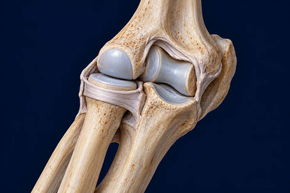
Structure notes · further context
- Humerus
- The arm bone carries the two distal humeral articular regions.
- Capitellum
- The rounded lateral humeral surface meets the superior radial-head dish.
- Trochlea
- The more medial spool-like humeral surface meets the ulnar notch.
- Radial head
- The small proximal radial head has a superior articular surface and an articular circumference.
- Annular ligament
- The pale retaining band wraps the radial-head circumference and connects with the ulnar radial-notch margins.
- Trochlear notch
- The larger trochlear notch cups the humeral trochlea. It is different from the smaller radial notch beside the radial head.
- Radial neck
- The narrower region continues below the radial head into its shaft.
- Ulna
- The proximal ulnar articulation continues into the more medial forearm bone.
Whole forearm · follow the same radius

Structure notes · further context
- Proximal joint
- The radial head meets the ulnar radial-notch setting near the elbow.
- Radius
- Follow this bone from its small proximal head to its broader distal end.
- Ulna
- The larger proximal ulnar region continues toward a smaller distal head.
- Interosseous tissue
- Thin fibrous tissue connects the shafts. Visible fibres do not map every named band.
- Distal radius
- The broad radial end participates in the wrist relationship and moves around the distal ulna.
- Ulnar head
- The smaller distal ulnar head retains its bone identity.
- Distal joint
- The ulnar notch of the radius meets the ulnar head at the second radioulnar joint.
Same anterior direction, right forearm, elbow above and wrist end below. The comparison changes the view, not the bone identities.
- BendHumeral trochlea ↔ ulnar notch. The larger shaped contact supports flexion and extension.
- Turn proximallyRadial head ↔ ulnar radial notch, retained by annular tissue.
- Turn distallyDistal radius ↔ ulnar head. Follow the whole forearm below.
Match the trochlea with the ulnar notch.
Read the larger medial articular pairing. Its geometry supports the dominant bending relationship between humerus and ulna.
Rings locate the compared structures; they do not measure pressure, force, rotation or muscle activation.
Sources for this mechanism: OpenStax · Selected synovial joints · NCBI/PubMed · Elbow annular ligament · Proximal radioulnar joint · anatomical study
02 / ONE REGIONAL COMPLEX
A hinge and a rotating neighbour share the same region.
Find humerus, radius and ulna before deciding which movement a contact permits.
The trochlea of the humerus meets the trochlear notch of the ulna. Beside it, the capitellum meets the top of the radial head. The radial-head circumference also meets the radial notch of the ulna. Three nearby contacts therefore join the same three bones in different ways.
The local articulations share a capsular setting, but their functions are not interchangeable. Elbow bending changes the relationship of arm and forearm; forearm rotation changes the radius–ulna relationship and the orientation carried toward the hand. A single word such as hinge does not describe every contact in the complex.
Read the partner on each side
The names humeroulnar, humeroradial and proximal radioulnar identify the two bones at a contact. Following those pairings prevents the radial-head collar from being confused with the humeral hinge.
Sources for this mechanism: OpenStax · Selected synovial joints · NCBI/PubMed · Elbow annular ligament
03 / BENDING AND CONTACT
The ulna cups the humeral trochlea; the radial head meets a different surface.
The large ulnar notch and the smaller radial head have different shapes and partners.
During flexion and extension, the forearm changes its angle relative to the humerus. The spool-like trochlea and the curved ulnar notch provide a strongly shaped articulation. The olecranon belongs to that proximal ulna; it is not the head of the radius.
The radial head has a shallow superior articular dish for the capitellum and an articular circumference for its ulnar relationship. It participates in the bending and rotating system, rather than functioning as a detached wheel. These surfaces carry contact through changing positions; a visible joint gap is not a measurement of cartilage or fluid thickness.
A hinge description is a useful first approximation
Living joint geometry includes non-perfectly circular surfaces and coupled small translations. This course identifies the dominant normal movement without assigning a perfectly fixed metal-hinge axis to a person.
Sources for this mechanism: OpenStax · Selected synovial joints · Proximal radioulnar joint · anatomical study
04 / A ROTATING HEAD, HELD IN PLACE
The annular ligament completes a retaining relationship around the radial head.
Find the pale band at the head circumference, then follow it toward the ulnar radial notch.
The annular ligament attaches around the margins of the radial notch of the ulna and wraps around the radial head. This ring-and-notch arrangement helps keep the head related to the ulna while allowing rotation. It is connective tissue, not an extra bone or a free bracelet around the shaft.
Stability is shared among articular shape, ligamentous structures, the interosseous system and active muscular contributions. An annular band in a picture does not mean that this one structure supplies all stability in every position. Experimental release of constraints has shown that their contributions depend on the tested direction and rotational setting.
Locate the retaining band and its radial-head context →
The top and the circumference do different jobs
The superior radial-head surface faces the capitellum. The surrounding radial-head surface participates in the proximal radioulnar relationship. Reading both avoids confusing the humeroradial contact with the entire forearm pivot.
Sources for this mechanism: NCBI/PubMed · Elbow annular ligament · Proximal radioulnar joint · anatomical study
05 / SURFACE AND SUPPORT
Cartilage permits contact; the capsule and ligaments constrain unwanted separation.
A smooth covering, a fibrous sleeve and a supporting band are different tissue arrangements.
Articular cartilage covers the load-bearing joint surfaces. A fibrous capsule encloses the local joint environment, with synovial tissue on its inner aspect supporting the fluid setting. The lining is not another layer spread over every articular-cartilage surface.
Medial ulnar collateral and lateral collateral structures reinforce the elbow in different directions. Their geometry and tension change with position and load. The close-up opens selected tissue to show the contacts and annular support; it does not turn every white strand into a separately certified collateral-ligament subdivision.
Structure is not one universal stiffness
Collagen organisation, attachment and current geometry affect a ligament’s behaviour. A static view cannot reveal all recruited fibres or determine a safe load. The cartilage and tendon courses provide the deeper tissue comparisons.
Sources for this mechanism: OpenStax · Selected synovial joints · Proximal radioulnar joint · anatomical study
06 / FOLLOW THE WHOLE FOREARM
The radius turns at the elbow and sweeps around the distal ulna.
Compare the two complete bone preparations; do not judge rotation from one isolated wrist position.
Supination · approximately parallel
Structure notes · further context
- Proximal joint
- The radial head meets the ulnar radial-notch setting near the elbow.
- Radius
- Follow this bone from its small proximal head to its broader distal end.
- Ulna
- The larger proximal ulnar region continues toward a smaller distal head.
- Interosseous tissue
- Thin fibrous tissue connects the shafts. Visible fibres do not map every named band.
- Distal radius
- The broad radial end participates in the wrist relationship and moves around the distal ulna.
- Ulnar head
- The smaller distal ulnar head retains its bone identity.
- Distal joint
- The ulnar notch of the radius meets the ulnar head at the second radioulnar joint.
Pronation · radius in front
Structure notes · further context
- Proximal joint
- The rotating radial head keeps its proximal relationship with ulna and humerus.
- Radius in front
- The crossing bone is the radius. The covered portion of the ulna is behind it, not fused into it.
- Ulna behind
- The ulnar shaft is visible again below the anterior radial crossing.
- Distal radius
- The broad distal radius has changed position; it has not become the ulna.
- Ulnar head
- The smaller distal head remains the end of the ulna.
- Distal joint
- The radial notch region follows the ulnar head during coupled forearm rotation.
Keep the small proximal head and the broad distal radius in view.
In the supinated exposure, the radius is lateral and approximately parallel to the ulna. The proximal radial head remains the small head; the distal radial end remains broad.
Both views face the same anterior direction, with proximal bones above. These native windows share one original bitmap. The hand is omitted; the explanation relates its orientation to the distal radius. Selecting a focus does not deform bones or claim measured motion.
In the supinated comparison, radius and ulna lie approximately parallel. During pronation, the radius changes its relationship to the ulna and passes anteriorly across it in this anterior view. The broad distal radius changes position while the small proximal radial head remains near its humeral and ulnar partners.
Both proximal and distal radioulnar joints participate. The hand is carried with the distal radius through the wrist relationship, so forearm rotation is different from simply bending the wrist. The ulna is a relatively stable reference in these preparations, but living tasks can also involve ulnar, humeral and shoulder movement.
A comparison is not a calibrated motion recording
These are two original anatomical states with the same stated viewing direction. They show topology and depth, not a measured angle, a perfectly fixed axis or the entire continuous trajectory. No bone is stretched or warped by the webpage interaction.
Sources for this mechanism: NCBI · Proximal radioulnar anatomy · Functional anatomy of the distal radioulnar joint · Proximal radioulnar joint · anatomical study
07 / THE SECOND END MATTERS
A broad distal radius meets a much smaller ulnar head.
Keep bone identity while changing position: the broad end does not become the ulna.
At the distal radioulnar joint, the ulnar notch of the radius articulates with the ulnar head. This reverses the proximal pattern, where the radial head meets the ulnar radial notch. Matching the names to the two ends makes the linked rotation easier to follow.
The triangular fibrocartilage complex and associated capsuloligamentous tissues contribute to distal stability and the ulnar-side wrist relationship. The forearm-bone comparison does not resolve all of these tissues. Its useful finding is the paired distal contact and the different radial and ulnar shapes, rather than an invented complete wrist reconstruction.
Compare both proximal and distal ends →
A forearm is more than two independent rods
Proximal and distal contacts, central connective tissue and muscle forces interact. Understanding this linked mechanical unit is more useful than imagining one bone spinning without any relationship to the other end.
Sources for this mechanism: NCBI · Distal radioulnar joint · Functional anatomy of the distal radioulnar joint
08 / CONNECT THE SHAFTS
The interosseous membrane is a connective system, not a third forearm bone.
Read the thin tissue between the shafts in the supinated preparation.
Fibrous tissue links the interosseous margins of the radius and ulna. Anatomical studies describe stronger bands together with thinner membranous areas, not one uniformly thick slab. Its organisation supports longitudinal stability, muscle attachment and transmission of load between the bones.
Many central fibres pass distally and toward the ulna from the radius, while other parts have different orientations. Position and loading affect their behaviour. The illustration locates the broad connective relationship; visible strands are not a counted reconstruction of all named bands or a measurement of tension.
What dissection and strain experiments establish
A primary anatomical series described distinct components in preserved specimens and then tested a smaller subset mechanically. Those observations support complexity and variable loading, not the idea that every visible fibre in an original atlas is a verified individual ligament.
Sources for this mechanism: Interosseous membrane · primary anatomy and function · Interosseous membrane · forearm biomechanics review
09 / AN ANTERIOR FORCE PATH
A radial attachment lets biceps contribute to both bending and turning.
Retain the established biceps preparation; compare its attachment with the ulnar triceps attachment.
Anterior route · radius

Structure notes · further context
- Biceps belly
- The anterior arm muscle contributes to flexion and supination.
- Distal tendon
- The tendon transmits muscle force across the front of the elbow toward the radius.
- Radial attachment
- This radial bone context differs from the ulnar triceps attachment.
Posterior route · ulna

Structure notes · further context
- Triceps tissue
- Posterior muscle supplies a major extension contribution.
- Triceps tendon
- The posterior connective path transmits force to the ulna.
- Olecranon region
- The proximal ulnar prominence receives the posterior attachment.
An anterior tendon force can contribute to flexion.
The selected biceps path crosses the front of the elbow and reaches the radius. Brachialis and brachioradialis add other contributions described in the retained regional lesson.
Distal biceps tendon reaches the radial tuberosity. Because the attachment is on the radius, its force can contribute to supination as well as elbow flexion. The contribution changes with forearm position, elbow angle and task. The proximal muscle heads and their shoulder origins are outside this local crop.
Brachialis reaches the ulna and is an important flexor; brachioradialis contributes from the lateral arm–forearm setting. Muscles can cooperate or remain active while another movement is controlled. The image shows a connective path across the joint, not an electrical recording of which muscles are currently active.
A tendon and an aponeurosis are not interchangeable names
The distal radial tendon attachment is distinct from the bicipital aponeurosis that fans into forearm fascia. The retained illustration teaches the selected radial force path rather than a complete fascial map.
Sources for this mechanism: Distal biceps insertion · anatomical study · OpenStax · Upper-limb muscles
10 / A POSTERIOR FORCE PATH
The triceps tendon reaches the ulna behind the bending relationship.
Compare anterior and posterior attachment routes before naming their action.
Triceps force is transmitted through the posterior tendon to the olecranon region of the ulna. An appropriate posterior moment contributes to extension, whereas the anterior flexor force paths contribute to flexion. The direction of external load also matters: a muscle can move, hold or brake a segment.
The long, lateral and medial triceps heads contribute to the distal system. The long head also crosses the shoulder, linking its circumstances to more than elbow angle alone. The retained local preparation does not show every origin, but it clearly separates muscle, distal tendon and ulnar attachment.
Antagonists can be active together
Flexors and extensors are not a pair of switches that must always exclude one another. Coactivation can contribute to control and stiffness while external forces and the task determine the resulting movement.
Sources for this mechanism: Triceps insertion onto the olecranon · anatomy · OpenStax · Upper-limb muscles
11 / TURNING HAS SEVERAL CONTRIBUTORS
Pronators and supinators act through different regional attachments.
Relate muscular force to the already visible radius–ulna geometry.
Pronator teres and pronator quadratus contribute to pronation. The distal quadratus links the distal forearm bones, while teres has a more proximal course toward the radius. Supinator wraps around the proximal radial setting, and biceps supplies another important supination contribution.
These broad action names are useful starting points. The effect of a pull depends on the current geometry, load and recruitment. Brachioradialis can help return the forearm toward a mid-position from different starting positions; this is different from declaring it the sole pronator or supinator.
The cross-section and the long view answer different questions
A transverse muscle view locates groups around the two shafts. The paired long views show how the bones change relationship from elbow to distal joint. Neither view alone displays every origin-to-insertion route.
Sources for this mechanism: NCBI · Proximal radioulnar anatomy · NCBI · Radial nerve anatomy · OpenStax · Upper-limb muscles
12 / CONNECTED LOAD SHARING
A force applied through the hand does not remain on one isolated bone.
Contact and connective tissues distribute load through a linked forearm.
The distal radius has an important relationship with the wrist and receives load from the hand–wrist unit. Interosseous connective tissue can transfer part of that load toward the ulna as forces pass proximally. Proximal contacts, distal restraints and muscle forces all contribute to the resulting mechanical setting.
The distribution depends on position, loading conditions and the integrity of the connected tissues. There is no universal percentage displayed by this atlas. Passive specimen experiments, active muscle behaviour and a person holding an object are related but different sources of evidence.
Moment and axial load are different quantities
A bending moment depends on a force and its perpendicular distance from an axis. Axial load follows another directional description. The established lever course lets you compare these ideas without treating an anatomical highlight as measured force.
Sources for this mechanism: Interosseous membrane · forearm biomechanics review · Functional anatomy of the distal radioulnar joint
13 / SUPPLY AND REGIONAL NEIGHBOURS
The same region carries movement commands, sensory traffic and blood.
Use the established nerve and vascular lessons for resolved routes.
Musculocutaneous supply reaches biceps and much of brachialis. Radial-nerve pathways supply triceps and brachioradialis, while its deep branch serves supinator. Median-nerve pathways serve the pronators, including the anterior interosseous route to pronator quadratus. These are broad normal relationships, not a complete root-level or variant map.
The brachial artery divides into radial and ulnar continuations in the proximal forearm setting, with recurrent and collateral vessels contributing around the elbow. The ulnar nerve passes behind the medial epicondylar region; radial and median pathways have different relationships. The bone preparations do not invent coloured strands as proof of these hidden routes.
A landmark does not identify every neighbouring structure
Finding an epicondyle or radial head helps orient the region. A precise nerve branch, vascular variant or compression site still needs its own anatomical evidence; this normal course provides no symptom diagnosis or self-test.
Sources for this mechanism: NCBI · Proximal radioulnar anatomy · NCBI · Radial nerve anatomy
14 / CONTROL AND EVIDENCE
A pose, a force and a muscle signal are different observations.
The nervous system coordinates movement using multiple sources of information.
Muscle length and tension-related information, joint and skin signals, and wider motor commands contribute to control. Changes in load can demand different recruitment even when the joint looks still. A selected anatomical state does not prescribe a sequence that every person’s muscles follow.
Anatomical preparations establish relationships. Imaging depends on the plane and acquisition; motion studies depend on reference frames; electromyography samples electrical activation rather than directly measuring tendon force. Read a source for the question it actually answers, not for an appearance of precision.
What this course adds and what it links
The chapter connects elbow contacts, both forearm pivots, connective load sharing and the existing regional muscle paths. Fine acquired imaging, named distal soft-tissue subdivisions, individual motion measurements and clinical decision-making require additional evidence; they are not silently certified by generated anatomy.
Sources for this mechanism: Proximal radioulnar joint · anatomical study · Interosseous membrane · primary anatomy and function · Functional anatomy of the distal radioulnar joint
Keep the bone, its partner and its job together.
Which contact is the proximal radioulnar joint?
The radial-head circumference and the radial notch of the ulna, with annular support; not simply the top of the radius against the capitellum.
Which distal end is broad in both positions?
The distal radius. The distal ulna has a smaller head; changing position does not swap bone identities.
Why is pronation different from wrist bending?
It involves coupled radius–ulna rotation along the forearm; the distal radius carries the wrist/hand relationship.
Is the annular ligament a free ring on the shaft?
No. It completes a retaining relation with the ulnar radial notch.
Does a pale interosseous sheet mean a third bone?
No. It represents a complex fibrous connective system, with different bands and membranous areas.
Can a still pose prove one muscle is inactive?
No. Force, external load and recruitment can change without an obvious change in position.
Sources and original-image identity
Normal anatomy source checks:22October2026. Existing FB017, FB028, FB032 and FB033 research guides coverage; no reference-book text or image is reproduced. Older specimen work is retained for supported anatomy, with its experimental limits.
- OpenStax · Selected synovial jointsReadable official elbow section: three local articulations, shared capsule and collateral/annular support. No copied figure, clinical manoeuvre or universal angle adopted.
- NCBI · Proximal radioulnar anatomyOfficial indexed normal rotation, muscular and arterial relationships. Distal biceps tendon and fascial aponeurosis remain distinct; a compressed excerpt combining these insertions is not adopted.
- NCBI/PubMed · Elbow annular ligamentReadable official excerpt supports the three articular pairings and radial-head support. Direct Bookshelf retrieval encountered a challenge. The proximal radioulnar pivot is distinguished from radiocapitellar contact; simplified classification wording is not treated as complete kinematics.
- Proximal radioulnar joint · anatomical studyReadable official primary abstract from ten cadaveric elbows: annular and interosseous constraints, position-dependent behaviour and radial-head shifting. No specimen measurements or surgical conclusions transferred to these pictures.
- NCBI · Distal radioulnar jointOfficial indexed normal distal radius/ulna and capsuloligamentous context. No imaging diagnosis, exact attachment footprint or treatment rule.
- Functional anatomy of the distal radioulnar jointReadable official abstract and indexed article context connect rotation, soft tissues and load distribution. Direct PMC retrieval challenged; no claim to a fresh complete methods or clinical review.
- Interosseous membrane · primary anatomy and functionReadable primary abstract: twenty preserved specimens, complex bands and membranous portions, with a smaller strain experiment. No angles, band counts, universal tension or specimen forces are assigned to the generated sheet.
- Interosseous membrane · forearm biomechanics reviewReadable official abstract supports longitudinal stability and force transfer. Repair recommendations and injury management are outside the normal teaching scope.
- Distal biceps insertion · anatomical studyRetained qualified Muscle source evidence for the radial attachment; not a fresh complete reread or measured footprint certificate.
- Triceps insertion onto the olecranon · anatomyRetained qualified Muscle source evidence for broad posterior ulnar attachment, without measurements or surgical interpretation.
- NCBI · Radial nerve anatomyOfficial indexed motor supply and deep-branch/supinator context. Exact branch-transition points and a complete nerve map are not inferred from the illustrations.
- OpenStax · Upper-limb musclesEstablished regional action framework; named actions additionally checked against independent anatomy and retained insertion evidence. Not an activation trace or exercise programme.
The joint close-up and paired rotation preparation are new original generated illustrations. The two force-path illustrations are retained qualified project artwork. These are not patient scans, acquired microscopy or dissection photographs. Native views, independent labels and default short names are shared by the body and detail reader; no language is burned into the base images.
Gross anatomy, visual teaching quality and label–mechanism coherence need separate checks. Generated fine fibres, exact band footprints, hidden nerves and individual motion angles are not certified. AI-assisted review and Owner visual standards are not physician review. This normal course supplies no diagnostic test, reduction manoeuvre, treatment or rehabilitation prescription.