The cornea and lens focus light onto the retina. Retinal cells change light into neural activity; the optic nerve carries their output toward brain relays and visual cortex.
Gold · incoming lightCyan · neural output
Anterior left, posterior right. Qualitative guides locate light entry, retinal conversion and ganglion-cell output; they are not measured rays or individual synapses. Light does not continue along the optic nerve.Structure notes · further context
Ciliary and zonular context
The ciliary region connects to lens-supporting zonular fibres.
Iris / pupil approach
The iris is in front of the lens; its opening controls light entry.
Lens
A transparent biconvex structure adjusts focusing power.
Vitreous cavity
Clear gel occupies the large space behind the lens.
Posterior nerve approach
Ganglion axons collect at the disc and leave as the optic nerve.
Corneal dome
The front window separates external tears from internal aqueous fluid.
Anterior aqueous chamber
Watery fluid lies between cornea and iris; it is separate from the vitreous behind the lens.
Inner retinal lining
Neural retina lies inside the vascular choroid and white sclera.
Chapter overview video: planned. No playable video or duration is currently available.
Front window → adjustable lens → neural lining
Follow light through a living optical organ.
Anterior is left, posterior is right in this selected cutaway. The front compartment and back gel space serve different roles.
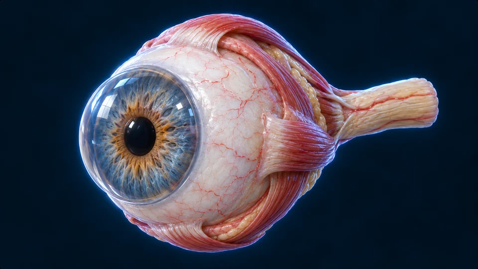
Compare the outside of the globe
Retained exterior context, not a complete six-muscle dissection. Fine vessels and texture are illustrative.Structure notes · further context
Iris
The coloured diaphragm regulates the pupil, the dark opening admitting light.
Optic nerve
Retinal ganglion-cell axons leave posteriorly toward the brain.
Sclera
The tough white outer coat supports the globe.
Cornea
A clear projecting dome is the first major refracting surface.
Locate the cornea before the iris, then the lens and the large vitreous cavity. The thin retina lines the back; its output leaves through a different structure, the optic nerve.
Go deeper · Anatomy and the optical route
Protected, yet open to light
Light enters the eye. The retina responds. The brain makes seeing possible. The globe is both an optical organ and a neural interface: clear surfaces focus an image, retinal circuits shape a message, and connected brain networks interpret it.
Each eye rests in a bony orbit. The orbit helps protect the globe; the eyelids and tear film protect its exposed surface. The clear cornea is the first major focusing surface. Behind it, the iris changes the pupil’s size, regulating how much light enters.
Three different structures at the front
The cornea bends incoming light. The pupil is an opening within the iris, not a lens. The lens adds adjustable focus behind the iris. The ciliary region changes its shape for near vision. The ciliary body connects to lens-supporting zonular fibres; the fine strands in the cutaway are illustrative.
An image on neural tissue
Follow light from the cornea and pupil, through the lens and vitreous body, to the retina lining the back of the eye. The retina receives the focused image. Ganglion-cell fibres leave at the optic disc and form the optic nerve. Optical images are inverted on the retina; vision is not a small observer reading that picture inside the head.
Clear tissues need support
The cornea and lens must transmit light, so they do not contain the dense blood-vessel networks seen in many other tissues. The tear film and surrounding fluids help support the front structures. Clarity and focusing shape are as important as allowing light to enter.
Coats · chambers · circulation
The wall and the fluid spaces are not the same layer.
A deliberately magnified window from the same retained eye illustration, not a newly generated specimen. Outside above, vitreous below; proportions are illustrative.Structure notes · further context
Sclera · outer coat
Tough connective tissue surrounds the globe and continues anteriorly as cornea.
Choroid · vascular coat
The vascular middle layer lies between sclera and neural retina.
Retina · inner lining
The neural lining faces the vitreous; photoreceptors lie toward its outer side.
Outer sclera/cornea supports and transmits; the vascular middle coat supports; inner retina detects. Aqueous circulates in the anterior segment, while vitreous is the larger gel-filled space behind the lens.
Go deeper · Coats, chambers and aqueous flow
Three coats
The white sclera is an outer protective coat and continues anteriorly as the clear cornea. The vascular choroid lies between it and the neural retina. The iris and ciliary body belong to the middle vascular coat. Retina is the inner neural lining, not the tough outer wall.
Aqueous and vitreous differ
Aqueous humour is watery fluid in the anterior segment. Vitreous is the gel-like material occupying the large cavity behind the lens. “Posterior chamber” also names the small aqueous space behind the iris and in front of the lens; it must not be confused with the much larger vitreous cavity.
Fluid is made and drained
The ciliary processes produce aqueous fluid. It passes from the posterior chamber through the pupil into the anterior chamber, with drainage mainly through the angle near the corneal–iris junction. Production and outflow help determine intraocular pressure. This explanation does not supply an individual pressure measurement or diagnose a drainage problem.
Tears are outside
The surface tear film is outside the cornea, whereas aqueous is behind it. They are distinct systems. The large gel-filled back cavity similarly does not drain through the puncta in the eyelids. Naming each compartment prevents an apparently simple “fluid route” from joining unrelated spaces.
Tissue → cells → signal
Light enters from one side. Neural output leaves the other way.
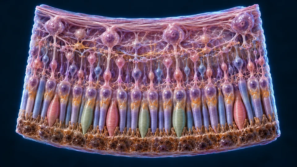
The vitreous-facing inner side is at the top of this enlargement. Photoreceptor segments lie toward the outer pigment epithelium below.
Gold guide downward: light toward outer photoreceptors. Cyan guide upward: broad neural information route back toward ganglion output. These are direction summaries, not one-to-one synapse traces. Generated tissue regions are illustrative, not authentic histology or measured cell identities.Structure notes · further context
Inner nerve-fibre context
Ganglion axons travel along the vitreous-facing side.
Ganglion-cell context
Retinal output cells generate action potentials toward the brain.
Bipolar and local circuit region
Bipolar and local interneuron pathways process photoreceptor responses.
Photoreceptor segment context
Rods and cones have light-sensitive outer segments toward the pigment epithelium.
Pigment epithelial context
The outer support layer lies beside the photoreceptor outer segments.
Retinal compartments and photoreceptor structure · Webvision source.
Rods and cones: distinguish structure from colour
Inner retinal side above; pigment epithelium below. Cylinder-like rod and tapered cone outer segments are compared at selected illustrative scales; the picture is not microscopy, a cell ratio or a wavelength colour map.Structure notes · further context
Rod · cylindrical outer segment
Disc-rich membrane contains light-sensitive pigment. Rod pathways support sensitivity in dim conditions.
Cone · tapered outer segment
Cone pathways support colour and fine detail in brighter conditions; colour here does not identify spectral sensitivity.
Inner segment · mitochondrial region
A separate, mitochondria-rich cellular region supports the outer segment.
Nuclear cell body
The nucleus lies in the cell body, separate from the mitochondria-rich inner segment.
Rod synaptic ending
This ending changes transmitter release to downstream retinal circuitry, not directly to visual cortex.
Cone synaptic ending
A broader terminal communicates with the retinal circuit; exact contacts are not reconstructed.
Pigment epithelial support
Pigmented cells and their apical processes support the photoreceptor outer segments.
Connecting cilium (illustrative)
A narrow connection joins inner and outer segments; it is not the neuronal axon to the synaptic ending.
Light reaches the outer photoreceptors after entering from the inner retinal side. Bipolar pathways and local circuits reshape responses before ganglion cells send action potentials toward the brain.
Go deeper · Phototransduction and retinal processing
Different detectors
The retina is light-sensitive neural tissue. Its rods and cones are photoreceptors; their responses enter retinal circuits before signals travel along the optic nerve. Rods work well in dim light. Cones support colour vision and fine detail in brighter light. Cone sensitivities overlap; colour is not read by one isolated “red pixel” cell.
A local circuit before the nerve
The broad route is photoreceptors through bipolar pathways to retinal ganglion cells. Horizontal and amacrine cells contribute local processing, and multiple ON/OFF pathways respond differently to light changes. Ganglion axons converge at the optic disc. The enlargement cannot verify each synapse or imply one detector always connects to one output cell.
Light does not simply excite every cell
Light approaches from the vitreous side before reaching outer photoreceptors. Activated photopigment changes intracellular signalling, reducing the open cGMP-gated channel current and hyperpolarizing the photoreceptor. Transmitter release changes, reshaping downstream activity. Photoreceptor responses are graded; ganglion cells generate the action potentials carried by the optic nerve.
Support next to the outer segments
Retinal pigment epithelium supports the photoreceptors, absorbs stray light and participates in outer-segment maintenance and visual-pigment recycling. The adjacent choroidal supply supports the outer retina, while retinal vessels support inner regions. A light-microscope field or a generated wedge cannot display every biochemical step.
Authentic acquired evidence
See a real retinal surface.
This fundus photograph is a separately sourced image of the back of the eye. It is not the generated retinal-cell enlargement.
Authentic fundus photography. Viewed from the front: image left corresponds to the person’s right; the optic disc is nasal, on image right. Source describes a healthy volunteer, not an independent diagnostic finding by AllHumanBody.Structure notes · further context
Retinal vessel context
Surface vessels are visible; this is not an individual vascular or diagnostic assessment.
Macular region
The central retinal region supports fine central vision; its foveal centre is not resolved as cells.
Optic disc
Axons leave here. No photoreceptors means a physiological blind spot.
Mikael Häggström · original photograph, 21 March 2012 · CC0 1.0. Proportional WebP resizing only; no crop, recolour, generated detail or diagnostic overlay. Public derivative metadata omitted.
The real photograph shows surface vessels and the optic disc, not individual rods or cones. Compare its nasal disc with the central macular region.
Go deeper · Macula, disc and the blind spot
Macula, fovea and detail
The macula is the central retinal region; its foveal centre supports the sharpest central vision. Dense cone sampling and specialised local organisation help distinguish fine detail. A fundus image shows a surface region, not individual cones or an exact cellular boundary.
Disc and blind spot
Ganglion axons travel along the inner surface and leave at the optic disc. The disc has no photoreceptors, creating the physiological blind spot. It is different from the macula or fovea. The brain combines available information, so this gap does not usually look like a permanent black patch in ordinary binocular viewing.
Refraction · accommodation
The ciliary muscle changes tension, not the light opening.
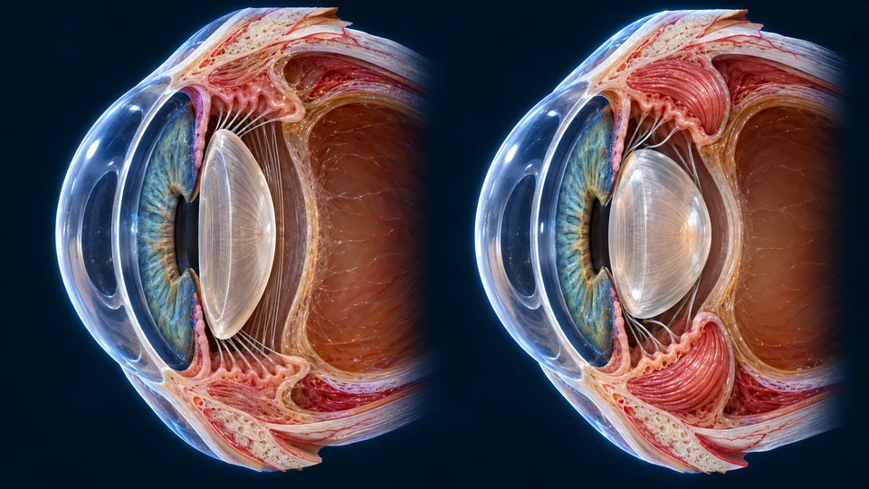
Separate anatomical preparations compare distant viewing and near viewing. The dimensions are illustrative, not measured optical power.
Distant: ciliary muscle is more relaxed → greater zonular tension → a flatter lens.
Gold arrows represent qualitative zonular pull, not fibre movement or measured force. Short dashed near arrows mean less tension, not a reversed pull. Cyan marks ciliary-ring narrowing. The original anatomy is not animated.Structure notes · further context
Near preparation · ciliary context
Contraction reduces the tension transmitted to the lens.
Distant preparation · zonules
Greater tension helps maintain a flatter lens.
Near preparation · zonules
Lower tension permits the lens to round up; the fibres do not pull it thicker.
Distant preparation · lens
A flatter lens has less focusing power than the near-view state.
Near preparation · lens
A rounder lens increases focusing power for a nearer object.
Distant · greater pull keeps the lens flatter
Gold is zonular pull; short dashed arrows mean less pull. Cyan is ciliary-ring narrowing. Contraction does not make the fibres pull the lens thicker.Structure notes · further context
Distant preparation · zonules
Greater tension helps maintain a flatter lens.
Distant preparation · lens
A flatter lens has less focusing power than the near-view state.
Near · contraction allows rounding
Gold is zonular pull; short dashed arrows mean less pull. Cyan is ciliary-ring narrowing. Contraction does not make the fibres pull the lens thicker.Structure notes · further context
Near preparation · ciliary context
Contraction reduces the tension transmitted to the lens.
Near preparation · zonules
Lower tension permits the lens to round up; the fibres do not pull it thicker.
Near preparation · lens
A rounder lens increases focusing power for a nearer object.
Distant rays need less lens adjustment
Whole-eye optical context, using the retained cutaway. Guides change with the selected object distance; the globe raster does not change lens shape. Compare the actual lens shapes in the preparations above. Not a measured ray trace or an eyesight test.Structure notes · further context
Retinal focus
The changed qualitative ray guide ends at the retinal lining, not the optic disc.
For near focus: ciliary contraction lowers zonular tension and permits a rounder lens. Pupil size changes the opening; it does not pull the lens thicker.
Go deeper · Refraction, accommodation and the pupil
Bring light into focus
The cornea provides most of the eye’s refraction. The lens fine-tunes focus so an image forms on the retina. For a near object, the ciliary muscle contracts and tension on the lens-supporting fibres decreases. The lens becomes rounder and more powerful. For distant viewing it is flatter. This adjustment is called accommodation.
Keep focus and pupil regulation separate
Changing lens curvature changes focusing power. Changing pupil size regulates the aperture and can influence image quality, but it does not pull the lens rounder. Convergence of the eyes and pupil constriction often accompany near viewing; they are related responses, not the same structure doing all three jobs.
When the focus misses
With accommodation relaxed, myopia brings distant rays to a focus in front of the retina; in hyperopia the optical focus would lie behind it. Accommodation can compensate for some hyperopia, so symptoms are not identical in every person.
When the lens changes less
Presbyopia reduces near focusing as the aging lens becomes less flexible. It differs from a long or short globe. Astigmatism makes focusing vary with direction rather than producing one simple sharp focus.
Normal-to-changed optical mechanisms · NEI source. Educational comparison, not a prescription, diagnosis or an eye examination.
Orbit · lids · tears · pupil
A protected surface still has to face the world.
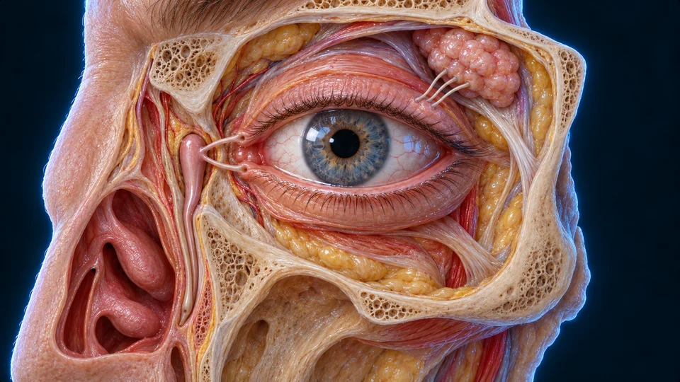
Selected frontal context of the person’s left eye: nasal is image left, lateral is image right. The gland and medial drainage are on opposite sides.
Generated selected lacrimal context, not CT or surgical anatomy. Canalicular dimensions, duct apertures and the exact inferior nasal opening are not independently resolved. The visible gross gland/drainage positions support the explanation.Structure notes · further context
Lacrimal gland region
A gland lies in the superior lateral orbit; ducts reach the conjunctival region.
Exposed eye and lids
Blinking spreads the tear film over the front surface.
Medial canalicular approach
Small channels from the lid margins lead toward the lacrimal sac.
Lacrimal sac context
The drainage sac lies near the medial orbital wall, beside the nose.
Nasolacrimal drainage approach
A descending duct carries drainage toward the nasal cavity.
The main gland is superolateral; drainage is medial. Blinking spreads fluid across the outside surface, toward puncta, canaliculi, the sac and the nasolacrimal duct.
Go deeper · Tear-film sources, drainage and protection
A film, not just a drop
Tears maintain a smooth moist front surface, help carry away debris and contribute protective components. The film includes watery, mucin-associated and lipid components from different sources; the main lacrimal gland is not the only contributor. Blinking distributes the film and supports drainage.
Production and drainage
The main lacrimal gland lies superiorly and laterally. Tear fluid spreads toward the medial side and enters small puncta in the lid margins, then canaliculi, lacrimal sac and nasolacrimal drainage toward the nasal cavity. This is external surface fluid, not fluid emerging from the vitreous.
The aperture has its own muscles
Circular sphincter muscle in the iris constricts the pupil; radial dilator muscle enlarges it. Parasympathetic control supports constriction, while sympathetic control supports dilation. The pupil is an opening, not a black anatomical plug. Its response involves connected sensory and autonomic pathways.
Layers of protection
Orbital bone surrounds much of the globe. Lids reduce exposure, eyelashes intercept debris, and blinking helps limit drying. These protections do not make the eye invulnerable. This chapter teaches normal mechanisms rather than personal eye-care or emergency treatment advice.
Muscle coordination · connected brainstem
Seeing clearly also means aiming and stabilising.
Anterior is image left in both preparations. The corrected superior-rectus point follows the globe-facing belly, not the thinner band above it. Oblique deep insertion boundaries are not fully exposed.Structure notes · further context
Lateral rectus
The foreground straight muscle pulls the eye outward; motor supply is CN VI.
Superior rectus · visible belly
This upper straight muscle contributes elevation, intorsion and adduction; motor supply is CN III.
Superior oblique · trochlea
Its tendon turns through a superomedial pulley before returning toward the globe; motor supply is CN IV.
Superior oblique · reflected tendon
The returned tendon attaches behind the globe equator; it does not continue to the posterior muscle origin.
Inferior oblique · anterior route
The short curved muscle begins at the anterior medial floor and passes under the globe, unlike the recti. Its hidden full insertion is not mapped here; motor supply is CN III.
View 1 · Straight muscles on the globe
Anterior is left. Follow superior rectus toward its anterior scleral attachment. The thinner upper band remains unlabelled because its full endpoint is hidden.Structure notes · further context
Lateral rectus
The foreground straight muscle pulls the eye outward; motor supply is CN VI.
Superior rectus · visible belly
This upper straight muscle contributes elevation, intorsion and adduction; motor supply is CN III.
View 2 · Different oblique routes
The pulley turns the superior-oblique tendon back toward the globe. Hidden complete posterior insertions cannot be confirmed from this exposure.Structure notes · further context
Superior oblique · trochlea
Its tendon turns through a superomedial pulley before returning toward the globe; motor supply is CN IV.
Superior oblique · reflected tendon
The returned tendon attaches behind the globe equator; it does not continue to the posterior muscle origin.
Inferior oblique · anterior route
The short curved muscle begins at the anterior medial floor and passes under the globe, unlike the recti. Its hidden full insertion is not mapped here; motor supply is CN III.
Reveal the two recti hidden on the far side
Other muscles are omitted for exposure. Anterior is left; orbital apex is right. This complementary preparation supplies the medial/inferior rectus labels rather than guessing at hidden bands in the other view.Structure notes · further context
Medial rectus
The exposed nasal-side rectus draws the globe toward the nose; motor supply is CN III.
Inferior rectus
The lower rectus contributes depression, extorsion and adduction; motor supply is CN III.
Optic nerve
The pale neural output is separate from the red muscle bellies.
Common-origin region
The four recti arise posteriorly near a common tendinous origin; its complete ring anatomy is not resolved here.
Actions in primary position; vertical effects vary with gaze
Muscle
Main actions
Motor nerve
Medial rectus
Adduction · toward the nose
III
Lateral rectus
Abduction · away from the nose
VI
Superior rectus
Elevation, intorsion, adduction
III
Inferior rectus
Depression, extorsion, adduction
III
Superior oblique
Intorsion, depression, abduction
IV
Inferior oblique
Extorsion, elevation, abduction
III
Origins, insertions and actions · Anatomy source. Levator raises the upper lid and is not one of these six globe movers.
Four recti pull from different sides of the globe; two obliques use different routes. The lateral rectus turns outward and medial rectus inward. Vertical and twisting actions depend on gaze position.
Go deeper · Six movers, their nerves and gaze stability
Six movers, coordinated actions
The superior, inferior, medial and lateral rectus muscles, plus superior and inferior oblique muscles, move each globe. Medial and lateral rectus are major inward and outward movers. Vertical and twisting actions depend on gaze position and combined muscle activity; a single band in an illustration cannot supply a complete movement map.
Separate nerves and eyelid movement
The lateral rectus receives the abducens nerve, the superior oblique the trochlear nerve, and the other four movers the oculomotor nerve. Levator palpebrae raises the upper lid; it is not one of the six globe movers. Brainstem networks coordinate both eyes rather than commanding each visible muscle independently.
Fast shifts, pursuit and fixation
Saccades rapidly redirect gaze; pursuit follows a moving target. Small adjustments occur during fixation. These movements place useful detail near the fovea and maintain a stable perceptual scene. They are physiological roles, not an eyesight or eye-movement examination here.
Head movement need not blur everything
Vestibular signals can drive compensatory eye movements through brainstem motor networks—the vestibulo-ocular reflex. This connects vision with balance and head movement. The exterior plate above provides only selected muscle context; a fully verified individual-muscle orbital atlas remains deeper scope.
Partial crossing · relay · perception
Each cerebral side receives an opposite half of the visual world.
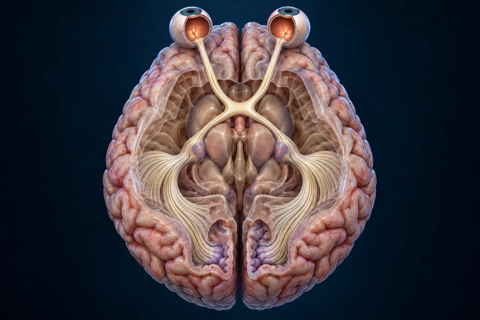
An opened superior view connects the eyes with the thalamic relay and medial occipital destination. Anterior is above; patient right is image left. Select a visual half-field to follow its correspondence guides.
Left visual field → right cerebral hemisphere (image left).
Original anatomical illustration. Patient right is image left. Dashed guides show correspondence, not measured individual axons. Gold: retinal axons to LGN; cyan: a different neuron's onward route after the relay. Exact fan subdivisions, Meyer’s loop and V1 borders are not resolved. Names and short meanings remain visible in both states.Structure notes · further context
Retinal output
Ganglion-cell axons leave each retinal surface; this is neural output, not a light ray.
Optic nerve
Before the chiasm, each nerve contains output from one eye.
Optic chiasm
Nasal retinal axons cross; temporal retinal axons stay on their own side.
Optic tract
After the partial crossing, each tract carries the opposite visual half-field from both eyes.
LGN · thalamic relay
Retinal axons relay to new neurons in the lateral geniculate nucleus.
Optic radiations
Axons of LGN relay neurons spread through cerebral white matter toward occipital cortex.
Medial occipital target region
Primary visual cortex lies around the calcarine sulcus. This opened region locates the destination, not a measured V1 boundary.
Both routes remain visible in words: left field → left-eye nasal retina plus right-eye temporal retina → right LGN and cortex; right field → right-eye nasal retina plus left-eye temporal retina → left LGN and cortex. A crossing changes sides; a relay changes neurons.
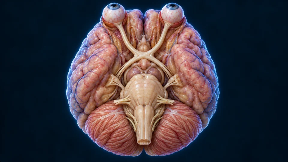
Generated gross inferior anatomical context. The X-like structure does not mean every fibre crosses. Exact tract courses, individual nuclei and optic radiations are not independently mapped; primary visual cortex is not labelled on the hidden medial occipital surface.Structure notes · further context
Optic nerve
A nerve carries output from each retina toward the basal chiasm.
Optic chiasm
Nasal retinal fibres cross; temporal retinal fibres remain on their own side.
Posterior optic-tract context
Beyond the chiasm, each tract carries information from the opposite visual half-field.
Retinal axons end at a relay in the LGN. New relay-neuron axons form the optic radiations; primary visual cortex lies on the medial occipital surface around the calcarine sulcus.
Go deeper · Partial crossing, visual fields and cortical processing
The retained broad route
Retinal output follows the optic nerve toward the brain. At the optic chiasm, some fibres cross; most image-forming visual information then relays through the thalamus on its way to visual cortex. The route is retina → optic nerve → chiasm → optic tract → lateral geniculate nucleus (LGN) → optic radiations → visual cortex.
Crossing is retinal, not “one whole eye”
Nasal retinal fibres cross, while temporal retinal fibres remain on the same side. Each optic tract therefore combines information from both eyes about the opposite visual half-field. Left eye and left visual field are different concepts. The main image does not map every visual-field fibre.
Binocular overlap helps depth
The eyes view much of the same scene from slightly different positions. Brain networks can use the disparity between these views as one depth cue. Motion, perspective and familiar size provide additional cues; depth perception is not a single chiasm junction comparing two photographs.
Vision involves processing
Primary visual cortex lies mainly on the medial occipital surface. Connected areas process properties such as form, colour and movement. Other retinal outputs contribute to light responses and timing functions, so not every retinal pathway is simply an image-forming LGN route.
The eye focuses light. Retinal cells detect it and reshape the message. The brain receives and interprets that information. Existing correct eye structure, retina, accommodation and partial-crossing knowledge is retained and expanded. Book records FB058–060 set the topic and multiscale teaching benchmark; original text and illustrations are not copied.
Targeted new structure and claim checks:16 October 2026. Source checks and independent AI review are not human medical review.
Existing qualified and newly produced generated atlas masters are visibly distinguished from the acquired fundus photograph. Fine tissue dimensions, cell colours/connections, fibre paths and drainage apertures are illustrative, not patient measurements. No authentic OCT, retinal histology or quantitative ray trace is supplied. Selected new orbital preparations do not certify every hidden attachment or surgical measurement. NCBI pages blocked by recaptcha are not claimed as newly read; prior evidence is retained in project history. Source checks and AI visual inspection are not human medical review. Physical-phone acceptance remains pending. No diagnosis, treatment tool, unavailable video or CMS publication-state change is implied.

 A different scale
 An adjustable lens
 Protection & drainage
 Vision needs a brain