Contact / loading
Ground ↔ heel / soft tissue ↔ hindfoot
Representative heel-first walking contact; timing varies.
REGIONAL ANATOMY · CONTACT & CONTROL
Precise touch. Adaptable support.
Follow small bones into moving joints, gliding tendons, active muscles and living contact tissues. Structure matters because a hand must manipulate and a foot must adapt to loading.
Start with the framework →Palmar hand, thumb to the left. Regional labels identify visible anatomy; bone counts and fine joint organisation are explained below.

READ THE CONTACT BEFORE THE LONG EXPLANATION
Compare the heel contact, shared support and heel-raised conditions. Bone, soft tissue and joint position change together; no pressure measurement is implied.
Contact · heel meets the ground

Read the contact surface and the framework above it. The departure view ends before swing; the lifted limb during swing is not shown. The three views compare representative heel-first contact conditions, not a measured sequence of one foot. Gross contact relationships were independently AI-inspected; fine tarsal boundaries, muscle identities and attachments are not certified. They do not measure pressure, percentage of stance, joint angles or equal-scale deformation. Each retains its own visible names and local numbering. Explore why toe extension is only part of the arch explanation →
Ground ↔ heel / soft tissue ↔ hindfoot
Representative heel-first walking contact; timing varies.
Ankle + joints + arches + muscles
Tissues deform and activity regulates support.
Forefoot / toes → unloading and repositioning
Propulsion and limb clearance have different tasks.
Supporting relationship map, not exact anatomical geometry or a measured time course. All steps are visible before highlighting.
Connect the foot comparison to the hand’s tissue coordination, arch support and heel’s internal compartments.
Teaching references: NLM · Selected normal gait organisation · NLM · Active arch regulation during walking/running · NLM · Primary dynamic arch experiment and model limits
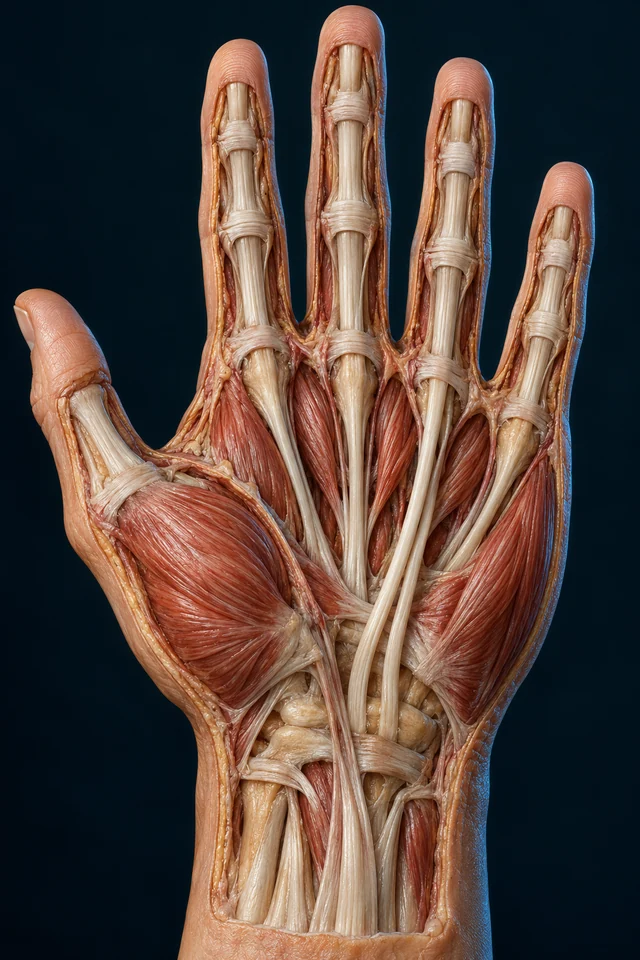 Inside the palm ↗Short muscles work with long tendons.
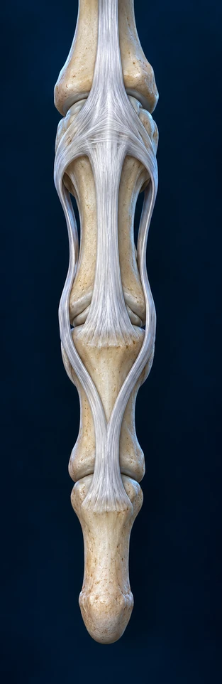 Opening the finger ↗One apparatus, different attachment levels.
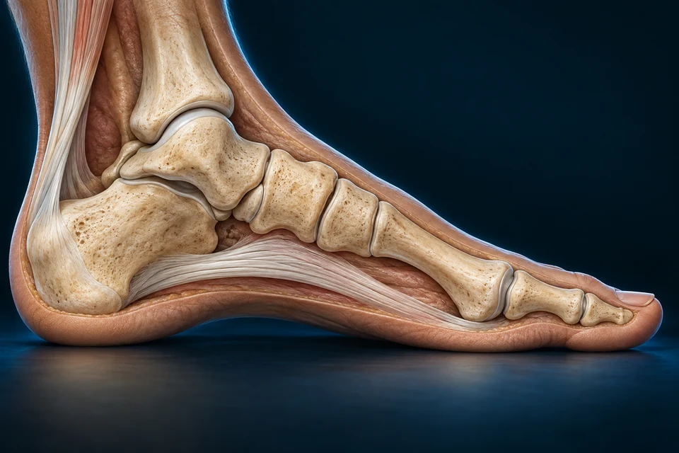 A supported arch ↗Bone, connective tissue and muscle interact.
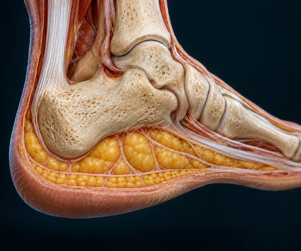 Beneath the heel ↗Living compartments help manage contact.
ORIENTATION / BONES / JOINTS
Locate the wrist, palm and digits in the opening plate. Then connect the conventional bone plan to the different motion available at each joint level.
Both regions arrange small bones, joints and connective tissues into multiple rays. Different proportions and muscle control make their tasks distinct.
Wrist → palm → digits
Positioning and sensation support manipulation.
Hindfoot → midfoot → forefoot
Contact adaptation and propulsion share a connected framework.
Muscle / tendon / joint / skin
Several tissues coordinate movement and feedback.
Teaching references: NCBI · Hand bones and normal variation · NCBI · Selected normal foot and ankle anatomy
The distal forearm meets a small-bone wrist, then metacarpals and digit segments. The thumb diverges from the palm instead of repeating the four longer fingers.
Teaching references: NCBI · Hand bones and normal variation · OpenStax8.2 · Upper-limb regional relationships
Scaphoid, lunate, triquetrum and palmar pisiform are conventionally described proximally; trapezium, trapezoid, capitate and hamate distally. Adjacent joints and ligaments distribute motion.
Thumb-side radius / scaphoid-lunate
The distal radius contributes to wrist articulation.
Articular disc / ligament relationships
Soft-tissue interfaces complete the ulnar-side relationship.
Two rows / intercarpal interfaces
Several small joints contribute to movement.
Teaching references: NCBI · Hand bones and normal variation · OpenStax8.2 · Upper-limb regional relationships
A conventional hand has eight carpals, five metacarpals and fourteen phalanges. Variable sesamoids and accessory bones require separate descriptions.
One metacarpal + two phalanges
One IP joint differs from the longer fingers.
Three phalanges each
MCP, PIP and DIP describe different levels.
Five metacarpal rays
Heads form knuckle context; bases meet the wrist.
Teaching references: NCBI · Hand bones and normal variation
Thumb positioning combines movement and rotation around its first carpometacarpal relationship, then adjustments at MCP and IP joints. The pad can face another digit during manipulation.
The first metacarpal meets the trapezium in a saddle-shaped interface. Thenar muscles, adductor and long tendons share actions; no single visible muscle explains every grip.
The static tissue view below is regional context, not an exact dynamic opposition frame.
Teaching references: NCBI · Intrinsic hand organisation · NCBI · Hand muscle context · OpenStax8.2 · Upper-limb regional relationships
MUSCLE BELLIES / FORCE-TRANSMITTING TENDONS
Red short-muscle regions lie beside pale long-tendon routes. A regional view helps separate where force is made from where it is transmitted.
Precision · regional thumb and short-muscle contextThe open palm locates tissues that cooperate in controlled pad contact; it is not a frame of an actual pinch.
Gold outlines locate the selected anatomical context; they are not measured contraction, tendon travel or a nerve route. All short names remain available without selecting a state.
Thumb pad ↔ selected fingertip
Position, sensation and joint control refine a small contact.
Fingers + thumb + palm around an object
Several rays adapt; intrinsic/extrinsic actions coordinate.
Forearm activity → tendon conditions
Stabilisation assists the task without one universal posture.
Supporting relationship map, not exact anatomical geometry or a measured time course. All steps are visible before highlighting.
Intrinsic muscles lie within the hand. Extrinsic muscle bellies originate in the forearm and send tendons into it; the two systems coordinate rather than compete.
Teaching references: NCBI · Intrinsic hand organisation · NCBI · Hand muscle context · NCBI · Long flexor tendons and sheaths
A pinch emphasises controlled contact between pads. A surrounding grip adapts digits and palm to an object; stabilising wrist activity changes tendon conditions.
Supporting task comparison, not a strength test, exercise prescription or ideal posture.
Teaching references: NCBI · Intrinsic hand organisation · NCBI · Hand muscle context
PALMAR WINDOW · PROXIMAL ABOVE / FINGERTIP BELOW
The superficial flexor divides into two slips. The deep flexor passes through their opening, crosses the distal interphalangeal joint and reaches the terminal bone.
FDS · the middle bone is the destinationFollow the superficial split to the sides of the middle phalanx, rather than continuing every tendon to the fingertip.

Gold outlines locate the selected anatomical context; they are not measured contraction, tendon travel or a nerve route. All short names remain available without selecting a state.
Supporting insertion map · lines show connections, not blood flow or measured tendon paths.
P · Proximal phalanx · M · Middle phalanx · D · Distal phalanx. Warm split: superficialis. Cool continuous route: profundus. Both routes are tendons; these colours do not encode blood oxygen.
Forearm → split around profundus → middle phalanx
Direct insertion provides PIP-flexion context in longer fingers.
Forearm → through split → distal phalanx base
Distal insertion provides DIP-flexion context.
Pollicis longus → distal thumb phalanx
Different two-phalanx and pulley pattern; no thumb superficialis split.
Supporting relationship map, not exact anatomical geometry or a measured time course. All steps are visible before highlighting.
In the four longer fingers, superficialis divides around profundus. Its slips reach the middle phalanx; profundus continues to the palmar base of the distal phalanx.
Teaching references: NCBI · Long flexor tendons and sheaths · NCBI · Superficialis normal route · NLM · Finger tendon relationships and variation
Different endpoints help explain PIP versus DIP flexion. The thumb uses flexor pollicis longus and a different pulley arrangement; it does not copy the finger superficialis split.
Teaching references: NCBI · Long flexor tendons and sheaths · NLM · Finger tendon relationships and variation · PubMed · Thumb pulley anatomy abstract
Fibrous bands restrain the flexor route close to bone. A synovial-lined relationship permits glide; tissue organisation and blood supply vary along the tendon.
Fibrous restraint beside bone
Limits separation of tendon from its route.
Gliding interface around tendon
Provides a low-friction sliding context.
Collagen-rich force transmission
Transfers tension from muscle to insertion.
Teaching references: NLM · Finger tendon relationships and variation · PubMed · Primary finger pulley dissection abstract · NCBI · Selected tendon matrix organisation
Teaching references: AO Surgery Reference · Normal flexor tendon mechanism · ASSH · Flexor tendon anatomy and mechanics
The supporting connection map simplifies the routes to compare destinations; the main atlas above provides their anatomical setting. It does not represent every fibre or the thumb pattern.
DORSAL FINGER · PROXIMAL ABOVE / TIP BELOW
Find the central attachment at the middle phalanx, then follow the side routes into one terminal tendon. This is the opposite surface from the flexor view above.
Extensor tendons, dorsal hood relationships, interossei and lumbricals coordinate digit posture. Intrinsic actions can flex MCP context while assisting IP extension.
The extensor apparatus is distributed across central and lateral relationships. This regional chapter does not claim a complete surgical hood or individual nerve-insertion atlas.
Fine retinacular fibre paths and a complete surgical extensor hood remain beyond this regional foundation.
Teaching references: NCBI · Intrinsic hand organisation · NLM · Finger tendon relationships and variation
Intrinsic tendons pass on the palmar side of the MCP axis and into the dorsal apparatus beyond it. This helps explain how intrinsic activity can flex MCP while contributing to IP extension. The forearm extensor can also contribute to IP extension; joint posture and coordinated tension matter. The drawing shows attachment relationships in one extended long finger, not a measured movement sequence or the thumb pattern.
The central slip reaches the middle bone, while the terminal tendon reaches the distal bone. Following their routes separates the PIP and DIP levels without treating the apparatus as three independent strings ending at the same place.
Teaching references: Matzon / Bozentka · Extensor anatomy (2010, ASSH-hosted) · Newport · Normal extensor relationships (1997, BSSH-hosted) · AO · Central-slip dorsal middle-base attachment
DORSAL FOOT · HALLUX TO THE LEFT
Follow the large hindfoot into tarsal connections, metatarsals and short toes. The upper ankle surface and the heel are different structures with different relationships.

Talus and calcaneus organise the hindfoot. Navicular, cuboid and cuneiform context links onward to metatarsals and short toe segments.
Teaching references: NCBI · Selected normal foot and ankle anatomy · OpenStax8.4 · Foot framework
Talar head relates to navicular, then cuneiform context. Calcaneus relates anteriorly to cuboid on the lateral side. The forefoot continues through several joints.
Talus → navicular → cuneiform context
Continues toward the medial metatarsals.
Calcaneus → cuboid context
Continues toward fourth/fifth metatarsals.
Hallux two / other toes usually three phalanges
Conventional foot count is seven tarsals, five metatarsals, fourteen phalanges.
Teaching references: NCBI · Selected normal foot and ankle anatomy · NCBI · Metatarsal organisation · OpenStax8.4 · Foot framework
Tibiotalar context mainly permits dorsiflexion and plantarflexion. Subtalar and other tarsal joints contribute to adapting the foot; toe-base motion adds another level.
The Lisfranc ligament complex includes a medial-cuneiform to second-metatarsal connection. This does not make the whole midfoot immobile or define every ligament. Fibula participates in ankle structure and some load transfer; it is not literally load-free.
No operative ligament map or joint-angle measurement is implied.
Teaching references: NCBI · Selected normal foot and ankle anatomy · NCBI · Metatarsal organisation
BONE SHAPE / CONNECTIVE TISSUE / ACTIVE CONTROL
The medial arch is a connected chain above plantar tissue. No single bone, ligament or muscle explains the whole foot's response to load.
The medial longitudinal arch is typically higher than the lateral one. Transverse curvature adds another direction; bone shape, joint motion and soft tissues interact.
Teaching references: NCBI · Selected normal arch organisation · NCBI · Arch muscles and connective support · OpenStax8.4 · Foot framework
Plantar aponeurosis spans beneath the foot. Plantar calcaneonavicular and other ligaments restrain regional relationships; intrinsic and extrinsic muscles modulate support.
Fascia / ligament context
Collagen-rich structures restrain and deform under load.
Several articulating regions
Geometry distributes forces through changing relationships.
Short plantar / long extrinsic actions
Active force changes support with the task.
Teaching references: NCBI · Selected normal arch organisation · NCBI · Arch muscles and connective support · NLM · Active arch regulation during walking/running
A supporting windlass model explains plantar tissue winding around metatarsal-head context. Under dynamic loading, tissue strain, muscle activity and joint motion prevent a simple one-to-one rule.
An unloaded toe-extension observation cannot predict every loaded walking arch. The plantar fascia has extensibility; the foot can absorb, store, return and dissipate energy through several structures.
No claim that toe extension always stiffens the foot or gives a complete gait explanation.
Teaching references: NLM · Normal windlass mechanism context · NLM · Primary dynamic arch experiment and model limits · NLM · Arch rise and task-dependent windlass contribution
Intrinsic plantar muscles and tendons arriving from the leg support toe position and arch behaviour. Interossei, lumbricals and longer flexor/extensor systems contribute at different scales.
Short muscles within the foot
Help adapt contact and regulate digit/arch conditions.
Muscles in the leg with foot tendons
Transmit force across ankle and foot context.
Neural activation / tissue deformation
Support changes with contact and movement demand.
Teaching references: NCBI · Arch muscles and connective support · NLM · Active arch regulation during walking/running · NCBI · Selected normal foot and ankle anatomy
A MOVING STEP / A LIVING CONTACT PAD
The heel pad sits beneath the calcaneus. Fibrous partitions restrain soft compartments, while skin forms the outer contact surface.
A representative heel-first walking step shifts contact through the foot before toe departure. Stance and swing overlap across the two limbs; speed and task change timing.
Qualitative normal walking sequence. No fixed phase percentages, running template or measured pressure/FPS data.
Teaching references: NLM · Selected normal gait organisation · NLM · Active arch regulation during walking/running · NLM · Primary dynamic arch experiment and model limits
Fibroadipose tissue under the calcaneus combines fat compartments with connective restraint. The arrangement changes shape under contact rather than behaving as free liquid.
Teaching references: NLM · Heel pad material context · PubMed · Primary heel retinacula dissection abstract · NLM · Primary normal heel-layer observations
CONTACT INFORMATION / CELLULAR MAINTENANCE
Sensory feedback helps regulate contact. Tendon, cartilage, bone, muscle and fat are maintained by different cells and matrices; the visible surface does not reveal every microscopic relationship.
Palmar and plantar receptors contribute information about contact, vibration and deformation. Muscle and tendon receptors contribute other signals; the nervous system combines feedback with goals.
Brain: sensory integration → · Cells & Tissues: coverings and connective tissues →
Teaching references: NCBI · Selected mechanoreceptor roles · NLM Neuroscience · Muscle spindle feedback context · NCBI · Intrinsic hand organisation
Tenocyte context supports collagen-rich tendon matrix; cartilage cells, bone cells, muscle fibres and adipose cells carry other tasks. No one generated cellular shape represents every tissue.
Collagen-rich fascicle context
Maintain extracellular matrix in a force-transmitting tissue.
Articular and bony context
Maintain very different matrices and mineral organisation.
Active short/long muscle context
Activation changes force while connective tissue transmits it.
Heel pad compartments and septa
Maintain soft fat and restraining matrix relationships.
Cells & Tissues: living tissue environments →
Teaching references: NCBI · Selected tendon matrix organisation · NLM · Primary normal heel-layer observations · NLM · Active arch regulation during walking/running
Vascular supply and diffusion contribute differently along tendons and their surrounding tissues. Synovial contact and connective coverings do not turn every tendon into a completely avascular cable.
Collagen-rich matrices deform over time and depend on living maintenance. The chapter explains normal organisation, not repair schedules, loading programmes or therapies.
Teaching references: NCBI · Selected tendon matrix organisation · NCBI · Long flexor tendons and sheaths
Cartilage, ossification centres and maturing joints contribute to adult form. Sesamoids and accessory structures vary; one image cannot classify age or determine normality for every person.
Changing material / joint relationships
Adult views omit much developmental cartilage context.
Accessory / sesamoid context
Conventional counts remain useful starting points.
Proportions and tissue adaptation
Function depends on more than an outline.
Teaching references: NCBI · Hand bones and normal variation · NCBI · Selected normal foot and ankle anatomy
Nervous control, muscle force, joint interfaces, skin and circulation support hand/foot function. Regional form links to whole-body tasks rather than living in isolation.
Brain: sensory integration → · Cells & Tissues: living tissue environments → · 3D Atlas: skeletal reference → · Muscles & Movement: regional force paths → · Cells & Tissues: coverings and connective tissues →
Teaching references: NCBI · Intrinsic hand organisation · NCBI · Arch muscles and connective support · NCBI · Selected mechanoreceptor roles
Contact tissue →Living skin and restrained soft compartments encounter the ground; sensory receptors are not individually identified in this cutaway.
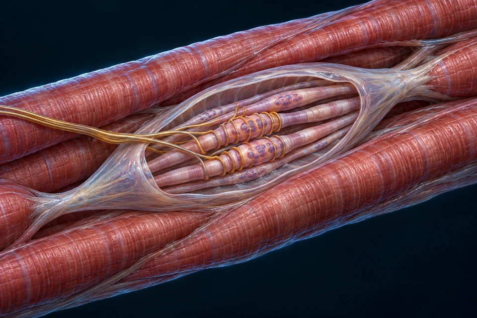 Muscle-position feedback →
The retained spindle teaching preparation adds the sensory tissue missing from a bone-only view.
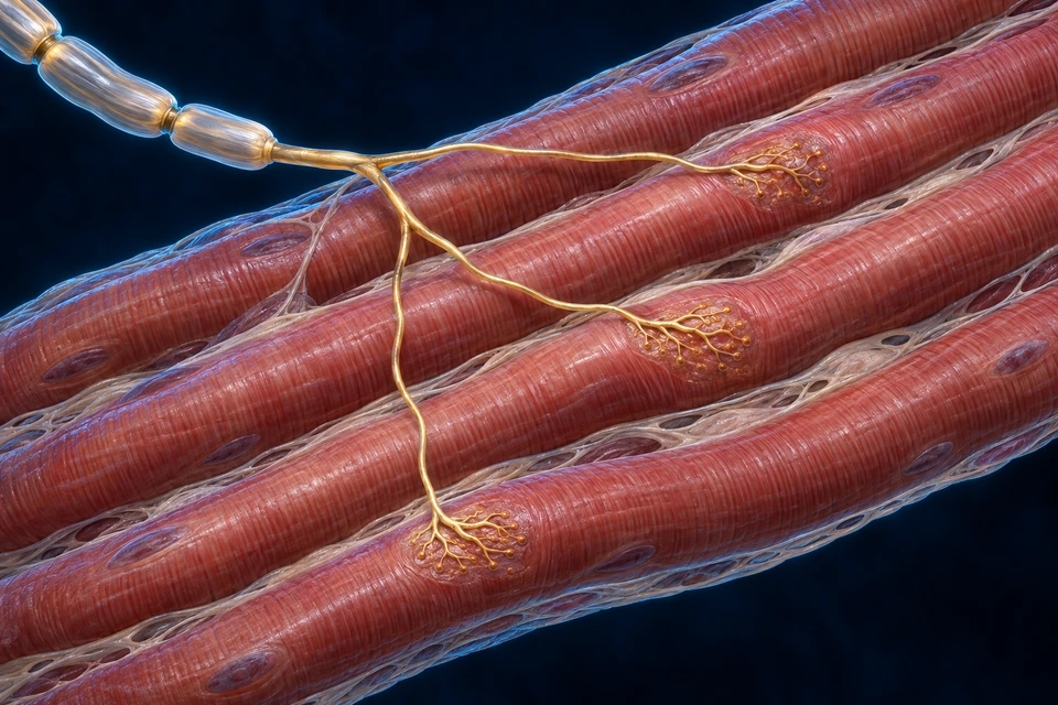 Motor output →
A motor neuron and its muscle fibres provide an output context, distinct from the sensory signals returning from tissues.
Skin deformation → sensory signals
Hairless skin contributes touch and vibration information.
Muscle / tendon receptors → feedback
Different sensors contribute length, change and force context.
Nervous system ↔ motor output ↔ tissues
Goals and multiple feedback sources coordinate movement.
Supporting relationship map, not exact anatomical geometry or a measured time course. All steps are visible before highlighting.
These are complementary sensory and motor contexts, not a reconstructed continuous cable. The selected emphasis does not assert a receptor location in the heel picture.
FOLLOW A QUESTION ACROSS STRUCTURES
A tendon transmits force from a muscle. Many long finger muscle bellies lie in the forearm, while shorter muscles lie within the hand. Following tissue type and attachment is more useful than treating every pale route as a muscle.
The superficial flexor splits and reaches the middle bone. The deeper route continues to the distal bone, crossing another joint on the way. On the dorsal side, the central and terminal extensor destinations also differ; matching endpoints does not make flexor and extensor apparatus identical.
An outline alone cannot establish function or diagnose a foot. Bone and joint relationships, connective tissue deformation, muscle activity and the task all matter. The supported pose and the heel-raised pose show different relationships, not a universal stiffness rule.
Skin information and muscle-position information contribute different feedback. Central circuits combine these with goals, while motor neurons activate muscle fibres. The heel cutaway shows contact tissues; it does not itself expose a complete sensory route.
Illustrations select a teaching view. Imaging and microscopy observe defined signals at specific scales; a rendered pore or coloured area is not observational evidence.
Regional orientation and visible relationships
Makes names and function readable at first glance.
Real signal or sampled tissue
Could add independent normal evidence with clear redistribution rights.
Defined measurements and experiments
Explains which model limits matter.
Teaching references: NLM · Finger tendon relationships and variation · NLM · Primary normal heel-layer observations · NLM · Primary dynamic arch experiment and model limits
This regional foundation covers manipulation, contact and tissue support. It is not a complete named muscle, nerve, vessel or ligament atlas.
Clinical disease, fracture comparison, diagnosis and treatment remain deferred. Fine generated anatomy is illustrative / not independently verified. Human medical review has not been performed.
No full-system completeness percentage, patient-specific anatomy, new geometry or database publication change.
Teaching references: NCBI · Hand bones and normal variation · NCBI · Selected normal foot and ankle anatomy
The existing whole-book inventory guides the hand/foot/arch scope (FB022) and tendon/mechanical connections (FB032). Original regional explanations are retained and reorganised. No book prose, illustrations or page layouts are reproduced. Detailed normal flexor and extensor routes are independently sourced additions; the extensor-hood detail is not claimed as an observed FB022 book illustration.
All main pictures in this reading are generated medical illustrations, not genuine microscopy, clinical imaging or measured mechanical data. Qualified existing Golden-family plates are preserved. The new flexor primary replaces an unreliable old insertion view; the complementary new dorsal extensor primary supplies an anatomical setting for a previously text-only explanation. Essential labels and explanations are separate from the images and remain visible.
Fine generated carpal/tarsal boundaries, ligament attachments, pulleys, individual muscle origins, nerve routes and cellular proportions are not independently established. A complete retinacular or surgical extensor-hood map, named vessels/nerves, authentic imaging examples and clinical comparisons remain outside this normal regional foundation. Human medical review has not been performed. Browser narrow-screen testing is separate from physical-phone acceptance. The older extensor references are used for normal anatomy, not as current treatment guidance.
The early walking comparison is an original text-free atlas preparation with separate labels and native source windows. It shows representative contact conditions, not measured pressure, a prescribed gait, a time series of the same foot or a new clinical test.
Content version: handfoot-eyes-standard-20261017-v1. Original knowledge and source masters remain preserved; the outdated future-visual note is replaced by a precise fine-detail scope. Educational anatomy; not diagnosis, surgery or a treatment programme.