Musculoskeletal / VISUAL EXPLAINER
How Do Cartilage Cells Get Nutrients Without Blood Vessels?
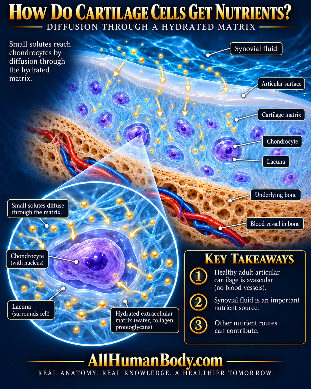
Healthy adult articular cartilage is avascular. Small solutes reach its chondrocytes by diffusion through hydrated matrix; synovial fluid is an important nutrient source at the articular surface, without being the only possible route.
READ THE IMAGE
What the illustration shows
Navy and cyan educational poster showing blue synovial fluid above a smooth pale articular surface. Violet chondrocytes occupy lacunae within luminous blue cartilage matrix. Gold solute dots and short arrows point into the matrix. Ivory underlying bone contains red and blue vessels, with none drawn inside cartilage. A circular enlargement shows a living chondrocyte, its lacuna and hydrated extracellular mesh. Three takeaways identify avascular cartilage, synovial nutrient supply and possible other routes. Gold AllHumanBody.com and complete slogan appear on a plain navy footer.
How to interpret this poster
- No vascular perichondrium covers the articular surface. Bone contribution is not universally zero.
- Cell spacing, mesh, wall layers and bony channels are conceptual, not measured adult-human histology, exact cartilage zones or a tidemark diagram.
- Solute symbols do not give molecular structure, concentration or diffusion depth. No exercise prescription or cartilage-regrowth promise.
- AI-assisted educational illustration; not physician certification, microscopy, a diagnosis or individualized medical advice.
- Fine annotations need enlargement; the full description provides an accessible text alternative.
- Display the full uncropped illustration; do not trim titles, legends or boundary annotations.
AI-assisted educational illustration, adapted into web-sized images without changing its composition. This is general education, not an individual diagnosis or treatment recommendation.
EVIDENCE / CONTEXT
Sources and what they support
- Histology Guide: Cartilage and Bone ↗
Cartilage avascularity and diffusion-based nutrition.
- Medical University of Warsaw: Structure of cartilage and bone ↗
Chondrocytes/lacunae, articular absence of perichondrium and synovial exchange.