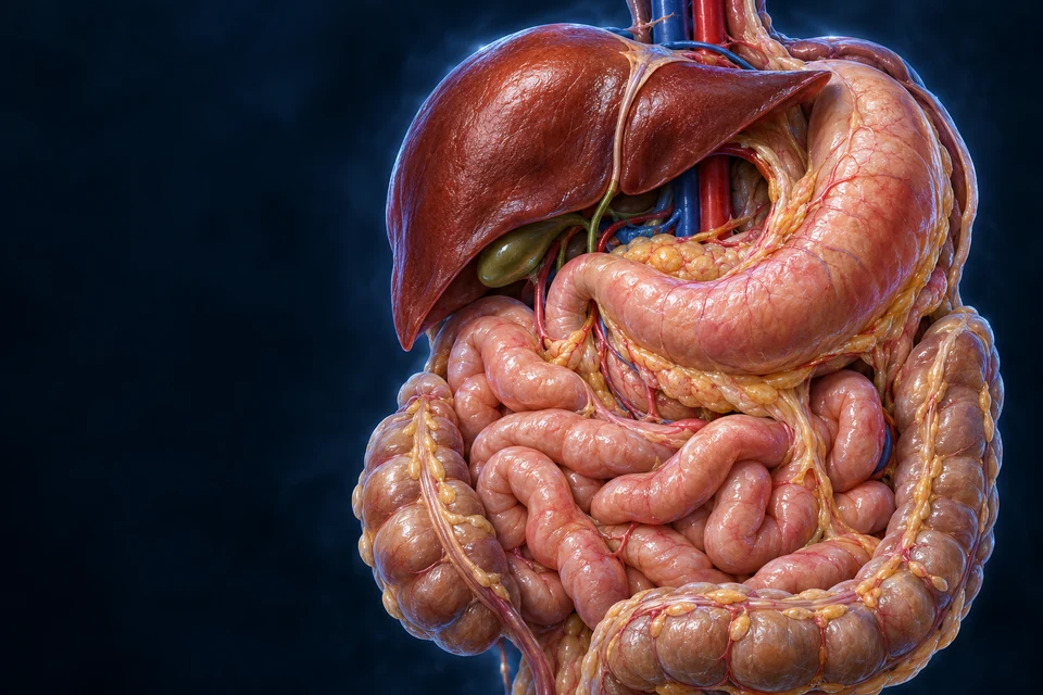
A meal passes through the tract. Secretions join it. Absorbed products leave by different tissue routes. Supporting membranes keep these mobile organs connected.
▷
Watch chapter overviewFilm planned · not yet available
Connected visceral anatomy · fine generated detail illustrative
The tract and its partners occupy different routes.
Abdominal organs share space, but a swallowed bolus does not pass through the liver, pancreas or gallbladder. Their secretions and blood-borne roles connect to the tract by different paths.
New original Golden-family text-free teaching preparation. Expanded regional orientation. Patient right is image left. Selected overlaps are opened for teaching, not a literal single-plane specimen. Hidden junctions, generated vascular detail, exact positions and proportions remain illustrative. Human medical review: Not performed.Structure notes · further context
Esophageal entry
The neck and thoracic journey is taught in Mouth & Swallowing. No sphincter state or full diaphragmatic opening is verified by this static preparation.
Liver
A swallowed bolus does not travel through liver tissue. Bile exits by ducts; venous nutrient delivery is a different route.
Gallbladder
It is not a passage through which all food travels. Its complete duct junction is hidden; this view does not invent a visible connection.
Stomach
Its muscular and glandular mechanisms are explained in the linked Stomach lesson. This view locates it in the whole system.
Pancreas
The gland lies largely behind the stomach. Selective exposure reveals context, not normal unobstructed anterior visibility or exact duct openings.
Duodenal region
Terminal duct arrangements vary. The expanded curved preparation does not establish a measured duodenal plane or hidden luminal continuity.
Small intestine
Jejunum and ileum are not reliably separated by external color. Their regional and cell-scale differences belong to the linked deep lesson.
Colonic frame
The colon surrounds central loops in this expanded view. Real bowel positions vary; generated haustra are not a specimen count.
One luminal journey passes through several regions
The esophageal route reaches the stomach, then the intestine. The liver and pancreas are not extra chambers through which a swallowed bolus passes.
Gold outlines locate broad organ contexts. The ordered route names give direction without inventing a continuous trace through hidden bowel loops or ducts. Oral, thoracic and final pelvic-outlet portions extend beyond this abdominal preparation. The rectum stores material before coordinated anal emptying; the whole route is named above.
The oral and thoracic portions lie beyond this preparation. Regional exposure helps orientation; it does not show every junction, vessel, duct or membrane.
A connected normal-function map, not a claim that all digestion happens in isolated sequential compartments or that every swallowed substance follows identical timing.
The liver produces bile; the gallbladder stores and concentrates it. Bile components aid lipid processing. They are not protein-cleaving enzymes, and the liver is not a food-lumen segment.
Preparing, digesting and absorbing are different jobs.
Movement brings food into contact with secretions and surfaces. Chemical processing changes molecules. A selective epithelial barrier determines what can enter tissue transport.
Food → smaller mixed pieces
Teeth and muscles change size, contact and distribution. This is mechanical preparation, not epithelial uptake.
Coordinated wall movement → onward transfer
Propulsion and mixing have different jobs. Contents can be redistributed locally; the route is not one unbroken gravity-driven fall.
Large food molecules → smaller products
Enzymes cleave suitable substrates. Bile assists lipid processing but is not itself a digestive enzyme.
Lumen → epithelial barrier → tissue transport
Digestion creates usable products; absorption moves selected substances across the lining. Membrane routes differ by substance.
Tissue transport → other organs
Portal blood links intestinal uptake to liver handling. Chylomicron-rich lymph initially reaches systemic blood by a different route.
Remaining contents → stool → coordinated emptying
Colon fluid handling and microbial activity modify residues. Elimination is distinct from renal excretion into urine.
Supporting process guide. Activities overlap and recur; this is not a fixed-duration anatomical animation. All basic explanations remain visible.
Parietal peritoneum lines the abdominal wall; visceral peritoneum covers organ surfaces. Opposed serosal surfaces bound a lubricated potential space, separate from the lumen inside the bowel.
Original generated teaching cutaway, not a patient image or micrograph. The bowel window is deliberately opened for teaching, not a natural perforation. External surfaces are locally separated for visibility: normally the interface contains only a thin lubricating film, not a large reservoir. The bowel lumen is inside its wall and is separate from this external interface. No fine mesothelial cells, membrane thickness, complete peritoneal reflections or exact wall-layer measurements are certified. Human medical review: Not performed.Structure notes · further context
Inside and outside are different compartments
The epithelium of the gut lining faces food and digestive fluid. The mesothelial surface of serosa faces the external lubricated interface. Similar words such as lining or membrane do not make these surfaces one shared channel.
A teaching window is not a normal hole
Removing a local piece of wall lets both compartments be seen at once. It must not be read as a natural opening between bowel lumen and peritoneal space.
A local interface is not a complete cavity atlas
The illustration does not map every reflection, recess, organ attachment or mesothelial cell. Continue to the retained mesenteric fan and omental apron for larger support relationships.
Why is the potential space not the food passage?
The bowel wall encloses a mucosal surface facing contents. Its outer visceral serosa faces the lubricated peritoneal interface. A local window exposes both relationships at once without creating a natural connection between them.
Mesenteric reflections carry connective tissue and supply routes between surfaces. They are not a second alimentary tube. Compare the mesenteric fold with the omental apron, then return to the epithelial uptake boundary.
Blood vessels, lymphatic vessels and nerves travel within the core of a mesenteric fold. The fold is not a pocket through which a swallowed bolus passes.
The free bowel edge can move while its supply remains connected to posterior support.
Supporting tissue relationship only. Not a histological section, scaled cell model or a literal count of all omental layers. The actual mesentery and omentum anatomy is shown in the main plates.
A membrane is living tissue
Mesothelial cells rest on a basal/supporting connective-tissue layer. Their surface participates in fluid and tissue interactions; it is not inert plastic. Fine microvilli and pores are not drawn or claimed verified here.
Behind the lining is another relationship
Kidneys lie behind the posterior peritoneal lining. Much of the pancreas and selected bowel regions also have posterior attachment relationships. Intraperitoneal does not mean food floats freely in a cavity, and retroperitoneal does not mean outside the abdomen.
The bowel can move while its supply stays connected.
A small-bowel mesenteric fold connects jejunal/ileal bowel with posterior support. Vessels, lymphatics and nerves travel within it; the bowel lumen remains a separate channel.
New expanded small-bowel mesenteric preparation. Root orientation, exact vessel branching, fat, nodes, nerves and microscopic sheet detail remain illustrative. Human medical review: Not performed.Structure notes · further context
Root region
The real attachment is oblique and three-dimensional. This spread preparation is not its surgical length, angle or vertebral-level map.
Mesenteric fold
Two surface sheets enclose connective tissue, vessels, lymphatics and nerves. They are not two channels for passing food.
Small-bowel edge
The loop is spread for visibility. Its surface and diameter are illustrative, not a literal isolated complete intestine or a jejunum-versus-ileum diagnostic feature.
Arterial branches
Broad superior-mesenteric supply is source-supported. Every drawn arcade and terminal branch is not independently validated.
Venous branches
Small-bowel venous return contributes to portal blood. Blue vessels do not drain food out of the bowel lumen; exact pairing and junctions are illustrative.
The fold connects a movable bowel edge with deeper support
A mesenteric root region connects with a broad fold reaching the bowel. The food passage remains inside the attached bowel, not inside the fatty sheet.
A focus locates an existing anatomical context. It does not measure vessel flow, prove hidden layer fusion or animate organ movement.
A vascular corridor
Jejunal and ileal arterial branches arise from superior-mesenteric supply, with branching connections toward the wall. Venous drainage contributes to portal return. Exact branch counts, arcades and variations are not established by this generated preparation.
Mobility differs by region
Mesenteric and mesocolic attachments differ around the tract. Transverse and sigmoid colonic support is not identical to the small-bowel fan; posterior relations elsewhere are more fixed. A single spread preparation cannot define every organ’s attachment.
The greater omentum descends from the greater-curvature region and relates to the transverse colon, lying in front of deeper bowel. Soft fat and vessels occupy its folds; their amount and appearance vary.
New original Golden-family text-free teaching preparation. Omental-layer view adds a different spatial relationship, not another complete tract map. The lower edge is lifted for teaching. Fine fat, vessel branches, exact fused layers and concealed attachments remain illustrative. Human medical review: Not performed.Structure notes · further context
Greater omentum
It extends from the greater-curvature/gastrocolic region and relates to the transverse colon. Individual fat amount and folding vary; it is not the small-bowel mesentery.
Stomach region
The greater curvature is related to the omental origin. This cropped plate is not an exhaustive gastric ligament or vessel map.
Transverse-colon region
Omental folds and attachment/fusion can obscure the full segment. Exact layer fusion and lumen route are not asserted from the generated view.
Bowel behind the apron
Omentum normally lies superficial to these loops. The lifted preparation must not be mistaken for a freely detached membrane.
Liver context
The lesser omentum linking liver and lesser gastric/proximal-duodenal regions is not directly exposed here; its omission does not imply absence.
Lifted tissue reveals bowel behind the covering
The greater omentum hangs anterior to deeper loops. A lifted lower edge reveals this front-to-back relationship; it is not a freely detached sheet.
A focus locates an existing anatomical context. It does not measure vessel flow, prove hidden layer fusion or animate organ movement.
Greater and lesser are different connections
The lesser omentum links liver with the lesser gastric/proximal-duodenal region. It is not an identical second apron. Its hidden connections are stated from sources, not invented from this view.
Not every concealed space is shown
The lesser sac lies behind the stomach and communicates with the larger peritoneal space. This plate does not establish its complete boundaries, foraminal relations, embryonic rotation or precise layer fusion. Those require a dedicated spatial lesson.
The blood/lymph distinction connects digestive uptake with liver handling and systemic delivery. It does not change the fact that all these substances first cross selected epithelial routes.
Selected context: epithelial entry and tissue blood
The existing qualified Small Intestine villus preparation is reused, not regenerated. Epithelial lining, tissue blood vessels and a central lacteal occupy different compartments. The tissue highlights locate entry contexts, not complete portal or thoracic-duct trajectories. Luminal residue remains outside the epithelial barrier. Fine generated cell identity, vessel junctions and relative scale remain illustrative; not acquired microscopy. Human medical review: Not performed.Structure notes · further context
Villus
Its core is supporting connective tissue, not an open digestive pipe.
Epithelial lining
Absorptive cells have apical microvilli. Junctions regulate passage between neighboring cells; the barrier is selective, not impermeable.
Lamina propria
It also contains immune and other supporting cells. A generated nucleus cannot establish a particular cell identity.
Blood vessels
The capillary network connects with larger vessels. Exact branch paths and red/blue vessel identities are illustrative.
Lacteal
Its tip is closed. It does not open into the lumen, and it does not carry every kind of absorbed nutrient.
Intestinal crypt
Crypts lie between villi. Stem-cell renewal and Paneth-cell roles are explained separately rather than guessed from generated cell colors.
Gold outlines locate tissue contexts, not particles, measured flow or a complete vessel network. For residues, the marked lining is the boundary: the residue is outside it, not inside the villus.
Why are blood and lymph different exits from the same lining?
The epithelial barrier comes before either tissue route. Many sugars and amino acids enter tissue blood and reach the liver through portal circulation. Many long-chain dietary lipids are packaged into chylomicrons and enter lacteals; these particles initially reach systemic venous blood through lymphatic return.
Different lipid products can use other routes, so the contrast is not a universal rule that every fat enters lymph. Neither tissue exit carries an intact swallowed meal. Continue into epithelial transport, portal handling or intestinal lymphatic return.
Many long-chain dietary lipids leave in chylomicrons. Newly secreted chylomicrons do not first enter the liver through portal blood. This is not a claim that every lipid uses only lymph.
Unabsorbed material, microbes and shed cells contribute to stool. Microbial products can also be absorbed; residue handling is not a second kidney.
Supporting destinations, not a scale drawing, complete metabolic network or measured meal transit. The routes remain visible; buttons emphasize a comparison.
Local neural circuits, signals to and from the central nervous system, and endocrine messages coordinate secretion and movement. They are cooperating influences, not a single brain-controlled on/off switch.
Before and during preparation
Sensory cues and oral handling can recruit secretory responses before nutrients are absorbed. Autonomic influence does not turn every organ into the same mechanical program.
Acid and nutrient arrival influence pancreatic secretion, biliary delivery and gastric emptying. Gut hormone and neural pathways connect the region to broader metabolic control.
Regional hormone/cell identities and mechanisms remain in the linked lessons. This supporting comparison avoids claiming every reflex, peptide or neural pathway is fully mapped.
Intestinal signals and pancreatic hormone responses help coordinate a meal with fuel use. The endocrine bloodstream route differs from digestive enzyme secretion.
Detailed serosal microscopy, complete peritoneal recesses/ligaments, developmental rotation and canonical CMS/atlas mapping remain unasserted. Clinical imaging, disorders, diagnosis and treatment stay in the later disease phase. No copied tissue specimen or patient scan is presented.
Tract versus accessory outputs, distinct digestive activities, broad regional anatomy, peritoneal surfaces and fold support, mesenteric supply, omental relationships, blood/lymph destinations and linked neural/endocrine coordination. Sources support teaching relationships, not every rendered pixel.
Explicit limits
AI-assisted visual/content production. Generated exact vessel branches, membrane thickness, fat, hidden connections, cells and organ proportions remain illustrative / not independently verified. Human medical review: not performed. Educational purpose only; not a surgical map, clinical guide or exhaustive digestive atlas.
English revision: digestive-system-english-2026-10-18-eyes-foundation-v1. Chapter identity: ahb_ch_digestive_system_english_v1. No canonical digestive CMS entity or chapter-specific3D is asserted. Migration physical-phone acceptance: Pending, non-blocking. Four retained qualified text-free masters and18 original independent labels remain in use. One new serosal-interface preparation adds eight semantic labels; the qualified Small Intestine villus and its six labels are reused for tissue destinations. Anatomy, language copy and focus marks remain separate. Supporting mechanisms remain editable English code; fine generated vessel branches, membrane thickness, fat and hidden connections remain illustrative / not independently verified.